Received from Hospital São Carlos, Fortaleza, CE, Brazil.
1. MD; Clinical Director ofHospital São Lucas
2. Anesthesiologist; Member of the Câmara Técnica de Anestesiologia do CRM/RS 3. Responsible for CET of Instituto Dr. José Frota (IJF), Fortaleza; Scientific Director of SAEC
4. R3 Anesthesiologist, IJF, Fortaleza 5. Medical Undergraduate Student
Submitted on September 10, 2010. Approved on November 23, 2011.
Correspondence to: Dr. Rogean Rodrigues Nunes
Avenida Comendador Francisco Ângelo, 1185, casa Dunas
60181500 – Fortaleza, CE, Brazil E-mail: [email protected] SCIENTIFIC ARTICLE
Influence of Total Intravenous Anesthesia, Entropy and
Laparoscopy on Oxidative Stress
Rogean Rodrigues Nunes
1, Fernando Squeff Nora
2, Danielle Maia Holanda Dumaresq
3,
Rute Maria Araújo Cavalcante
4, Amanda Antunes Costa
5,
Lara Moreira Mendes Carneiro
5,
Julio Cesar Garcia de Alencar
5,Flávia Pereira Fernandes Cardoso
5Summary: Nunes RR, Nora FS, Dumaresq DMH, Cavalcante RMA, Costa AA, Carneiro LMM, Alencar JCG – Influence of Total Intravenous Anesthesia, Entropy, and Laparoscopy on Oxidative Stress.
Background and objectives: Recent studies have correlated postoperative mortality with anesthetic mortality, especially with the depth of anes-thesia and systolic blood pressure (SBP). The aim of this study is to evaluate the effects of the depth of total intravenous anesanes-thesia (TIVA) using remifentanil and propofol, performed with monitoring of response entropy (RE) on blood concentrations of oxidative stress markers (TBARS and glutathione) during laparoscopic operations.
Method: Twenty adult patients, ASA I, BMI 20-26 kg.m-2, aged 20 to 40 years, were randomly distributed into two groups: Group I underwent anesthetic-surgical procedure with RE maintained between 45 and 59, and Group II underwent anesthetic-surgical procedure with RE between 30 and 44. In both groups, the remifentanil and propofol infusion was controlled by the effector site (Es), adjusted to maintain RE desired values (Groups I and II) and always assessing the suppression rate (SR). Patients were evaluated in six periods: M1 (immediately before anesthesia), M2 (before tracheal intubation [TI]), M3 (5-minutes after TI), M4 (immediately before pneumoperitoneum [PPT]), M5 (1-minute after PPT), and M6 (1-hour after the operation). The following parameters were assessed at all times: SBP, DBP, HR, RE, SR, TBARS, and glutathione.
Results: We found increases in TBARS and glutathione in M5, both in Group I and Group II (p < 0.05), with higher values in Group II, and SR in three patients in Group II, immediately after PPT.
Conclusions: Increased markers in Group I (M5) suggests an increase in anaerobic metabolism (AM) in the splanchnic circulation while the highest values seen in Group II (GII > GI in M5, p < 0.05%) suggest interference of another factor (deep anesthesia) responsible for the increase in AM, probably as a result of increased autonomic nervous system depression and minor splanchnic self-regulation.
Keywords: Entropy; Intravenous Anesthesia; Laparoscopy; Oxidative Stress.
©2012 Elsevier Editora Ltda. All rights reserved.
INTRODUCTION
Recent studies suggest that postoperative mortality may be influenced by anesthetic management during surgery, particu-larly control of anesthetic depth and systolic blood pressure. Major surgeries, severe trauma, sepsis, respiratory distress syndrome, ischemia and reperfusion (laparoscopic proce-dures) are important components of acute inflammation and
represent a new challenge for anesthesiologists when manag-ing drugs and anesthetic techniques 1. Many animal and human
studies have shown a decrease in endogenous antioxidants in oxidative stress conditions (oxidant and antioxidant imbalance favoring the former, i.e., a chemical-biological state in which production of reactive oxygen species exceeds the antioxidant capacity), particularly in ischemia-reperfusion injury and, more recently, systemic inflammatory response syndrome (SIRS). Excessive production of reactive oxygen species is one of the mechanisms founded in the pathogenesis of inflamma-tory reactions in response to trauma, surgery, sepsis, organ transplants, burns, and ischemia and reperfusion (I/R) 2,3. In
aerobic systems, the balance between oxide-reducing agents (such as ROS) and antioxidant defense system is essential 3-5.
These agents are endogenously generated as a direct result of O2 metabolism, and in non-physiological conditions such as
cell exposure to xenobiotics that cause incomplete reduction of O2. For protection, the cell has a two-line defense system.
vitamin E, a membrane structural antioxidant, the antioxidants are mostly located intracellularly. Other molecules, such as al-pha and beta-carotene, ubiquinol, and cysteine, also remove free radicals 4,6. This study aim was to evaluate the effects of
pneumoperitoneum (ischemia-reperfusion injury) on oxidative stress and lipid peroxidation and the influence of adequate an-esthesia with total intravenous anan-esthesia (TIVA), monitored by EEG on in vivo concentrations of oxidative stress markers and lipid peroxidation (glutathione and TBARS).
METHOD
A prospective, randomized study, conducted after approval by the Clinical Research Ethics Committee and signing of in-formed consent.
Case report
We evaluated 20 female patients who underwent videolaparo-scopic operations for oophorectomy or myomectomy, physical status ASA I, aged between 20 and 50 years, and body mass index between 22 and 26. Patients were randomly assigned into two groups of 10 individuals before induction of anesthe-sia: Group I (more superficial anesthesia), TIVA to maintain response entropy (RE) between 45 and 59. Group II (deeper anesthesia), TIVA to maintain RE between 30 and 44.
Equipment used
1) Entropy Module; EEG signal is collected from the fron-totemporal region and treated by Shannon’s equation 7
(H = - pk log pk), where pk is the probability of a
dis-crete k event, resulting in two types of analyzes: a) state entropy (SE), which is the evaluation of cerebral cortex electrical activity (0.8-32 Hz); and b) response entropy (RE), which analyzes frequencies from 0.8 to 47 Hz (containing both cortical and subcortical EEG components). The frontal muscles activation may indi-cate inadequate subcortical component (bulb-pontine region). This way, there is the possibility of assessing anesthetic depth with a mixed-index; however, the time window of response entropy is lower than the state entropy, allowing a more rapid adjustment of anesthetic components 8,9. In addition to these data,
the equipment also evaluates the presence of burst-suppression (BS), which may be indicative of cerebral hypoperfusion in the absence of other factors, such as deep hypothermia, too deep anesthesia or both.
The assembly used was unilateral referencial, with the ex-plorer electrode positioned at FT10 (frontotemporal region) and the reference electrode at Fpz (frontopolar) (Figure 1). This de-termines that the EEG tracing obtained is single channel (left or right, depending on the frontotemporal electrode position).
2) Infusion pump with control target (plasma and effector site) for remifentanil and propofol using the following pharmacokinetic models: Minto for remifentanil and Marsh for propofol; 3) Two channels electrocardiogram – DII and V5; 4) Pulse oximetry; 5) Capnography and capnometry; 6) Automatic non-invasive blood pres-sure; 7) Air heater forced convection heat; 8) Specific material for plasma markers collection; 9) Thermom-eter with nasopharyngeal sensor.
Preoperative evaluation
All patients underwent clinical and laboratory evaluation pre-operatively.
Anesthesia technique
None of the patients received premedication. All patients were subjected to the effects of the same surgical anesthetic and technique, maintaining a pneumoperitoneum pressure of 12 mm Hg and flow of 3 mL.min-1. After venipuncture in the
right upper limb, all patients received an infusion of saline so-lution NaCl 0.9% (2 mL.kg-1 for fast replacement and 6 mL.kg-1
for replacement of losses during surgery). Anesthesia was induced with concurrent intravenous propofol infusions per-formed with the aid of infusion pumps controlled by the effec-tor site (Es), with initial target of 4 µg.mL-1 and remifentanil,
Es, with initial target of 4 ng.mL-1 until the response entropy
(RE) value reached 40. If RE did not reach 40, the Es con-centration of remifentanil would be increased by 0.5 ng.mL-1
until achieving a RE of 40. At this point, the Es concentration of remifentanil would be fixed and orotracheal intubation
formed (OTI). In Group I, propofol and remifentanil infusions were adjusted to maintain a RE of 45-59. In Group II, infu-sions of propofol and remifentanil were adjusted to maintain a RE of 30-44. In both groups, pre-oxygenation with 100% oxygen was performed under mask 5 minutes before induc-tion of anesthesia until immediately before tracheal intubainduc-tion. Neuromuscular block was not used because it may interfere with the processed EEG values 10. Management of
anesthe-sia maintenance was performed according to the guidelines specified in Chart 1.
However, RE values within specified limits (Groups I and II) were adjusted as follows: determining the degree of hypnosis by RE and assessing the analgesia by systolic blood pres-sure (SBP), which should not exceed 20% of baseline value (M1) for the upper limit, nor fall below 80 mmHg for the lower
limit. Thus, after OTI, propofol was titrated in concentrations sufficient to maintain the specified values of RE for Groups I and II, and remifentanil was adjusted according to the concen-tration required to maintain PAS within the variations specified in this protocol. Changes in propofol concentrations were per-formed by 0.5 µg.mL-1, with subsequent variations performed
only after achieving the concentration at the effector site, pro-vided by the infusion pump. The same procedure was applied to remifentanil, with variations by 0.1 ng.mL-1. Heart rate with
variations greater than ± 25% from baseline (M1) were con-sidered clinically significant. After tracheal intubation, respira-tory rate was adjusted to maintain PETCO2 between 35 and 40
mmHg, FiO2 35%, with a tidal volume of 8 mL.kg-1. Ventilation
was performed in a circle system with CO2 reabsorber.
1. Assess stimulus level 2. Check anesthetic equipment 3. Consider increasing hypnotic 4. Consider increasing analgesic 5. Consider anti-hypertensive
1. Assess neuromuscular blockade 2. Assess stimulus level
3. Consider increasing analgesic 4. Consider anti-hypertensive
1. Consider anti-hypertensive 2. Assess stimulus level 3. Consider reducing hypnotic 4. Consider increasing analgesic
1. Assess stimulus level 2. Consider increasing hypnotic 3. Consider increasing analgesic
1. Monitoring
1. Consider reducing hypnotic 2. Consider reducing analgesic
1. Consider hemodynamic support 2. Consider increasing hypnotic 3. Consider reducing analgesic
1. Assess other causes
2. Consider hemodynamic support
1. Consider reducing hypnotic 2. Consider reducing analgesic 3. Consider hemodynamic support 4. Assess other causes
High
High
High Desirable
Desirable
Desirable Low
Low
Low Deep
Adequate Superficial
Clinical signs Clinical profile Index* (EEG-based) Strategy
instability
* Always evaluate suppression rate: if non-zero, treat causes.
For clinical study purposes and statistical analysis, six times were assessed: M1, admission to the surgical center; M2, im-mediately before tracheal intubation; M3, 5-minute after tra-cheal intubation; M4, immediately before pneumoperitoneum installation; M5, 1-minute after the end of pneumoperitoneum; and M6, 1-hour after the operation. At each time, the following variables were recorded: systolic and diastolic blood pressure, heart rate, hemoglobin peripheral saturation, expired carbon dioxide concentration, and nasopharyngeal temperature. Plas-ma Plas-markers of oxidative stress (TBARS and glutathione) were determined in venous blood samples at the following times: M1, M2, M3, M4, M5, and M6. The awakening time was considered as the time elapsed from cessation of anesthetic agents to RE greater than or equal to 90. The duration of the operation was considered as the time elapsed from skin incision to the end of the dressing and the duration of anesthesia from the start of remifentanil infusion until extubation.
In all patients, the nasopharyngeal temperature was main-tained between 35°-36°C with the aid of forced-air convection thermal blanket. The maximum value for Trendelenburg posi-tion was considered at 15°. EEG data were measured using a specific device (processed EEG), with sensor recommended by the manufacturer at the following points: FT10 (signal cap-ture of frontotemporal region), FP2 (artifact elimination), and Fpz (reference), coupled to an analog-digital converter. Data were computed after impedance test performed by the device and subsequent reading. At the end of the procedure, the same respiratory rate was maintained and administrations of remifen-tanil and propofol interrupted. We also evaluated the time of hospital discharge, interval between tracheal extubation, and satisfactory Romberg, with the latter performed by asking the patient to remain standing up, still, with feet close together, and eyes closed. The test is considered satisfactory when the pa-tient can maintain this posture for one minute. This test was performed every 15 minutes, starting 10 minutes after the pa-tient could maintain the sitting position without assistance.
Venous blood collection for plasma markers measurement
Samples were collected by venous catheter (cephalic vein), using 10 mL disposable syringe by the method of two syringes.
Determination of the concentration of thiobarbituric acid reactive substances (TBARS)
The evaluation of lipid peroxidation (indicating cell damage) was performed by reaction with thiobarbituric acid.
Determination of glutathione concentration
The determination of glutathione concentration is based on the reaction of 2-nitrobenzoic acid with the free thiol, yielding a mixed disulfide plus 2-nitro-5-thiobenzoic acid. Measurement of the product reaction formation is performed in a Beckman spectrophotometer by reading the absorbance at 412 nm.
Evaluation criteria
The data obtained from hemodynamic, autonomic, pharmaco-dynamic, metabolic or oxidative parameters were compared both between times in the same group and between groups at equivalent times.
Statistical analysis
We used the analysis of variance according to the repeated measures model with two factors of classification (group and time) and Tukey’s test for comparison of measurements of time within the group and group within time, considering as statistically significant p < 0.05.
RESULTS
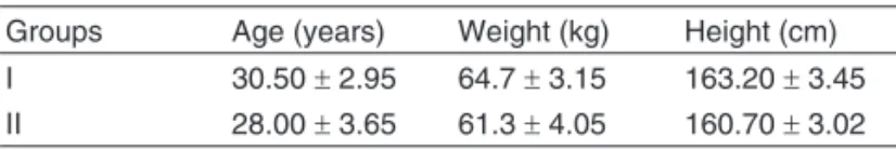
Both groups were homogeneous regarding age, BMI, physical condition, height (Table I) and duration of pneumoperitoneum (Table II). Duration of anesthesia in Group I was 118.00 ± 6.10 minutes and in Group II 114.00 ± 5.22 minutes (p > 0.05). Duration of operation in Group I was 82.80 ± 4.81 minutes and 75.40 ± 7.20 minutes in Group II (p < 0.05). Awakening time in Group I was 7.70 ± 1.24 minutes and 10.20 ± 0.90 minutes in Group II (p < 0.05) (Table III). All patients could be extubated immediately after awakening. The time of hos-pital discharge was 362.40 ± 14.80 minutes in Group I and 430.50 ± 17.81 minutes in Group II (p < 0.05), values con-siderednot clinically relevant (Table III). SBP, DBP, and HR values are shown in Table IV and did not exceed the limits established in the protocol.
Regarding ER, the values at M1 and M6 are significantly dif-ferent from M2, M3, M4, and M5, both in Group I and Group II (p < 0.05) (Table V). As for intergroup assessment, there is significant difference between measurements regarding the in-teraction effect of time-groups for significance levels less than 5% at the following times: M2, M3, M4, and M5 (Figure 2).
Regarding suppression rate, which in this study represents deleterious cellular changes in CNS, there were three cases in Group II immediately after pneumoperitoneum.
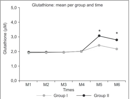
Analysis of oxidative stress markers showed, with respect to glutathione, significant differences between M4 and M5, comparing the GI and GII alone. Analysis of groups within each time showed significant different at M5 and M6 (p < 0.05) (Table VI, Figure 3).
Regarding TBARS variable, which is related to lipid per-oxidation, there was a significant increase in both Group I and Group II between M4 and M5, with the highest values recorded in Group II, and analysis of both groups within each time showed significant differences at M5 and M6, with higher values seen in Group II (Table VII, Figure 4).
Table II – Duration of Pneumoperitoneum by Group (Mean ± SD)
Pneumoperitoneum Groups N Minimum Maximum Mean ± SD
Duration (min) I 10 60 70 61.90 ± 2.50
II 10 56 64 62.40 ± 3.32
Table III – Duration of Anesthesia, Duration of Operation, Awakening Time, and Time of Hospital Discharge by Group (Mean ± SD) Groups Duration of Anesthesia
(min)
Duration of Operation (min)
Awakening Time (min)
Time of Hospital Discharge (min)
I 118.00 ± 6.10 82.80 ± 4.81 7.70 ± 1.24 362.40 ± 14.80
II 114.00 ± 5.22 75.40 ± 7.20* 10.20 ± 0.90* 430.50 ± 17.81*
* p < 0.05.
Table IV – Heart Rate (HR), Systolic Blood Pressure (SBP), and Diastolic Blood Pressure (DBP) by Group (Mean ± SD)
Time Groups HR SBP DBP
Mean ± SD (beats.min-1) Mean ± SD (mm Hg) Mean ± SD (mm Hg)
M1 I 77.00 ± 7.13 110.70 ±9.24 75.70 ± 4.32
II 73.70 ± 7.07 111.90 ±5.07 74.20 ± 3.79
M2 I 66.70 ± 4.47 94.30 ± 5.21 61.00 ± 4.27
II 63.00 ± 3.97 95.80 ± 8.48 67.00 ± 3.46
M3 I 69.10 ± 2.77 96.00 ± 9.35 69.00 ± 2.98
II 65.40 ± 2.46 97.60 ± 8.97 65.80 ± 4.21
M4 I 67.30 ± 4.21 95.20 ± 7.76 61.00 ± 3.09
II 62.30 ± 3.47 100.30 ± 8.64 62.60 ± 3.81
M5 I 71.10 ± 5.51 104.00 ± 9.64 71.60 ± 3.84
II 63.40 ± 3.92 106.20 ± 10.08 70.70 ± 2.98
M6 I 79.90 ± 3.84 114.90 ± 7.40 79.60 ± 3.95
II 82.80 ± 7.33 111.40 ± 8.17 78.50 ± 3.27
Figure 2 – RE and its Evaluation between Times.
* p < 0.05 for M2-M5, between GI and GII. RE: response entropy.
Group I Group II
M1 M2 M3 M4 M5 M6
Times
RE: mean per group and time
*
*
*
*
120
100
80
60
40
20
0 RE Table V – Electroencephalogram: RE (Mean ± SD)
Groups Time N Mean ± SD
M1 10 96.60 ± 3.80*
M2 10 52.20 ± 2.10
I M3 10 53.40 ± 1.90
M4 10 55.70 ± 2.30
M5 10 52.40 ± 2.60
M6 10 94.70 ± 3.50*
M1 10 98.20 ± 3.40*
M2 10 36.20 ± 3.60
II M3 10 38.40 ± 3.40
M4 10 36.10 ± 3.90
M5 10 39.30 ± 2.30
M6 10 96.90 ± 2.70*
DISCUSSION
The metabolic response to trauma is an increasingly impor-tant issue when general anesthesia is studied as a predictor factor of postoperative outcome. The predictors of intraopera-tive morbidity and mortality are divided into three categories: predictors related to the associated comorbidities, predictors attributable to the operation itself, and predictors associated with the anesthetic 11. Currently, the risk of anesthesia
dur-ing the immediate perioperative period seems to be rather small 12. However, little is known about the effects of
anes-thetic on long-term results. Although no lasting benefit has been linked to specific anesthetic so far, studies suggest that local anesthesia may improve survival in certain patient groups 13. Monitoring anesthetic depth is now possible thanks
to the use of EEG digital signal processing techniques 14.
Al-though no technology, including pulse oximetry, has proven to reduce mortality, it has been suggested that anesthetic depth monitoring allows the use of exact dosages of anes-thetics and thus to reduce the cardiovascular effects arising from overdoses 15. A study by Monk et al. 16 suggests that
intraoperative anesthetic management, particularly depth and blood pressure control, may influence the mortality assessed in up to one year. The independent association of cumula-tive time of deep anesthesia with mortality in one year was a new finding in this study. Lindholm et al. 17 evaluated data
from bispectral index to assess the effects of monitoring on incidence of intraoperative awakening and showed that a cumulative BIS time of less than 45 was associated with an increased risk of death in evaluations up to two years after operation.Another recent study, analyzing long-term mortal-ity, showed that the absence of a BIS less than 40 (deep
an-Table VI – Glutathione Variable (Mean ± SD)
Groups Time N Mean ± SD
M1 10 1.90 ± 0.15
M2 10 1.93 ± 0.14
I M3 10 1.94 ± 0.11
M4 10 2.02 ± 0.08
M5 10 2.42 ± 0.14*
M6 10 2.18 ± 0.05
M1 10 1.94 ± 0.14
M2 10 1.96 ± 0.14
II M3 10 1.94 ± 0.10
M4 10 2.00 ± 0.08
M5 10 3.07 ± 0.19*
M6 10 2.80 ± 0.18
* p < 0.05. Intragroup assessment.
Figure 3 – Glutathione: Intergroup Analysis. *p < 0.05 for M5 and M6, between GI and GII.
5,0
Glutathione (
µ
M)
4,0
3,0
2,0
1,0
0,0
Group I Group II
M1 M2 M3 M4 M5 M6
Times
Glutathione: mean per group and time
*
*
Tabela VII – TBARS Variable (Mean ± SD)
Groups Time N Mean ± SD
M1 10 0.02 ± 0.01
M2 10 0.02 ± 0.01
I M3 10 0.02 ± 0.01
M4 10 0.03 ± 0.01
M5 10 1.66 ± 0.46*
M6 10 0.72 ± 0.10
M1 10 0.02 ± 0.01
M2 10 0.02 ± 0.01
II M3 10 0.02 ± 0.01
M4 10 0.02 ± 0.01
M5 10 5.10 ± 0.68*
M6 10 2.60 ± 0.60
* p < 0.05. Intragroup assessment.
Figure 4 – TBARS: Intergroup Analysis. *p < 0.05 for M5 and M6, between GI and GII.
6.0
TBARS (nmol/mL)
5.0
4.0
3.0
2.0
1.0
0.0
M1 M2 M3 M4 M5 M6
Times
Group I Group II
*
*
esthesia) was associated with improved survival and reduced morbidity 18. A study of comatose patients with ischemic
en-cephalopathy who underwent emergency operation showed that data collected from BIS were better predictors than clini-cal judgment in identifying patients with a good chance of recovery 19. Monitoring the depth of anesthesia using EEG
produces data clinically useful, as routine practice results in large variation in anesthetic dose and patient’s response 20.
Elderly patients or patients with several comorbidities require lower anesthetic doses than younger and healthier patients, which can be detected by EEG monitoring 21. Previous studies
probably failed to detect the effects of general anesthesia on long-term outcome because they focused on the type of anes-thesia administered rather on quantity or effect of anesthetics on brain. Even when inhaled or total intravenous anesthesia is compared, it is necessary to maintain doses and concentra-tions of both agents equivalent to have the same anesthetic depth with both techniques. EEG monitoring is a non-invasive method that uses a specific algorithm that has a direct cor-relation with the adequacy or depth of anesthesia. Although universally used, the clinical signs to evaluate this adequacy are not reliable 22. Clinical signs, such as blood pressure and
heart rate, have widely dispersed probability indices to diag-nose the possible superficiality of general anesthesia. Clinical signs are sensitive but less specific (Pk ranging from 0.6 to 0.9), which can result in intraoperative awakening even in the absence of tachycardia and hypertension. Therefore, several equipments have emerged aiming at improving the manage-ment of intraoperative anesthetic drugs, some of them directly measuring cerebral cortical activity (hypnosis), subcortical ac-tivity, or both 23. Taken into account that anesthetics and
anes-thetic techniques themselves are able to change significantly cytokine profiles 16,24, the findings of an association between
the depth and duration of anesthesia and mortality in one year raises the plausible hypothesis of a process mediated by cy-tokines. It is possible that prolonged deep anesthesia change the inflammatory response in high-risk patients, predisposing them to adverse outcomes. Glantzounis et al. 25 found that free
radicals are generated at the end of laparoscopic procedures possibly as a result of the ischemia-reperfusion phenomenon induced by inflation and deflation of pneumoperitoneum. How-ever, patients in this study had a normal postoperative period, despite increased liver enzymes and lipid peroxidation. Other studies found a lower inflammatory response in videolaparo-scopic operations compared to conventional colorectal opera-tions 26,27. Although a minor surgical trauma, hemodynamic
changes determined by pneumoperitoneum involve both car-diac output and splanchnic perfusion, mainly intra-abdominal pressure over 15 mm Hg 28,29. Videolaparoscopy procedures
then became models of ischemia-reperfusion, where pneumo-peritoneum installed during laparoscopy produces a condition similar to abdominal compartment syndrome 30. In healthy
pa-tients, increased intra-abdominal pressure of 10 to 15 mm Hg decreases the stomach blood flow by 54%, jejunum by 32%, colon by 44%, liver by 39%, parietal peritoneum by 60%, and duodenum by 11%. Splanchnic blood flow typically represents 29% of the cardiac output and remains reduced throughout the
inflation time 31. Perfusion of splanchnic system is performed
through three major arteries (celiac artery and superior and inferior mesenteric arteries) and have a wide distribution of adrenergic receptors.
Experimentally, changes in visceral blood flow occur rapidly, even with moderate increases in intra-abdominal pressure 32.
Gastric mucosal hypoperfusion may persist during postopera-tive period because the recovery of splanchnic circulation trig-gered by the effects of pneumoperitoneum occurs more slowly than the systemic circulation 33. Decreased intestinal
perfu-sion is proportional to the values of intra-abdominal pressure. Reduction of splanchnic circulation perfusion is measured by mechanical compression of mesenteric vessels and vaso-constriction secondary to vasopressin secretion 34. Increased
intra-abdominal pressure and decreased blood flow may dam-age endothelial and Kupffer cells, while the head-up tilt posi-tion is related to decrease in total hepatic blood flow 35. To
avoid significant decreases in liver and splanchnic blood flow, intra-abdominal pressure should be maintained between 8 and 10 mmHg and even smaller values than these 31. Studies
relat-ing the depth of anesthesia based on EEG-ER and oxidative stress markers were not found in literature. However, Shimo-gai et al. 36 described a significant reduction in BIS value when
there was deflation of a pneumatic tourniquet on the lower limb of patients undergoing orthopedic surgery, speculating that the transient reduction of BIS may have been caused by di-rect toxicity of substances, such as hypoxanthine, produced during ischemia. In the present study, we found in Group II episodes of suppression in three patients, which may reflect direct cellular damage from substances released after pneu-moperitoneum (PPN) deflation, in glial cells, and oligodendro-cytes 37. These neural cells require antioxidant defenses to
cope with the continuous supply of reactive oxygen species (ROS) produced in the brain during aerobic metabolism. Oxi-dative stress is greatly increased during anaerobic respiration that occurs during ischemia and reperfusion. In culture, oligo-dendrocytes seem particularly sensitive to oxidative stress 37.
Some of the reasons proposed for the high vulnerability of oli-godendrocytes, compared to other cells, include high lipid and iron content and reduction of antioxidant enzymes or limited substrates 38. Thorburne et al. 39 found that oligodendrocytes
have low stocks of glutathione, which may have reflected the presence of suppression rate in Group II of our study, with no hemodynamic changes outside the clinically accepted stan-dard. For this study, only the depth of anesthesia remained altered, which resulted in increases in blood markers of oxida-tive stress (TBARS and glutathione). These increases were statistically more significant in Group II (M5), influenced by the adequacy of anesthesia in this model of ischemia-reperfu-sion. Studies show that the autonomic nervous system activ-ity is dose-dependent depressed by inhaled anesthetics 40,41.
Therefore, because autonomic nervous system is significantly modulated by the baroreceptor system (reflex mechanisms), the effects of inhaled anesthetics on efferent system also de-pend on the baroreflex arch integrity. Halothane, enflurane, and isoflurane 42,43 depress the baroreflex control of heart rate
hav-ing the least effect. Similar responses in reflex control of heart rate have been shown with sevoflurane and isoflurane 42,44,45.
It has been observed dose-dependent depression of the reflex control of sympathetic outflow relatively equivalent for sevoflu-rane, isoflusevoflu-rane, and desflurane. Anesthesia with more super-ficial levels, i.e., 0.5 MAC has little effect on baroreceptor reflex function and this may be very important in patients with com-promised clinical state. Opioids and benzodiazepines have only minimal effects on baroreceptor reflex function, which combined with low concentrations of inhaled agents may pre-serve these reflexes 46,47. In the present study, with the use
of TIVA (propofol and remifentanil) in both groups, there was significant increase of oxidative stress markers in Group II after pneumoperitoneum, showing that deeper anesthesia contrib-uted to a greater oxidative stress or to a lesser control of it. In-hibition of the baroreceptor reflex activity occurs as a result of central nervous system depression, associated with changes in the afferent impulses, weakening of the efferent autonomic nervous system activity, reductions in ganglionic transmission, and responses of target organs. In this case, we observed as a complicating factor both the installation of pneumoperito-neum, with increased oxidative markers in Group I, and deep anesthesia, as in Group II showed significant increases during M5 compared to Group I at same time, showing that in this model of ischemia-reperfusion, deep anesthesia further con-tributed to exacerbate oxidative stress. According to Olshan et al. 48 and Gribbin et al. 49, the action of volatile anesthetics
on baroreceptor reflexes, which exert major modulating effect on ANS, may promote significant changes in elderly patients with autonomic dysfunction or in those with essential hyper-tension, diabetes mellitus, or heart failure. Moreover, there is
increased secretion of antidiuretic hormone (ADH) stimulated by the pneumoperitoneum, which exerts a vasoconstrictor ef-fect in the superior mesenteric artery, further compromising splanchnic perfusion 34. These findings show that the
pneumo-peritoneum, with the pressures used, generated an increase in lipid peroxidation, as well as in the application of glutathione (with initial increase of this natural antioxidant), showing higher oxidative stress in response to splanchnic ischemia when RE was maintained between 45 and 59. In Group II, it was also observed (RE between 30 and 44 with deeper anesthesia) that the oxidative stress markers increased significantly in M5, more in GII than GI (p < 0.05), showing that deeper TIVA with remifentanil and propofol is a significant deleterious factor, in addition to pneumoperitoneum, and has contributed signifi-cantly to increase the oxidative stress.
In conclusion: 1) CO2 pneumoperitoneum, within the
pres-sures used (12 mm Hg), resulted in significantly higher oxida-tive responses, indicating significant cell damage. 2) Accord-ing to this study protocol, TIVA with remifentanil and propofol to maintain RE at lower values (deeper anesthesia) was as-sociated with more intense oxidative responses in videolap-aroscopic procedures, representing a deleterious factor in this model of ischemia-reperfusion.
REFERENCES
01. Sherwood ER, Toliver-kinsky T – Mechanisms of the inflammatory re-sponse. Best Pract Res Clin Anaesthesiol, 2004;18:305-405. 02. Fink MP – O papel das citocinas como mediadores da resposta
infla-matória. Em: Townsend JR CM, Beauchamp RD, Evers BM, Mattox KL – Sabiston: Tratado de Cirurgia, 2ª Ed, Elsevier: São Paulo, 2005, pp 45-66.
03. Weigand MA, Horner C, Bardenheuer HJ, Bouchon A – The systemic inflammatory response syndrome. Best Pract Res Clin Anaesthesiol, 2004;18:455-475.
04. Wilson JX, Gelb AW – Free radicals, antioxidants, and neurologic in-jury: possible relationship to cerebral protetion by anesthetics. J Neu-rosurg Anesthesiol, 2002;14:66-79.
05. Ferreira ALA, Matsubara LS – Radicais livres: conceitos, doenças re-lacionadas, sistema de defesa e estresse oxidativo. Rev Ass Med Br, 1997;43:61-68.
06. Aldemir O, Celebi C, Cevik K, Duzung E – The effects of propofol on free radical prodution after tournequet induced ischaemia-rep-erfusion injury during knee arthroplasty. Acta Anaesthesiol Scand, 2001;45:1221-1225.
07. Shannon CE – A mathematical theory of communication. Bell Syst Tech J, 1948;27:379-423.
08. Viertïo-oja H, Maja V, Särkela M et al. – Description of the entropyTM algorithm as applied in the datex-ohmeda s/5TM entropy module. Acta Anaesthesiol Scand, 2004;48:154-161.
09. Bein B – Entropy. Best Pract Res Clin Anaesthesiol, 2006;20: 101-109.
10. Nunes RR, Cavalcante SL et al. – Influência do bloqueio neuromuscu-lar desponeuromuscu-larizante nas entropias. São Paulo Med J, 2007;125-126. 11. Fleisher LA, Anderson GF – Perioperative risk: How can we study the
influence of provider characteristics?. Anesthesiology, 2002;96:1039-1041.
12. Arbous MS, Grobbee DE, van Kleef JW – Mortality associated with anaesthesia: A qualitative analysis to identify risk factors. Anaesthe-sia, 2001;56:1141-1153.
13. Rasmussen LS, Johnson T, Kuipers HM – Does anaesthesia cause postoperative cognitive dysfunction? A randomized study of regional versus general anaesthesia. Acta Anaesthesiol Scand, 2003;47:260-266.
14. Glass PS, Bloom M, Kearse L – Bispectral analysis measures seda-tion and memory effects of propofol, midazolam, isoflurane, and alfen-tanil in healthy volunteers. Anesthesiology, 1997;86:836-847. 15. Buhre W, Rossaint R – Perioperative management and monitoring in
anesthesia. Lancet, 1997;362:1839-1846.
16. Monk TG, Saini V, Weldon BC, Sigl JC – Anesthetic management and one-year mortality after noncardiac surgery. Anesth Analg, 2005;100:4-10.
17. Lindholm ML, Träff S, Granath F et al. – Mortality within 2 years after surgery in relation to low intraoperative bispectral index values and preexisting malignant disease. Anesth Analg, 2009;108:508-512. 18. Leslie K, Myles PS, Forbes A et al. – The effect of bispectral index
monitoring on long-term survival in the B-ware trial. Anesth Analg, 2010;110:816-822.
19. Myles PS, Daly D, Silver A, Chan MTV – Prediction of neurological outcome using bispectral monitoring in patients with severe
ischemic-hypoxic brain injury during emergency surgery. Anesthesiology, 2009;110:1106-1115.
20. Guignard B, Cost C, Menigaux C, Chauvin M – Reduced isoflurane consumption with bispectral index monitoring. Acta Anaesthesiol Scand, 2001;45:308-314.
21. Katoh T, Sato S – Influence of age on hypnotic requeriment, bispec-tral index, and 95% specbispec-tral edge frequency associated with sedation induced by isoflurane. Anesthesiology, 2000;92:55-61.
22. Nunes RR – Componentes da atividade anestésica: uma nova visão. Rev Bras Anestesiol, 2003;53:145-149.
23. Nunes RR, Almeida MP, Sleigh JW- Entropia espectral: um novo mé-todo para adequação anestésica. Rev Bras Anestesiol, 2004;54:404-422.
24. Kudoh A, Katagai H, Takazawa T, Matsuki A – Plasma proinflamma-tory cytokine response to surgical stress in elderly patients. Cytokine, 2001;15:270-273.
25. Glantzounis GK, Tselepis AD, Tambaki AP et al. – Laparoscopic sur-gery-induced changes in oxidative stress markers in human plasma. Surg Endosc, 2001;15:1315-1319.
26. Leung KL, lai PBS, Ho RLK et al. – Systemic cytokine response after laparoscopic-assisted resection of rectosigmoid carcinoma. Ann Surg, 2000;231:506-511.
27. Schwenk W, Jacobi C, Mansmann U, Böhn B, Müller JM – Inflam-matory response after laparoscopic and conventional colorectal re-sections - results of a prospective randomized trial. Langenbeck’s Ar-chives Surgery, 2000;385:2-9.
28. Gerges FJ, Kanazi GE, Jabbour-khoury SI – Anesthesia for laparos-copy: a review. J Clin Anesth, 2006;18:67-78.
29. Gutt CN, Oniu T, Mehrabi A et al. – Circulatory and respiratory com-plications of carbon dioxide insufflation. Dig Surg, 2004; 21:95-105. 30. Morgan JR GE, Mikhail MS, Murray MJ- Clinical Anesthesia, 4th Ed,
McGraw-Hill: San Francisco, 2006, pp 725-741.
31. Schilling MK, Redaelli C, Signer C et al- Splanchnic microcirculatory change during CO2 laparoscopy. J Am Coll Surg, 1997;184: 378-382. 32. Schafer M, Sagesser H, Reichen J et al- Alterations in hemodynam-ics and hepatic and splanchnic circulation during laparoscopy in rats. Surg Endosc, 2001; 15: 1197-1201.
33. Koivusalo AM, Kellokumpu I, Ritkari S et al.- Splanchnic and renal deterioration during and after laparoscopic cholecystectomy: a com-parison of the carbon dioxide pneumoperitoneum and the abdominal wall lift method. Anesth Analg, 1997;85: 886-891.
34. Ishizaki Y, Bandai Y, Shimomura K et al.- Changes in splanchnic blood flow and cardiovascular effects following peritoneal insufflation of carbon dioxide. Surg Endosc, 2004;7: 420-423.
35. Bendet N, Morozov V, Lavi R et al.- Does laparoscopic cholecystec-tomy influence peri-sinusoidal cell activity?. Hepatogastroenterology, 1999;46:1603-1606.
36. Shimogai M, Iranami H, Yamazaki A et al. - Transient but profound reduction of bispectral index values after tourniquet deflation: did the BIS detect an alteration of brain electrocortical activity?. Anesth An-alg, 2006; 103: 1613-1614.
37. Dewar D, Underhill SM, Goldberg MP- Oligodendrocytes and isch-emic brain Injury. Journal of Cerebral Blood Flow & Metabolism, 2003; 23: 263-274.
38. Juurlink BH, Thorburne SK, Hertz L - Peroxide-scavenging deficit un-derlies oligodendrocyte susceptibility to oxidative stress. Glia, 1998; 22: 371-378.
39. Thorburne SK, Juurlink BH- Low glutathione and high iron govern the susceptibility of oligodendroglial precursors to oxidative stress. J Neu-rochem, 1996; 67: 1014-1022.
40. Seagard JL, Hopp FA, Bosnjak ZJ et al.- Sympathetic efferent nerve activity in conscious and isoflurane-anesthetized dogs. Anesthesiol-ogy, 1984; 61: 266-270.
41. Seagard JL, Hopp FA, Donegan JH et al.- Halotane and the carotid sinus reflex: evidence for multiple sites of action. Anesthesiology, 1982; 57:191-202.
43. Kotrly KJ, Ebert TJ, Vucins EJ et al. - Human baroreceptor control of heart rate under isoflurane anesthesia. Anesthesiology, 1984;60:173-179. 44. Tanaka M, Nishikawa T- Arterial baroreflex function in humans
anaes-thetized with sevoflurane. Br J Anaesth, 1999; 82: 350-354. 45. Ebert T, Muzi M, Lopatka C- Neurocirculatory responses to
sevoflu-rane in humans: a comparison to desflusevoflu-rane. Anesthesiology, 1995; 83: 88-95.
46. Ebert TJ, Kotrly KJ, Madsen KS et al.- Fentanyl-diazepam anesthesia with or without N2O does not attenuate cardiopulmonary baroreflex-mediated vasoconstrictor responses to controlled hypovolemia in hu-mans. Anesth Analg, 1988;67:548-554.
47. Kotrly KJ, Ebert TJ, Vucins EJ et al.- Effects of fentanyl-diazepam-nitrous oxide anaesthesia on arterial baroreflex control of heart rate in man. Br J Anaesth, 1986; 58:406-414.
48. Olshan AR, O’connor DT, Cohen IM et al.- Baroreflex dysfunction in patients with adult-onset diabetes and hypertension. Am J Med, 1983; 74: 233-242.