Received from the Instituto Nacional de Cardiologia/Ministério da Saúde (INC/MS), Brazil.
1. Ms.C. in Health from Universidade Federal de Juiz de Fora; Anesthesiologist of INC/MS; Professor of Anesthesiology of UNIPAC-JF
2. Medical Student; UNIPAC-JF
Received on July 21, 2010. Approved on January 17, 2011.
Correspondence to:
Dr. Marcello Fonseca Salgado Filho Rua Alexandre Visentin, 100 Jardim do Sol
36061530 – Juiz de Fora, MG, Brazil E-mail: [email protected] SCIENTIFIC ARTICLE
Comparison between the Hemodynamic Parameters of
Rigid Laryngoscopy and Lighted Stylet in Patients with
Coronariopathies
Marcello Fonseca Salgado Filho
1, Victor Hugo Cordeiro
2, Suzana Mota
2, Marina Prota
2,
Marina Natalino Lopez
2, Renzo A. de Lara
2Summary: Salgado Filho MF, Cordeiro VH, Mota S, Prota M, Lopez MN, Lara RA – Comparison between the Hemodynamic Parameters of Rigid Laryngoscopy and Lighted Stylet in Patients with Coronariopathies.
Background and objectives: Anesthesiologists are responsible for airway management whenever they assume the anesthesia of a patient. In this study, we compare the hemodynamic parameters of rigid laryngoscopy and lighted stylet in patients with coronariopathies.
Patients and methods: This randomized clinical trial included 40 patients undergoing myocardial revascularization assigned into two groups: lighted stylet and rigid laryngoscope. Besides time of tracheal intubation in each group, heart rate, mean arterial pressure, changes in ST seg-ment, and central venous pressure were evaluated during patient preparation, 1 minute and 5 minutes after anesthetic induction, and 1 minute after tracheal intubation.
Results: Both groups were homogenous regarding demographic data. Time of tracheal intubation in the rigid laryngoscope group (24 ± 5 sec) was lower than that of the lighted stylet group (28 ± 7 sec), but without significance. Heart rate showed a reduction in both groups during anesthetic induction (p < 0.05), but 1 minute after tracheal intubation the heart rate increased to levels close to baseline levels in both groups (p > 0.05). In the rigid laryngoscope group mean arterial pressure increased after tracheal intubation to levels close to those observed during patient preparation (p > 0.05), while in the lighted stylet group mean arterial pressure remained below baseline levels (p < 0.05). Central venous pressure increased on both groups at all times (p < 0.05).
Conclusions: It was possible to observe that both techniques are safe for tracheal intubation in patients with coronariopathies. However, lighted stylet has fewer repercussions on mean arterial pressure.
Keywords: Laryngoscopes; Intubation, Intratracheal; Coronary disease; Hemodynamics.
[Rev Bras Anestesiol 2011;61(4): 447-455] ©Elsevier Editora Ltda.
INTRODUCTION
A difficult airway is often a cause of concern for anesthesio-logists, and hypoxemia is the most feared complication 1. Se-veral airway access techniques have been described, some for use in elective procedures and other for emergency situ-ations 2. Lighted stylet is a safe and effective intubation tech-nique designed to guide tracheal intubation. It eliminates the need of direct laryngoscopy and is especially useful in the ma-nagement of difficult airway; however, its clinical application is not limited only to difficult airway 3,4.
Direct laryngoscopy with rigid laryngoscope is the tracheal intubation technique used more often worldwide due to the ease of learning and good exposure of anatomical structures of airways. However, it triggers an important sympathetic sti-mulus during the procedure 5. Some studies have proposed the use of lighted stylet as an alternative technique of airway access in patients who need special attention regarding the hemodynamic repercussions of laryngoscopy and tracheal intubation 4,6, as in high risk patients the hemodynamic res-ponse to tracheal intubation may be harmful 4-6.
Patients with coronary artery disease are among the group with the greatest risk of cardiovascular collapse during anes-thetic induction and tracheal intubation 4. Intubation-induced adrenergic release 5 leads to greater cardiac oxygen con-sumption, which can lead to acute myocardial infarction. The-refore, in these patients it is important to use the tracheal intu-bation technique with lower hemodynamic stimulation 6,7.
cardiovascu-SALGADO FILHO, CORDEIRO, MOTA ET AL.
448 Revista Brasileira de Anestesiologia
Vol. 61, No 4, July-August, 2011 lar effects of laryngoscopy and tracheal intubation 8,9.
Beta-blockers are another group of drugs commonly used; althou-gh they do not decrease adrenergic release secondary to la-ryngoscopy they can block its actions on the cardiac muscle, avoiding reflex tachycardia secondary to laryngoscopy and intubation 10.
The objective of the present study was to evaluate the he-modynamic response during tracheal intubation with lighted stylet and rigid laryngoscope in patients with coronariopathies undergoing myocardial revascularization.
METHODS
This study was approved by the Ethics Committee in Human Research of INC/MS, according to Helsinki declaration, and registered on Clinical Trials/FDA. All patients signed an infor-med consent.
A randomized clinical trial was undertaken with 40 patients admitted to the Instituto Nacional de Cardiologia/Ministério da Saúde (INC/MS), aged between 45 to 79 years; scheduled for elective myocardial revascularization; spontaneous interest in participating in the clinical study; and on beta-blockers. Pa-tients with a history of myocardial infarction; valvulopathies, pulmonary hypertension, cardiac tamponade, thyroid disea-ses, diabetes; ejection fraction lower than 45%; emergency surgeries; reoperation; concomitant surgeries; atrial fibrilla-tion; right or left branch block; patients with clinical criteria of difficult airway; more than one attempt to intubate or ≥ 1 mi-nute to do it; patients with body mass index greater than 30; and those who did not want to participate in the study were excluded.
After signing the informed consent, a computer randomly assigned the patients into two groups of 20 individuals: Rigid Laryngoscope group (RL) – #4 Macintosh blade, and Lighted Stylet group (LS) – Trachlight (Laerdal Medical Inc., Armonk, NY). All patients underwent anesthetic induction and tracheal intubation performed by a single anesthesiologist.
In the operating room, venous cannulation was performed with a 14G catheter with infusion of 10 mL.kg-1 of Ringer’s lac-tate, and midazolam (0.05 mg.kg-1) was used as premedica-tion in all patients. Patients were monitored with cardioscope on D2 and V5 derivations, analysis of the ST segment, in-vasive blood pressure (IBP), central venous pressure (CVP), pulse oximeter, capnography, bispectral index (BIS) (Datex-Ohmeda® S/5 Aspire, Anesthesia Machine; Helsinki, Finland, 2006), and precordial stethoscope.
Patients were pre-oxygenated for 3 minutes with 100% oxygen before anesthetic induction in both groups with eto-midate (0.3 mg.kg-1), cisatracurium (0.2 mg.kg-1), and fentanyl (7 µg.kg-1). After anesthetic induction patients were ventilated for 5 minutes with 100% oxygen, using the BIS between 40 and 60 as a reference. Patients were intubated according to the group they were assigned to, and maintenance was achie-ved with sevoflurane, up to 2 MAC, fentanyl (5 µg.kg-1.h-1), and cisatracurium (0.2 µg.kg-1.h-1), maintaining BIS between 40 and 60.
Anthropometric data were collected during pre-anesthetic evaluation, and hemodynamic data (heart rate, mean arterial pressure, and central venous pressure) were collected after monitoring, which was considered the baseline level; 1 minute after anesthetic induction; 5 minutes after anesthetic induc-tion; and 1 minute after tracheal intubation.
Because bilateral tests were used and 5% levels of signifi-cance were considered, with samples of n = 20 in each group, and statistical value t = 2.09, the maximal margin of error expected for mean arterial pressure (S = 18) was 12 mmHg; heart rate (S = 15), 10 bpm; and central venous pressure (S = 6), 4 mmHg.
The software SPSS, version 14 for Windows, was used for statistical analysis, and values lower than 5% were conside-red statistically significant. Parametric data were analyzed by the Student t test; non-parametric data by the Mann-Whitney test; and categorical data by the Chi-square test.
Results are presented as mean ± standard deviation. Patients’ data collected during the study were confidential, under the vigilance of the Ethics and Research Committee of the Instituto Nacional/Ministério da Saúde of the city of Rio de Janeiro.
RESULTS
Both groups were homogenous, without statistically signifi-cant differences regarding demographic data and preopera-tive evaluation of ejection fraction and number of coronary arteries with indication for revascularization (Table I).
It was observed an intubation time in the lighted stylet group of 28 ± 7 seconds with a minimal time of 18 seconds and maximal of 40 seconds. In the rigid laryngoscope group, the minimal time was 14 seconds and the maximal was 47 seconds with a mean of 24 ± 5 seconds, with no statistically significant differences between groups.
Only one patient in the lighted stylet group showed a 2.0 mm change in ST segment 5 minutes after anesthetic in-duction and 1 minute after tracheal intubation. Changes in ST
Table I – Assessment of Demographic Data, Preoperative Evaluation of Ejection Fraction, and the Number of Coronary Arteries Involved
Group Group p
LS RL
Age (years) 58.8 ± 7.4 60.6 ± 7.6 0.64 Height (m) 1.63 ± 8 1.65 ± 10.7 0.49 Weight (kg) 70 ± 5.4 65.5 ± 15 0.27 Ejection fraction (%) 63 ± 11.5 62.2 ± 12.6 0.82 Cardiac catheterization
(# of arteries involved)
2.7 ± 0.7 3.2 ± 0.6 0.1
segment were not observed in the RL group during the stu-dy. A significant difference was not observed between groups (p > 0.05).
Analysis of heart rate in the lighted stylet group showed a reduction 1 minute after anesthetic induction (58.8 ± 9 bpm) and 5 minutes after anesthetic induction (57.7 ± 10 bpm) com-pared to baseline levels (65 ± 10 bpm), which was statistically significant (p < 0.05). After tracheal intubation, an increase in heart rate (66.6 ± 14 bpm) close to baseline levels was observed, but it was not statistically significant (p < 0.05). A reduction in heart rate was observed in the rigid laryngoscope group 1 minute (58.9 ± 7 bpm) and 5 minutes (57.7 ± 9 bpm) after anesthetic induction (p < 0.05) that increased after tra-cheal intubation (63.3 ± 15 bpm) close to baseline levels (63.8 ± 8 bpm) without statistically significant difference (p > 0.05) (Table II).
Table II – Assessment of Heart Rate
Heart rate (bpm) Group Group
LS RL
Post-preparation 65 ± 10 63.8 ± 8 1 minute after anesthetic induction 58.8 ± 9 * 58.9 ± 7** 5 minutes after anesthetic induction 57.7 ± 10* 57.7 ± 9* 1 minute after intubation 66.6 ± 14 63.3 ± 15
Results expressed as mean ± SD; LS: lighted stylet; RL: rigid laryngoscope group; *Student t test; p < 0.05 when compared with baseline levels.
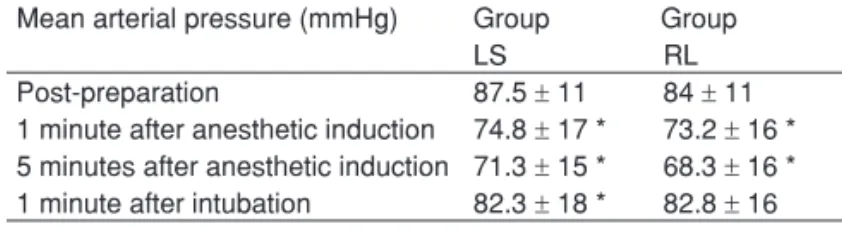
In both groups, a fall in mean arterial pressure was ob-served 1 minute (lighted stylet: 74.8 ± 17 mmHg; rigid laryn-goscope: 73.2 ± 16 mmHg) and 5 minutes (lighted stylet: 71.3 ± 15 mmHg; rigid laryngoscope: 68.3 ± 16 mmHg) af-ter anesthetic induction, which showed a statistically signi-ficant difference when compared to baseline levels (lighted stylet: 87.5 ± 11 mmHg; rigid laryngoscope: 84 ± 11 mmHg) (p < 0.05). Mean arterial pressure 1 minute after intubation in the lighted stylet group (82.3 ± 18 mmHg) remained below baseline levels (87.5 ± 11 mmHg), which was statistically sig-nificant (p < 0.05). The same was not observed in the rigid laryngoscope group (82.8 ± 16 mmHg) when compared to ba-seline levels (84 ± 11 mmHg) (p > 0.05) (Table III).
Table III– Evaluation of Mean Arterial Pressure Mean arterial pressure (mmHg) Group Group
LS RL
Post-preparation 87.5 ± 11 84 ± 11 1 minute after anesthetic induction 74.8 ± 17 * 73.2 ± 16 * 5 minutes after anesthetic induction 71.3 ± 15 * 68.3 ± 16 * 1 minute after intubation 82.3 ± 18 * 82.8 ± 16
Results expressed as mean ± SD; LS: lighted stylet; RL: rigid laryngoscope; *Student t test; p < 0.05 when compared to baseline levels.
When the central venous pressure was analyzed, an in-crease was observed in both groups 1 minute (lighted sty-let: 8.1 ± 4 mmHg; rigid laryngoscope: 7.9 ± 5 mmHg) and 5 minutes (lighted stylet: 7.4 ± 3 mmHg; rigid laryngoscope:
8.1 ± 5 mmHg) after anesthetic induction, and 1 minute after tracheal intubation (lighted stylet: 8.4 ± 5 mmHg; rigid laryn-goscope 9.1 ± 5 mmHg) when compared to baseline levels (li-ghted stylet: 3.9 ± 3 mmHg; rigid laryngoscope: 5.5 ± 4 mmHg), with a p < 0.05 for both groups at all times (Table IV).
Table IV –Evaluation of Central Venous Pressure Central venous pressure (mmHg) Group Group
LS RL
Post-preparation 3.9 ± 3 5.5 ± 4 1 minute after anesthetic induction 8.1 ± 4* 7.9 ± 5* 5 minutes after anesthetic induction 7.4 ± 3* 8.1 ± 5* 1 minute after intubation 8.4 ± 5* 9.1 ± 5*
Results expressed as mean ± SD; LS: lighted stylet; RL: rigid laryngoscope; *Student t test; p < 0.05 when compared with baseline levels.
DISCUSSION
In the present study, it was observed that tracheal intubation was faster with rigid laryngoscopy (24 ± 5 sec) than with li-ghted stylet (28 ± 7 sec). Heart rate showed a reduction in both groups during anesthetic induction and remained stable after tracheal intubation. Mean arterial pressure also showed a reduction in both groups during induction, but it increased after tracheal intubation in the rigid laryngoscope group. Cen-tral venous pressure increased during anesthetic induction and after tracheal intubation in both groups.
This study has some limitations because inflammatory me-diators and adrenergic hormones, such as adrenaline, nora-drenaline, cortisol, and interleukins were not investigated to correlate with clinical findings. Patients were not monitored with a pulmonary artery catheter, and for this reason we do not have data on systolic volume, cardiac output, or periphe-ral vascular resistance. Forty patients were evaluated, which might lead to discussions on the need to investigate a greater number of patients.
Tracheal intubation may be indicated in any situation in which it is necessary to maintain a patent and safe airway. The term appeared in anesthesia in the 18th century 1, but only in 1943 Macintosh developed a laryngoscope blade ca-pable of visualizing the vocal cords 11. In 1957, the first des-cription of the lighted stylet use was made, when Macintosh described the use of a guide-wire with distal illumination to facilitate tracheal intubation 12. The development of laryngos-copes with different models and blades, including models with fiberoptics, allowedthe placement of a tracheal tube with in-creased security 2.
stu-SALGADO FILHO, CORDEIRO, MOTA ET AL.
450 Revista Brasileira de Anestesiologia
Vol. 61, No 4, July-August, 2011 dies directed to this patient profile is very low, and therefore
a consensus on the choice of the best intubation technique does not exist.
Ainsworth et al. 13 performed a study of 200 patients easy to intubate according to Cormack and Lehane criteria. In their study, 87.5% of patients were intubated in the first attempt and 99% in up to three attempts. In our study, 100% of pa-tients were intubated in the first attempt for both techniques.
When the time of tracheal intubation between laryngos-copy and lighted stylet was compared we observed that ri-gid laryngoscopy was faster (24 ± 5 sec) than lighted stylet (28 ± 7 sec) although a statistical significant difference was not observed. In a study of 950 patients, Hung et al. 14 obser-ved that the lighted stylet had a shorter time of intubation, a result different from ours. However, Ellis et al. 15 reported si-milar results to the present study, which showed no statistical significant difference between groups.
In this study only one patient in the lighted stylet group had an ST segment change of 2 mm during anesthetic induction and tracheal intubation, but with no hemodynamic repercus-sions, and any increase in CPK-MB or troponin T.
Through meta-analyses, Figueiredo et al. 10, Ferringo et al. 16, and Zangarillo et al. 17 demonstrated that the increase in heart rate is the most undesirable hemodynamic change in patients with coronariopathies due to increased oxygen consumption, and it is this oxygen consumption that determines a greater risk of intraoperative myocardial ischemia seriously compro-mising the prognosis.
When we analyzed the heart rate, we observed a statisti-cally significant reduction 1 minute and 5 minutes after anes-thetic induction, both in the rigid laryngoscope and lighted sty-let groups. When laryngeal structures were stimulated during tracheal intubation an increase in heart rate was observed 1 minute after tracheal intubation 5. However, this increase in heart rate does not surpass baseline levels. This increase in heart rate was not greater, most likely due to the use of beta-blockers, which in association with adequate doses of opioids 8,9, was responsible for the stability and patient safety during intubation with both techniques.
An increase in mean arterial pressure was observed in both groups 1 minute after tracheal intubation. However, this increase was more significant in the rigid laryngoscope group than in the lighted stylet group when both groups were
com-pared to their baseline levels. Even using adequate doses of fentanyl for anesthetic induction and with a smaller intuba-tion time in the rigid laryngoscopy group, when we introduced the laryngoscope blade, it triggered a greater adrenergic sti-mulus than that of the lighted stylet as was demonstrated by Takahashi et al. 6. Thus, there was increased peripheral vas-cular resistance leading to increased mean arterial pressure in the rigid laryngoscope group 5,6.
Patients with myocardial infarction, valvulopathies, pulmo-nary hypertension, pericardial effusion, and ejection fraction lower than 45% were excluded from this study. The adminis-tration of crystalloids was similar in both groups. Therefore, we used the central venous pressure as an indirect indicator of cardiac function, recognizing the limitations of this method in the analysis of cardiac output 18,19. Griffin et al. 18 demons-trated in their study on myocardial dysfunction that the incre-ase in central venous pressure may be associated with the down regulation of cardiac beta-1 receptors, due to the pre-operative use of beta-blockers, and that, in association with anesthetic drug-induced myocardial depression, leads to a reduction in cardiac inotropism and consequent reduction in cardiac output 18. When this negative inotropism is associated with an increase in pre-load triggered by tracheal intubation 5, a more marked reduction in left ventricular systolic volume, and consequent increase in central venous pressure is ob-served 18,19.
Even using cardiostable drugs during anesthetic induction (etomidate, fentanyl, and cisatracurium) an increase in central venous pressure was observed in the rigid laryngoscope and lighted stylet groups 1 minute and 5 minutes after anesthetic induction 19.
One minute after intubation, we observed a greater incre-ase in central venous pressure, probably due to anesthetic drug-induced myocardial depression associated with an in-crease in peripheral vascular resistance triggered by stimula-tion of laryngeal structures during tracheal intubastimula-tion 5,18,19.
REFERÊNCIAS / REFERENCES
01. Mcewen W – Clinical observations on the introduction of tracheal tu-bes by the mouth instead of performing tracheotomy or laryngotomy. BMJ, 1880;122-4;163-165.
02. Macintosh RR – A new laryngoscope. Lancet, 1943;241(6233):205. 03. Macintosh R, Richards H – Illuminated introducer for endotracheal
tubes. Anaesthesia, 1957;12(2):223-225.
04. Cho J, Chung HS, Chung SP, Kim YM, Cho YS – Airway scope vs. Macintosh laryngoscope during chest compressions on a fresh cada-ver model. Am J Emerg Med, 2010 ;28(6):741-744.
05. Mort TC – Emergency Tracheal Intubation: Complications As-sociated with Repeated Laryngoscopic Attempts. Anesth Analg, 2004;99(2):607-613.
06. Ellis DG, Stewart RD, Kaplan RM, et al. – Success rates of blind
oro-tracheal intubation using a transillumination technique with a light sty-let. Ann Emerg Med, 1986;15(2):138-142.
07. Davis L, Cook-Sather SD, Schreiner MS – Lighted Stylet Tracheal Intubation: A Review. Anesth. Analg, 2000;90(3):745-756.
08. Nishiyama T, Misawa K, Yokoyama T et al. – Effects of Combining Midazolam and Barbiturate on the Response to Tracheal Intubation: Changes in Autonomic Nervous System. Journal of Clinical Anesthe-sia, 2002;14(5):344-348.
09. Takahashi S, Mizutani T, Miyabe M et al. – Hemodynamic Responses to Tracheal Intubation with Laryngoscope versus Lightwand Intubat-ing Device (Trachlight®) in Adults with Normal Airway. Anesth Analg,
2002;95(2):480-484.
10. Montes FR, Giraldo JC, Betancur LA et al. – Endotracheal intuba-tion with lightwand or a laryngoscope results in similar hemodinamic variations in patients with coronary artery disease. Can J Anaesth, 2003;50(8): 824-828.
11. Friedman PG, Rosenberg MK, Lebonbom-Mansour M – A compari-son of light wand and suspension laryngoscopic intubation techniques in outpatients. Anesth Analg 1997;85(3):578-582.
12. Knight RG, Castro T, Rastrelli AJ et al. – Arterial blood pressure and
heart rate response to lighted stylet or direct laryngoscopy for endo-tracheal intubation. Anesthesiology, 1988;69(2):269-272.
13. Dahlgren N, Messeter K – Treatment of stress response to laryngos-copy and intubation with fentanyl. Anaesthesia, 1981;36(11):1022-1026.
14. Martin DE, Rosenberg H, Aukburg SJ et al. – Low-dose fentanyl
blunts circulatory responses to tracheal intubation. Anesth Analg, 1982;61(8):680-684.
15. Figueredo E, Garcia-Fuentes EM – Assessment of the efficacy of esmolol on the haemodynamic changes induced by laryngoscopy and tracheal intubation: a meta-analysis. Acta Anaesthesiol Escand, 2001;45(8):1011-1022.
16. Zangrillo A, Turi S, Crescenzi G et al. – Esmolol reduces periopera-tive isquemia in cardiac surgery: A meta-analysis of randomized con-trolled studies. J. Cardiothoracic Vasc. Anest, 2009;23(5):625-632 17. Feringa HH, Bax JJ, Boersma E et al. – High dose B-blockers and tight
heart rate control reduce miocardial ischemia and troponin T release in vascular surgery patients. Circulation, 2006;114(1 Suppl):344-349.
18. Ainsworth QP, Howells TH – Transilluminated tracheal intubation. Br J Anaesth, 1989;62(5):494-497.
19. Hung OR, Pytka S, Morris I et al. – Clinical trial of a new
light-wand device (Trachlight) to intubate the trachea. Anesthesiology, 1995;83(3):509-514.
20. Ellis ET, Jakymec A, Kaplan RM, et al. – Guided orotracheal
intuba-tion in the operating room using a lighted stylet: a comparison with direct laryngoscopic technique. Anesthesiology, 1986;64(6):827-836. 21. Griffin MJ, Hines RL – Management of Perioperative Ventricular
Dys-function. J Cardioth and Vascul Anesth, 2001;15(1):90-106.
22. Mekis D, Kamenik M – Influence of body position on hemodynamics
in patients with ischemic heart disease undergoing cardiac surgery. Wien Klin Wochenschr, 2010;122(Suppl 2):59-62.
Resumen: Salgado Filho MF, Cordeiro VH, Mota S, Prota M, Lopez MN, Lara RA – Evaluación de los Parámetros Hemodinámicos entre la Laringoscopia Rígida y el Estilete Luminoso en Pacientes con Co-ronariopatías.
Justificativa y objetivos: El anestesiólogo está en contacto con el manejo de la vía aérea siempre que aplica una anestesia. En este estudio, estamos evaluando los parámetros hemodinámicos entre el laringoscopio rígido y el estilete luminoso en pacientes con corona-riopatías.
Pacientes y métodos: Este ensayo clínico randomizado fue llevado a cabo con la participación de 40 pacientes sometidos a la revascula-rización del miocardio, y divididos en dos grupos: estilete luminoso y laringoscopio rígido. Se evaluaron la frecuencia cardíaca, la presión arterial promedio, alteraciones del segmento ST y la presión venosa central durante la preparación del paciente, 1 minuto después de la inducción anestésica, 5 minutos después de la inducción anestésica y 1 minuto después de la intubación traqueal, además del tiempo de intubación traqueal en cada grupo.
Resultados: Los grupos fueron homogéneos con relación a los da-tos demográficos. El tiempo de intubación traqueal para el grupo la-ringoscopio rígido (24 ± 5 seg), fue menor que en el grupo estilete luminoso (28 ± 7 seg), sin embargo con p > 0,05. La frecuencia cardí-aca se reduce en los dos grupos durante la inducción (p < 0,05), sin embargo, 1 minuto después de la intubación, la frecuencia cardíaca aumentó alcanzando valores próximos al momento de la preparaci-ón en los dos grupos (p > 0,05). En el grupo laringoscopio rígido la presión arterial promedio aumentó después de la intubación traqueal para valores próximos al momento de la preparación del paciente (p > 0,05), mientras que en el grupo estilete luminoso la presión arte-rial promedio quedó por debajo de los valores basales con p < 0,05. La presión venosa central aumentó en ambos grupos durante todos los momentos (p < 0,05).
Conclusiones: En este estudio, pudimos observar que ambas técni-cas son seguras para la intubación traqueal en pacientes con coro-nariopatías. Sin embargo, el EL presenta una menor repercusión en la presión arterial promedio.
Descriptores: ENFERMIEDAD: Cardíaca; EQUIPOS: