Article
ISSN 0102-695X
http://dx.doi.org/10.1590/S0102-695X2011005000225 Received 25 Jan 2011 Accepted 19 Aug 2011 Available online 5 Dec 2011
Ehrlich solid tumor development
Ana Flávia C. Ribeiro,
1Thalita C. Telles,
2Vany P. Ferraz,
3Elaine M. Souza-Fagundes,
4Geovanni D. Cassali,
5Andréa T.
Carvalho,
6Marilia M. Melo
*,21Departamento de Ciências Agrárias e Ambientais, Universidade Estadual de Santa Cruz, Bahia, Brazil,
2Departamento de Clínica e Cirurgia, Escola de Veterinária, Universidade Federal de Minas Gerais, Brazil,
3Departamento de Química, Instituto de Ciências Exatas, Universidade Federal de Minas Gerais, Brazil,
4Departamento de Fisiologia, Instituto de Ciências Biológicas, Universidade Federal de Minas Gerais, Brazil,
5Departamento de Patologia Geral, Instituto de Ciências Biológicas, Universidade Federal de Minas Gerais, Brazil,
6Laboratório de Biomarcadores de Diagnóstico e Monitoração, Centro de Pesquisas René Rachou-Fundação Oswaldo Cruz, Brazil.
Abstract: The aim of this study was to investigate the effect of Arrabidaea chica
(Humb. & Bonpl.) B. Verl., Bignoniaceae, extracts on Ehrlich solid tumor development in Swiss mice. Leaves of A. chica were extracted with two distinct solvents, ethanol and water. The phytochemical analysis of the extracts indicated different classes of
secondary metabolites like as anthocyanidins, l avonoids, tannins and saponins. Ethanol
(EE) and aqueous (AE) extracts at 30 mg/kg reduced the development of Ehrlich solid tumor after ten days of oral treatment. The EE group presented increase in neutrophil
count, α1 and β globulin values, and decrease of α2 globulin values. Furthermore, EE
reduced the percentage of CD4+ T cells in blood but did not alter the percentage of inl ammatory mononuclear cells associated with tumor suggesting a direct action of EE
on tumor cells. Reduced tumor development observed in AE group was accompanied by a lower percentage of CD4+ T lymphocytes in blood. At the tumor microenvironment,
this treatment decreased the percentage of CD3+ T cells, especially due to a reduction
of CD8+ T subpopulation and NK cells. The antitumor activity presented by the AE is possibly related to an anti-inl ammatory activity. None of the extracts produced toxic
effects in animals. In conclusion, the ethanol and aqueous extracts of A. chica have
immunomodulatory and antitumor activities attributed to the presence of l avonoids,
such as kaempferol. These effects appear to be related to different mechanisms of action for each extract. This study demonstrates the potential of A. chica as an antitumor agent
coni rming its use in traditional popular medicine.
Keywords:
Arrabidaea chica
Bignoniaceae flavonols Ehrlich tumor antitumor activity
Introduction
Cancer is one of the leading causes of mortality worldwide. Many of the chemotherapeutic agents used in the treatment of cancer injure rapidly dividing normal cells presenting substantial side effects. Therefore, the quest for effective anticancer drugs is an active research i eld. The plant kingdom is a potential source of chemicals with antitumor and cytotoxic activities (Kim et al., 2005). New drugs generated from secondary metabolites of plants represent an alternative source of anticancer agents, which frequently seem to be more effective and/or less toxic (Ma & Wang, 2009).
Arrabidaea chica (Humb. & Bonpl.) B. Verl., Bignoniaceae, is a shrub plant widespread from South Mexico to central Brazil, particularly in the Amazon basin (Correa, 1931; Takemura et al., 1995). The leaves of the plant have been traditionally used by Brazilians Indians as dye in ritual body painting, to protect the skin against the sunlight and to repel insects (Chapman et al, 1927). Very little is known about the chemical constitution of the leaves (Barbosa et al., 2008). Earlier studies of A. chica
determined, ±10.2 mg/g and 0.06 mg/g, respectively (Silva et al., 2007). More recently the main classes of secondary metabolites in the ethanol extract was detected: anthocyanidins, anthocyanins, antraquinone, cathechins, organic acids, reducing sugars, steroids, xanthones, tannins, lavanonols and lavanone (Barbosa et al., 2008).
Nowadays A. chica is widely used in the popular medicine for wound healing, treatment of inlammation, intestinal colic, blood dysfunction, uterine inlammation and leukemia (Zorn et al., 2001; Jorge et al., 2008; Barbosa et al., 2008). Despite the wide use very little is known about the pharmacological properties of its extracts (Barbosa et al., 2008). The anti-inlammatory effect was already demonstrated in vivo (Oliveira et al., 2009) and
in vitro, with the inhibition of the transcription factor NF-kB (Zorn et al., 2001). Also, healing and antioxidant activities were reported, possibly related to the presence of anthocyanidins (Jorge et al., 2008). Antitumor activity of the ethanol extract (25 μg/mL) was observed in vitro
against Jurkat and HL60 cell lines (Ribeiro et al., 2010). Therefore, based on the empirical ethnopharmacological use of Arrabidaea chica crude extract as an antitumor agent associated with the strong cytotoxic activity in vitro (Ribeiro et al., 2010) and the lack of in vivo studies with this plant and its constituents, this study examined the antitumor activity of the ethanol and aqueous extracts of A. chica on the Ehrlich solid tumor model.
Material and Methods
Plant material
Leaves of Arrabidaea chica (Humb. & Bonpl.) B. Verl., Bignoneaceae, with the common names: Pariri, Crajiru, Carajuru or Carajiru were collected in Maringá (State of Paraná, Brazil). The plant material was identiied in Campinas-SP, Brazil, where a voucher specimen is deposited and registered under the number UEC 145.956 (Herbário da Universidade de Campinas).
Preparation of extracts
The leaves of A. chica were shade dried at room temperature, powdered, and extracted (40 g) with distillated water (0.5 L) originating a dark red crude extract. This extract was fractionated according to Matos (1988). Briely, the pH was basiied with ammonium hydroxide (pH 11.0). A solution of n-hexane and ethyl acetate (1:1) (0.5 L) was added to the crude extract leading to two distinct phases, one organic and one aqueous. After separation, hydrochloric acid (pH 1.5) and a solution of hexane: acetyl acetate (1:1) (0.5 L) were added to the organic phase leading to a new organic and a new aqueous phase. This last aqueous phase was concentrated under a
rotary vacuum evaporator (65 ºC) originating the aqueous extract used in the experiment (AE). The pH was basiied (pH 7.0) and the extract was submitted to lyophilization (1.6 g) and preserved in a freezer at -20 ºC until further use.
The ethanol extraction was performed as previously described (Leite et al., 2006) with slight modiications. Briely, the ethanol extract (EE) was prepared by maceration of dry leaves (100 g) for exhaustive percolation with ethanol at room temperature. After iltration, the solvent (ethanol) was evaporated under reduced pressure to yield 1.8 g of a viscous red brown extract (yield, 1.8%). After, this extract was submitted to exhaustive extraction with hexane under relux in a water-bath for 2 h, resulting in a hexane-insoluble fraction used in the experiment (EE) (1 g).
Phytochemical analysis
Chemical tests to detect the main classes of secondary metabolites were carried out in aqueous extract (AE) using classical speciic color reactions (Matos, 1988).
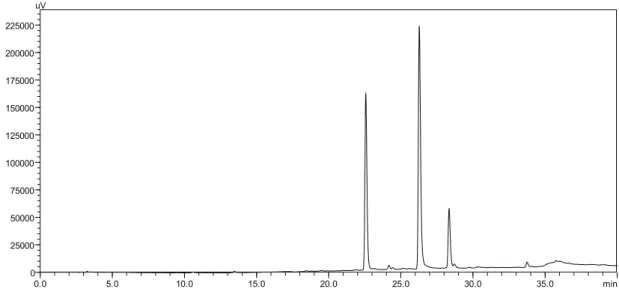
The ethanol extract (EE) was dissolved in methanol (1 mg/mL) and iltered through Whatman paper ilter and the iltrate was used for HPLC analysis. Chromatographic separation was performed on a Shimadzu® liquid chromatographic system equipped with
a LC-10AT-vp solvent delivery system, SPD M-10 AVP photo diode array detector, and Rheodyne 7725i injector with 5 μL loop. A Phenomenex GEMINI C18 column (25cm×4.6mm i.d., 5 μm) was used for the separation. A mixture (75:25 v/v) of phosphate buffer (25 mM%) and acetonitrile was used as the mobile phase. It was delivered at a low rate of 1.0 mL per min with detection at 360 nm. The retention time of kaempferol was found to be 28.34 min. The injection volume of the EE extract was 50 μL. Analysis was performed at room temperature.
Tumor cells
Ehrlich ascites carcinoma (EAC) cells were supplied by the Laboratório de Patologia Geral, Instituto de Ciências Biológicas, Universidade Federal de Minas Gerais, Brazil. The cells were maintained in vivo in Swiss albino mice by intraperitoneal transplantation. EAC cells aspirated from the peritoneal cavity of mice were washed with saline and injected subcutaneously to develop solid tumor.
Antitumoral activity
Universidade Federal de Minas Gerais, Brazil (CETEA/ UFMG) (Protocol 155/2009).
Fifteen female Swiss Albino mice with eight weeks of age were injected with EAC cells (2.5 x 10⁶),
subcutaneously in the left footpad to obtain the solid tumor. These animals were divided into three groups (n=5) and treated with aqueous extract (Group I), ethanol extract (group II) and distilled water (Group III).
The aqueous extract (AE) or the ethanol extract (EE) (30 mg/kg body weight) were suspended in distilled water (300 µL) and administrated to the animals orally, with the help of an intragastric catheter, once daily for ten days, beginning three days after the tumor inoculation. The selection of the dose was based on the work of Queiroz et al. (2008) who studied the antitumor properties of Tabebuia avellanedae, plant of the same family as A. chica. The control group received 300 µL of water by gavage, as describe to groups AE and EE.
Tumor growth was evaluated by measuring the inoculated footpad thickness with a graduate micrometer (Mitutoyo, model 7301, graduation 0.01 mm, range 0-10 mm, accuracy ±0.02 µm) as described (Silva et al., 2004; Silva et al., 2006; Pinto et al., 2009). This procedure was done at every 48 h. On the 10th day of treatment,
i.e., on the 12th day of tumor development, all mice were
euthanized. The heart, lungs, kidney, liver, spleen, the inoculated footpad and the ipsilateral popliteal lymph node were collected, ixed in 10% buffered formalin and conventionally embedded in parafin. The four micrometers sections were stained in hematoxilin and eosin for histological examination.
Blood samples were collected by retro-orbital puncture for hematological evaluation before the euthanasia. Total white blood cell (WBC), red blood cell (RBC) and haemoglobin (Hb) were determined by standard method using cell diluting luid and haemocytometer (CELM DA 500®, Barueri-SP, Brazil). The packed volume cell (PVC) was measured in a microhematocrit centrifuge (Microspin, São Paulo, Brazil) with capillary tubes. The mean corpuscular volume (MCV), mean corpuscular haemoglobin (MCH) and mean corpuscular haemoglobin concentration (MCHC) were calculated. Blood smears were stained by the May-Grünwald-Giemsa technique to obtain differential leukocyte count (Thrall, 2004).
Plasma protein concentration was measured using a refractometer (Ningbo Utech International CO Ltda, model 301). Plasmatic protein fractions were obtained by electrophoresis using an agarose gel electrophoresis system (CELM SDS-60, Brazil). A 1.0 μL sample of plasma was applied to the gel, which was exposed to 100V for 32 min. The gel was stained (Ponceau S, Vetec Química Fina Ltda, Duque de Caxias-RJ, Brazil), ixed and dried. Bands were scanned and measured using the software Celm SE-250. Percentage and absolute values (g/dL) for the proteins fractions were determined on the basis of total protein
concentration. The albumin/globulin ratio was calculated as albumin/(a1+a2+b+y globulins).
Analysis of the mononuclear leukocyte subpopulations in blood and tumor tissue by low cytometry
In this protocol the number of five types of mononuclear leukocytes on the blood and tumor tissue were evaluated: TCD4, TCD8 (CD3 PerCP, CD4 FITC, CD8 PECY5) and B lymphocytes (CD5 PerCP, CD19 FITC) and natural killer (NK1.1 FITC) cells. Total blood was collected and 50 μL were incubated with a monoclonal antibody labeled with fluorescein. The tumor tissue was squashed in RPMI-1640 (5 mL) media and filtrated on stainless steel gaze to obtain a single cell suspension. The cell suspension was kept on ice for 5 min and washed twice in RPMI-1640. After centrifugation (10000 x g, 10 min, 4 oC) the supernatant
was ressuspended in 150 μL. Subsequently 30 μL of the supernatants were collected and incubated with a monoclonal antibody labeled with fluorescein. The protocols, antibodies and reagents employed were as per the manufacturer’s recommendations (Sigma). The percentage of labeled cells was obtained by analysis of 10000 events with a FACSCalibur flow cytometer (Becton Dickinson. Palo Alto, CA, USA) using the Cell-Quest software.
Statistical Analysis
All data were checked for normality. Statistical signiicance between groups was performed by the application of one-way analysis of variance ANOVA followed by SNK (cv<25%) or Duncan (cv>25%) test when data were normally distributed. Kruskal-Wallis or Mann-Whitney U tests were used when data were not normally distributed. The difference was reliable with
p<0.05. Data were expressed as means±SD.
Results and Discussion
In order to evaluate if Arrabidaea chica (Humb. & Bonpl.) B. Verl., Bignoneaceae, ethanol or aqueous extracts demonstrate antitumoral effect against a solid tumor, mice received intraplantar injection of 2.5 x 10⁶
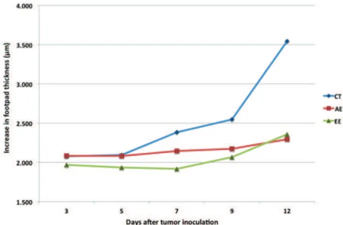
EAC cells. Measures of mice paws showed a continuous tumor growth in all experimental groups. Both groups treated with AE and EE showed similar patterns of tumor development - a steady growth without the occurrence of peaks during the experimental period, while the control group presented a peak seven days after the inoculation (Table 1).
In 12th day, the EE group presented a smaller
The growth curve of the Ehrlich tumor presents an exponential behavior from the third day post inoculation until day 30th (Dagli et al., 1992a). The control group
showed signiicant differences in tumor growth between days 7th and 12th. The growth rate of Ehrlich tumor in mice
differs in the site of implantation and in the form. In the solid form occurs a proliferation peak after the 7th day of
implantation (Silva et al., 2006) like demonstrated with control group in our experiment.
There was a signiicant reduce in the tumor growth in mice treated with AE and EE at 12th day, different from
control group (Figure 1).
Table 1. Measurement of Ehrlich solid tumor growth in mouse
footpad (μm).
Group Day of tumor development
3 5 7 9 12
Control 2.068±0.08 aA
2.092±0.06 aA
2.307±0.18 aAB
2.451±0.46 aAB
3.237±0.75 aB
AE 2.108±0.10 aA
2.053±0.08 aA
2.097±0.13 aAB
2.290±0.33 aAB
2.495±0.51 aB
EE 1.968±0.14 aA
1.935±0.13 aA
1.918±0.11 bA
2.067±0.22 aA
2.357±0.47 bA *Data was presented as mean±standard deviation. Values with different
letters are signiicantly different (p<0,05). Lowercase to columns and uppercase to lines.
Figure 1. Measurement of Ehrlich solid tumor growth (μm)
in the footpad of mice treated with Arrabidaea chica aqueous extract (AE) and ethanol extract (EE) during twelve days.
Cancer is a disease of misguided cells that have high potential of excess proliferation without apparent relation to the physiological demand of the process. It is the second largest cause of death in the world. Of all the available anticancer drugs used in the last decades, 40% were natural products per se or natural product derived, with another 8% being natural product mimics (Newman et al. 2003). The greatest recent impact of plant-derived drugs is observed in the area of antitumor research, where compounds such as taxol, vinblastine, vincristine, and camptothecin have dramatically
improved the effectiveness of chemotherapy against some of the most dreaded cancers (Rates, 2001). Hence, there is a great potential for the development of anticancer drugs from the essentially untapped reservoir of the plant kingdom. A large number of plants possessing anticancer properties have been documented (Gupta et al., 2004, Aoki et al., 2005, Kim et al., 2005).
Earlier studies carried out in our laboratory have shown potent cytotoxic activity of A. chica ethanol extract (Ribeiro et al., 2010). Based on this observation, in the present study, the aqueous and ethanol extracts were evaluated for its in vivo antitumor properties. These results could indicate either a direct cytotoxic effect of A. chica extracts on tumor cells or an indirect local effect, which may involve macrophage activation and vascular permeability inhibition. The direct strong cytotoxic effect of ethanol extract was already demonstrated in the in vitro
experiments against Jurkat and HL60 cell lines (Ribeiro et al., 2010) and in the present study, against Ehrlich solid tumor.
Preliminary phytochemical investigation of the ethanol extract of A. chica revealed the presence of phenolics, tannins, lavanols, anthocyanins, organic acids and reducing sugars. The antifungal and trypanocidal activities of A. chica ethanol extract appear to be related to the presence of quinones. Flavonoids could also be involved in the trypanocidal activity, since plants synthesized them in response to microbial infection (Barbosa et al., 2008).
We believed that the antitumoral activity of
A. chica might be due to the presence of anthocyanins, lavonoids or both of them. Our HPLC study of the EE extract in comparison to the standard kaempferol indicated the presence of this compound (Figure 3). Barbosa et al. (2008) isolated other two lavonoids from the ethanol extract of A. chica besides kaempferol: 4’-hydroxy-3,7-dimethoxy lavone and vicenin-2. During the last decade, natural antioxidants, particularly phenolics, have been under very close scrutiny as potential therapeutic agents against a wide range of ailments including cancer, inlammatory diseases and also aging. The medicinal actions of phenolics is mostly ascribed to their antioxidant capacity, free radical scavenging, chelation of redox active metal ions, modulation of gene expression and interaction with the cell signaling pathways (Soobrattee et al., 2005).
Most chemotherapeutic agents induce collateral effects. Direct myelotoxicity is often observed in patients undergoing anticancer treatments. Hence, the evaluation of toxicity can be accomplished by blood count evaluation (Perez et al., 2005). None of the animals bearing Ehrlich Tumor (control group) and treated with A. chica extracts (AE or EE) showed alterations in total erythrocytes count, haemoglobin concentration, packed volume cell and haematimetric indices (Table 2).
0.0 5.0 10.0 15.0 20.0 25.0 30.0 35.0 min 0
25000 50000 75000 100000 125000 150000 175000 200000 225000 uV
Figure 2. HPLC of standard lavonols (rutin RT= 22.56 min; quercetina RT= 26.28; kaempferol RT=28.34 min).
17.5 20.0 22.5 25.0 27.5 30.0 32.5 min
0 25000 50000 75000 100000 125000 150000 175000 200000 uV
Figure 3. HPLC of standard lavonols (pink line) and EE extract (black line).
Table 2. Mean, standard deviation and statistical analysis results of erythrogram in mice bearing Ehrlich solid tumor treated with different extracts of Arrabidaea chica.
Haemogram Control AE EE
RBC/μl 8300,000±600,000 a 8700,000±600,000 a 8600,000±800,000 a
Haemoglobin (g/dL) 12.7±0.9 a 13.3±0.7 a 14.2±0.4 a
Packed volume cell (%) 43.5±3.3 a 45.0±2.2 a 46.5±1.7 a
MCV (fL) 53.3±0.7 a 52.1±11.5 a 54.2±4.4 a
MCH (%) 15.3±0.3 a 15.4±0.3 a 16.6±1.3 a
MCHC (g/dL) 29.3±0.9 a 29.6±0.7 a 30.6±0.5 a
Data was presented as mean±standard deviation. Means with the same letters in the same line are not signiicantly different
(p≤0,05).
marrow. Verçosa Junior et al. (2004) reported discreet anemia (RBC=7.23±1.74/µL; haemoglobin=10.7±2.55g/ dl; PVC=30,86±7.78%) and leucopenia (2.96±1.22/µL) in mice bearing solid Ehrlich tumor. Also, it might be inferred that the extracts tested did not produce toxic
effects in animals. Another report showed that the hidroalcoholic extract of A. chica with concentrations as high as 5g/kg had no acute toxic effects in mice (Catargenes, 2009).
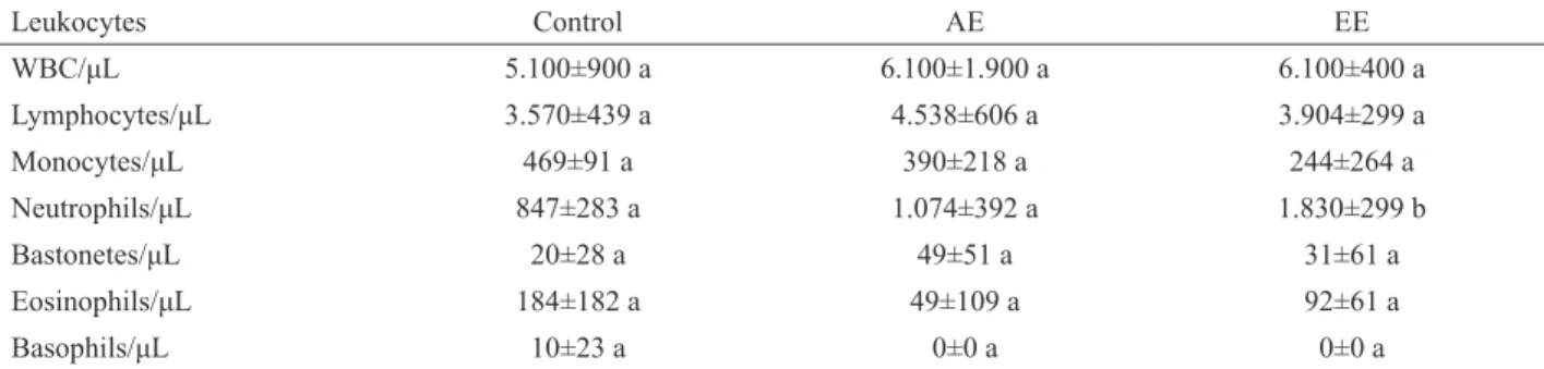
between groups. However, the ethanol extract (EE) caused a signiicant increase in neutrophils count (Table 3).
Neutrophils play an important role in tumor angiogenesis releasing chemokines that cause endothelial cell invasion and vessel formation. Other studies showed that potent activation of iniltrating macrophages and granulocytes could lead to tumor destruction and tumor rejection (Albini et al., 2005).
In our study we observed that the reduction in the size of the tumor in the EE group was accompanied by an increase in the number of neutrophils in blood. Also, this group presented a larger number of neutrophils in the histology of the tumor. Bergami-Santos et al. (2004) showed that neutrophils from Swiss mice are cytotoxic against Ehrlich tumor cells but are not eficient in controlling tumor growth. The treatment with AE, which also reduced tumor growth, did not alter the number of neutrophils in blood or differ from control group in concern to the morphology of the tumor.
Tumor growth initiates a myriad of functional and phenotypic changes in macrophages and T-cells in association with alterations in cytokine synthesis and responsiveness (Walker et al. 1994). Several types of tumor also express receptors for granulocyte-macrophage colony stimulating factors or produces and uses granulocyte– macrophage colony stimulating factors as an autocrine growth factor (Fu et al., 1991).
Phagocytes, particularly macrophages and neutrophils, play a vital role in both the innate and the acquired immunity, exerting a key role in host defense against various infectious agents and tumor growth (Natan et al, 1983). One of the most important characteristics of these cells is their capacity to migrate into inlammatory sites (Spector & Ryan, 1979). Animal model studies have shown that a variety of tumor cells can produce factors that impair inlammatory responses, thus allowing tumor growth in vivo. Alternatively, the tumor cells can stimulate macrophage suppressor activities in host cells (Elgert et al., 1998). In addition to the deicient accumulation of phagocytes at the inlammatory sites, several other cellular parameters have been shown to be altered in the tumor-bearing state, in particular the formation of granulocyte–
macrophage colonies in response to CSF (Baum & Fisher, 1972).
The treatment with ethanol extract (EE) caused an increase in α1 and β-globulins values compared to control group (Table 4). Increases in α-globulins can be accompanied by increased β-globulins. For example, α1-antitrypsin has antiprotease activity designed to inhibit proteases released by phagocytes and other cells of the immune system to minimize damage to normal tissues.
Acute phase proteins like β-lipoprotein and complement C3 were located in the β1 region, and transferrin and IgM were located in the β2 region. The increase of β-globulins values in the group treated with A. chica ethanol extract is likely to have happened due to the increase of IgM. The fact that lavonols were identiied in
A. chica ethanol extract raises the question of whether these substances could be responsible for an immunomodulatory effect.
Production of acute phase proteins (APP) is controlled by cytokines, with the pro-inlammatory cytokines interleukin-1, interleukin-6, and tumor necrosis factor-α released from the site of pathogenic or inlammatory damage stimulating the production of the APP. The mechanism of production involves cytokine receptor, signaling pathways, and induction of mRNA for the APP, which is released 6 to 12 h after stimulation. The liver is the main site of synthesis of the APP, but there have been recent reports of non-hepatic tissues such as lung, adipocyte, and intestine increasing expression of mRNA for APP following stimulation (Kaneko, 2008).
The treatment with ethanol extract (EE) caused a decrease of α2-globulins values when compared to control group. Haptoglobin and α2-macroglobulin were identiied in the α2 fraction. Haptoglobin has scavenging activities and binds metabolites released from cellular degradation so they can reenter host metabolic processes rather than be utilized by pathogen. Therefore, another question is raised: whether the ethanol extract have caused a decrease in the tumoral area.
Tumor morphology was similar in all groups. The tumor cells formed solid pattern or cords invading the muscle and bone tissue. The tumor cells showed moderate
Table 3. Mean, standard deviation and statistical analysis results of leukogram in mice bearing Ehrlich solid tumor treated with different extracts of Arrabidaea chica.
Leukocytes Control AE EE
WBC/μL 5.100±900 a 6.100±1.900 a 6.100±400 a
Lymphocytes/μL 3.570±439 a 4.538±606 a 3.904±299 a
Monocytes/μL 469±91 a 390±218 a 244±264 a
Neutrophils/μL 847±283 a 1.074±392 a 1.830±299 b
Bastonetes/μL 20±28 a 49±51 a 31±61 a
Eosinophils/μL 184±182 a 49±109 a 92±61 a
Basophils/μL 10±23 a 0±0 a 0±0 a
pleomorphism. There were groups of large and oval cells with abundant vacuolated faintly basophilic cytoplasm with indistinct borders. Oval basophilic nucleus was observed with multiple evident nucleoli. Other times the tumor cells were smaller, spindle shaped with basophilic cytoplasm. There were a moderate number of mitotic igures, some of them being atypical.
The inlammatory iniltrate was predominantly lympho-histiocytic moderate to mild with few neutrophils, except for the EE group, which presented lympho-histiocytic iniltrate with large amount of neutrophils. All groups presented extensive areas of necrosis scattered trough tumor tissue with discrete areas of hemorrhage.
Metastases were not seen in any of the organs studied (heart, lung, liver, spleen, kidney), or in the popliteal lymph nodes. The lymph nodes were enlarged with reactive hyperplasia of lymphoid follicles and active germinal centers.
The occurrence of metastases is not common in Ehrlich tumor. Exception is made to metastases to regional lymph nodes especially after the seventh day of tumor inoculation (Dagli, 1992a). In the present experiment there was no presence of metastasis in popliteal lymph nodes in all groups (including control group). Once started, neoplastic progression is usually accompanied by increased genetic instability. This may generate the phenotypic diversity manifested by different tumor cell clones (Dagli, 1992b).
Flow cytometric immunophenotyping of blood lymphocytes
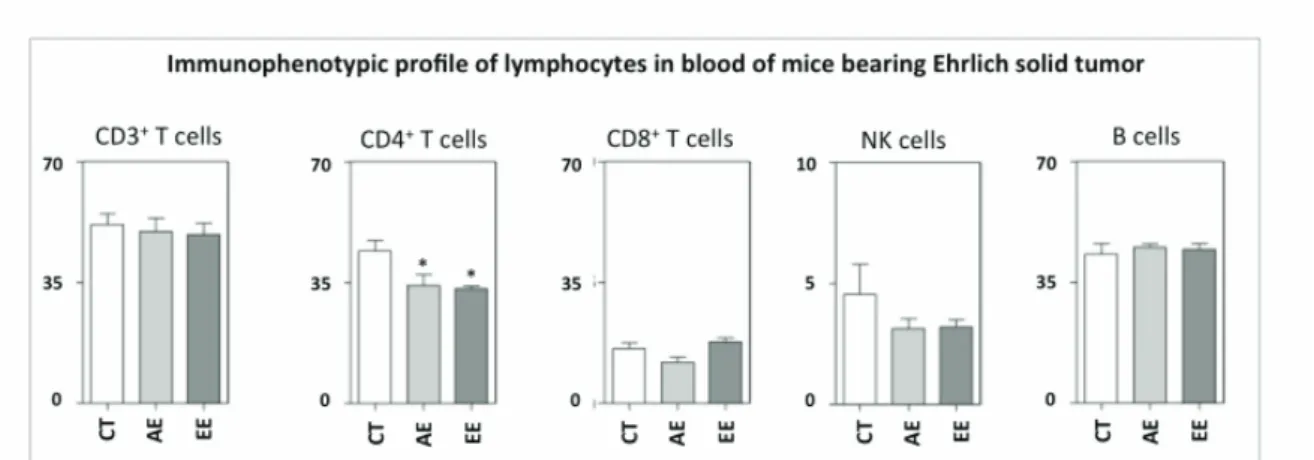
There are no reports on literature that demonstrate the cell types involved in the inhibition of Ehrlich solid tumor growth. In this study we evaluated mononuclear cells in the blood of ive animals per group. The frequency of NK cells, TCD3 and B-Lymphocyte as
well as CD4 and CD8 T cell subpopulations is shown in Figure 4.
The percentage of CD4 T lymphocytes in blood was reduced by the treatments with EE and AE of A. chica. There were no changes in the percentages of CD8 T cells subpopulation, B-lymphocytes or NK cells (Figure 4). Further studies are necessary to clarify the mechanisms involved in the immunomodulatory regulation presented by the extracts over CD4 T cells subpopulation.
Flow cytometric immunophenotyping of tumor iniltrating lymphocytes
Determination of the cell types involved in the inhibition of a tumor is important because it allows one to infer whether the inhibition of the tumor growth is associated with a certain cell population and/or with the direct effect of the treatment on these cells (Santos et al., 2004). In this study we evaluated eight tumors: ive from the control group, two from the AE group and two from the EE group. Three animals from the EE and three animals from the AE group were excluded from this study after the veriication of insuficient number of inlammatory cells in the gate of lymphocyte population. Interestingly, all animals excluded were treated with A. chica extracts.
The frequency of CD3 T cells and B-Lymphocyte as well as CD4 and CD8 T cell subpopulations is are shown in Figure 5. The ethanol extract did not cause alteration on the percentage of tumor iniltrating lymphocytes subpopulations. Signiicant differences were observed between AE and control group. Data analysis demonstrated that the AE group had signiicantly lower percentage of CD3 lymphocytes (p<0.05) associated with the reduction of the CD8 T cell subpopulation without changes in CD4 T lymphocyte subpopulation. Also, the treatment with aqueous extract caused a reduction in NK
Table 4. Mean, standard deviation and statistical analysis results of electrophoretic protein in mice bearing Ehrlich solid tumor treated with different extracts of Arrabidaea chica.
Control AE EE
Total protein 6.2±0.7 a 5.5±0.3 a 5.7±0.3 a
albumin (%) 48.8±6.6 a 50.0±7.3 a 50.7±7.3 a
albumin (g/dL) 3.0±0.6 a 2.8±0.4 a 2.9±0.4 a
α1-globulin (%) 8.3±1.4 a 10.6±3.5 ab 14.1±2.6 b
α1-globulin (g/dL) 0.5±0.1 a 0.6±0.2 ab 0.8±0.2 b
α2-globulin (%) 28.2±7.7 a 20.1±3.4 ab 12.4±3.1 b
α2-globulin (g/dL) 1.7±0.5 a 1.1±0.3 ab 0.7±0.2 b
β-globulin (%) 10.9±1.5 a 16.1±6.5 ab 19.8±2.2 b
β -globulin (g/dL) 0.7±0.1 a 0.9±0.3 ab 1.1±0.2 b
δ-globulina(%) 3.8±2.1 a 3.3±1.7 a 3.0±1.4 a
δ-globulin (g/dL) 0.2±0.1 a 0.2±0.1 a 0.2±0.1 a
albumin/globulin ratio 1.0±0.2 a 1.0±0.2 a 1.1±0.3 a
cells population. These indings suggest that A. chica
aqueous and ethanol extracts have immunomodulatory and antitumor activities attributed to the presence of lavonoids, such as kaempferol identiied by High Performance Liquid Chromatography.
The role of inlammatory and immune responses in tumor development is unclear. Leukocyte iniltration of tumors has been described as playing either inhibitory or stimulatory roles in tumor growth (Bergami-Santos et al., 2004). Data suggest that AE antitumor action is related to its anti-inlammatory potential and the modulation of the immune system. The aqueous extract of A. chica caused a reduction in tumor growth associated with a reduction in CD8 T cells and NK cells. According to the literature, NK cells seems to be devoid of an important role in the resistance against Ehrlich ascites tumor (Gil et al, 1990). Apparently the same occurs here, suggesting that other nonspeciic inlammatory mechanism might be relevant in tumor resistance. The Ehrlich tumor cells lack the
expression of major histocompatibility antigens. This characteristic probably excludes a major role for cytotoxic T lymphocytes during tumor development, indicating that cellular immunity is not the main mechanism of host reaction to this tumor (Bergami-Santos et al., 2004).
The role of CD4 T cells in tumor immunity is not totally clear. Interestingly, the percentage of CD4 T cells iniltrated in tumor tissue in AE and EE group remained similar to the control group (17.85±2.72; 13.51±5.20; 9.54±5.82 respectively) despite the lower number present in blood. These results could suggest that CD4 T cells probably took part in the antitumor immunity against Ehrlich cells mediated by the production of cytokines like TNF-α (tumor necrosis factor-alpha) and INF-γ (interferon-gamma).
Most likely the two extracts tested here exhibit different mechanisms of action. The ethanol extract inhibited Ehrlich solid tumor development without changing mononuclear cell population iniltrating in the
Figure 4. Immunophenotypic profile of lymphocyte in blood of mice bearing Ehrlich solid carcinoma. Lymphocyte populations were identified by flow cytometric immunostainning as described in Material and Methods. Data were expressed as percentage of positive cells within gated lymphocytes. Significant differences at p<0.05 are highlighted by asterisk.
tumor tissue. On the other hand, the morphologic study showed a large number of neutrophils iniltrating the tissue. It was already demonstrated that EE has potent cytotoxic effect against leukemic human cell lines (Ribeiro et al., 2010). This extract induces DNA fragmentation in Jurkat and HL60 cell lines as assessed by low citometry assays (data not shown). Possibly the antitumor effect demonstrated here is also related to the induction of apoptosis of tumor cells. We do not discard the possibility that neutrophils exert cytotoxic effects against EAC cells contributing to the inhibition of tumor growth.
The mechanism of action of extracts on tumor cells should be elucidated in a detailed manner in the future. The association between cytokines and tumor growth inhibition has been described (Silva et al., 2002). It is possible that the antitumor effect of A. chica extracts is associated with the action of cytokines possessing antitumor properties. Whether cytokines may be involved in this process is currently under study in this laboratory.
Conclusions
Data showed that Arrabidaea chica (Humb. & Bonpl.) B. Verl., Bignoneaceae, aqueous extract (AE) and ethanol extract (EE) reduced the solid tumor growth in mice. No adverse side effects due to treatment were observed. Our indings may contribute to a better understanding of the beneicial effects of this folk remedie.
Acknowlegments
The authors would like to thank Fundação de Amparo à Pesquisa do Estado de Minas Gerais for inancial support and Conselho Nacional para o Desenvolvimento Cientíico e Tecnológico for the scholarship to AFCR. The authors thank the Centro de Pesquisas René-Rachou Fiocruz for use of its facilities.
References
Aoki K, Yamakuni T, Yoshida M, Ohizumi Y 2005. Ephedorae herba decreases lipopolysaccharide-induced cyclooxgenase-2 protein expression and
NF-κB-dependent transcription in C6 rat glioma cells. J Pharmacol Sci 98: 327-330.
Albini A, Tosetti F, Benelli R, Noonan DM 2005. Tumor
Inlammatory Angiogenesis and Its Chemoprevention. Cancer Res 65: 10637-10641.
Barbosa WLR, Pinto LN, Quignard E, Vieira JMS, Silva Jr JOC, Albuquerque S 2008. Arrabidaea chica (HBK) Verlot: phytochemical approach, antifungal and trypanocidal activities. Rev Bras Farmacogn 18: 544-548.
Baum M, Fisher B 1972. Macrophage production by the bone marrow of tumor bearing mice. Cancer Res 32: 2813-2817.
Bergami-Santos PC, Mariano M, Barbuto JAM 2004. Dual role of polymorphonuclear neutrophils on the growth of Ehrlich ascites tumor (EAT) in mice. Life Sci 75: 245-255. Catargenes MSS 2009. Investigação dos efeitos tóxicos e
hipertensivo de Arrabidaea chica Verlot (Bignoniacea). 29f. Tese (Doutorado em Produtos Naturais e Sintéticos Bioativos) - Universidade Federal da Paraíba, João Pessoa, PB.
Chapman E, Perkin AG, Robinson R 1927. The coloring matters
of carajuruna. J Chem Soc: 3015-3040.
Côrrea MP 1931. Dicionário das plantas úteis do Brasil e das espécies cultivadas. V2. Rio de Janeiro: Ministério da Agricultura.
Dagli MLZ, Soma M, Guerra JL, Saldiva PHN 1992a. Lymphatic
dissemination in neoplasia: determination of nuclear volume and DNA content of primitive and regional lymphnode Ehrlich tumor cells. Braz J Vet Res Anim Sci 29: 267-271.
Dagli MLZ, Guerra JL, Saldiva PHN 1992b. An experimental
study on lymphatic dissemination of the solid Ehrlich tumor in mice. Braz J Vet Res Anim Sci 29: 97-103.
Elgert KD, Alleva DG, Mullins DW 1998. Tumor-induced
immune dysfunction: the macrophage connection. J Leukococyte Biol 64: 275-290.
Fu YX, Watson GA, Kasahara M, Lopez DM 1991. The role of
tumor-derived cytokines on the immune system of mice bearing a mammary adenocarcinoma: I. Induction of regulatory macrophages in normal mice by the in vivo administration of rGM-CSF. J Immunol 146: 783-789.
Gil J, Alvarez R, Vinuela JE, Morales JGR, Bustos A, De la Concha EG, Subiza JL 1990. Inhibition of in vivo tumor growth by a monoclonal antibody recognizing tumor cell surface carbohydrates. Cancer Res 50: 7301-7306.
Gupta M, Mazumder UK, Sambath Kumar R, Sivakumar T,
Vamsi MLM 2004. Antitumour activity and antioxidant status of Caesapinia bonducella against Ehrlich ascites carcinoma in Swiss albino mice. J Pharmacol Sci 94: 177-184.
Jorge MP, Madjarof C, Ruiz ALTG, Fernandes A T, Rodrigues
RAF, Sousa IMO, Foglio MA, Carvalho JE 2008. Evaluation of wound healing properties of Arrabidaea chica Verlot extract. J. Ethnopharmacol 118: 361-366. Kaneko JJ, Harvey DW, Bruss WL 2008. Clinical biochemistry
of domestic Animals, 6.ed. San Diego: Academic Press, 916p.
Kim JB, Koo HN, Joeng HJ, Lyu YS, Park SG, Won JH, Kim
YK, Hong SH, Kim HM 2005. Induction of apoptosis by
Korean medicine Gagam-whanglyunhaedoktang through
activation of caspase-3 in human leukemia cell line, HL-60 cells. J Pharmacol Sci 97:138-145.
Leite JPV, Oliveira AB, Lombardi JA, Filho JDS, Chiari E. 2006. Trypanocidal activity of triterpenes from Arrabidaea triplinervia and derivatives. Bio Pharm Bull 29: 2307-2309.
evolutionary perspective. Drug Discov Today 14: 1136-1142.
Matos FJA 1988. Introdução a Fitoquímica Experimental, Editora UFC, Fortaleza, Brasil.
Natan CF, Murray HW, Wiebe ME, Rubin BY 1983.
Identiication of interferon-gamma as the lymphokine
that activates human macrophage oxidative metabolism and antimicrobial activity. J Exp Med 158: 670-689.
Newman DJ, Cragg GM, Snader KM 2003. The inluence of
natural product upon drug discovery. Nat Prod Rep 17: 215-234.
Oliveira DPC, Borráz MRL, Ferreira LCL, López-Lozano JL
2009. Atividade antiinlamatória do extrato aquoso de Arrabidaea chica (Humb. & Bonpl.) B. Verl. sobre o edema induzido por venenos de serpentes amazônicas.
Rev Bras Farmacogn 19: 643-649.
Perez RR, Lobo e Silva MAM, Varzim FLSB, Oliveira SB, Hucke EETS 2005. A ação do decanoato de nandrolona (Deca-durabolin) sobre parâmetros hematológicos e proteína total plasmática de ratos (Rattus rattus) com depressão medular induzida após administração de sulfato de vincristina (Oncovin). Ciência Rural 35: 589-595.
Pinto FCH, Menezes GB, Moura SAL, Cassali GD, Teixeira MM,
Cara DC 2009. Induction of apoptosis in tumor cells as a mechanism of tumor growth reduction in allergic mice.
Pathol Res Pract 205: 559-567.
Queiroz MLS, Valadares MC, Torello CO, Ramos AL, Oliveira AB, Rocha FD, Arruda VA, Accorci WR 2008. Comparative studies of the effects of Tabebuia avellanedae bark extract and B-lapachone on the hematopoietic response of tumour-bearing mice. J Ethnopharmacol 117: 228-235.
Rates SM 2001. Plants as source of drugs. Toxicon 39: 603-613.
Ribeiro AFC, Melo MM, Cassali GD, Ferraz VP, Cardoso GMM,
Telles TC, Souza-Fagundes EM 2010. Antileukemic potential of crude extracts of Arrabidaea chica. XII International Congress of Toxicology. Barcelona, Spain, Abstract Adenda, p.46.
Santos MMV, Silva RJ, Silva MG, Fecchio D 2004. Subpopulations
of mononuclear leukocytes associated with inhibition of Ehrlich ascites tumor growth by treatment with Bothrops jararaca venom. Med Inlamm 13: 29-32.
Silva RJ, Silva MG, Vilela LC, Fecchio D 2002. Cytokine proile
of Ehrlich ascites tumor treated with Bothrops jararaca
venom. Med Inlamm 11: 197-201.
Silva AE, Serakides R, Ferreira E 2004. Efeito do hipotireoidismo no tumor de Ehrlich sólido em camundongos fêmeas castradas e não castradas. Arq Bras Endocrinol Metab 48: 867-874.
Silva AE, Santos FGA, Cassali GD 2006. Marcadores de
proliferação celular na avaliação do crescimento do tumor sólido e ascítico de Ehrlich. Arq Bras Med Vet Zootec 58: 658-661.
Silva EM, Souza JNS, Rogez H, Rees JF, Larondelle Y 2007.
Antioxidant activities and polyphenol contents of ifteen
selected plant species from the Amazonian region. Food Chem 101: 1012-1018.
Soobrattee MA, Neergheen VS, Luximon-Ramma A, Aruoma OI, Bahorun T 2005. Phenolics as potential antioxidant therapeutic agents: Mechanism and actions. Mutat Res 579: 200-213.
Spector WG, Ryan GB 1979. Mononuclear phagocytes. Oxford: Blackwell.
Takemura OS, Ilnuma M, Tosa H, Miguel OG, Moreira EA, Nozawa Y 1995. A lavone from leaves of Arrabidaea chica f. cuprea. Phytochemistry 35: 1299-1300.
Thrall MA 2004. Veterinary hematology and clinical chemistry.
Philadelphia: Lippincott Williams & Wilkins.
Verçosa-Junior D, Melo MM, Dantas-Barros AM, Gomes AMI, Silva Junior PGI, Lago EP 2004. Quadro hematológico
e peso do baço de camundongos com tumor de Ehrlich na forma sólida tratados com Agaricus blazei. Rev Bras Farmacogn 14: 32-34.
Walker TM, Burger CJ, Elgert K 1994. Tumor growth alters T cell and macrophage production of and responsiveness to granulocyte–macrophage colony-stimulating factor: partial deregulation through interleukin-10. Cell Immnunol 154: 342-357.
Zorn B, Garcia-Pineres AJ, Castro V, Murillo R, Mora G, Merfort
I 2001. 3-Desoxyanthocyanidins from Arrabidaea chica. Phytochemistry 56: 831-835.
*Correspondence
Marilia Melo
Departamento de Clínica e Cirurgia, Escola de Veterinária,
Universidade Federal de Minas Gerais Caixa Postal 567, 30123-970, Belo Horizonte-MG, Brazil