* Corresponding author.
E-mail: taofeek.ajiboye@alhikmah.edu.ng (T.O. Ajiboye).
0102-695X/$ - see front matter © 2014 Sociedade Brasileira de Farmacognosia. Published by Elsevier Editora Ltda. All rights reserved. http://dx.doi.org/10.1016/j.bjp.2014.10.010
Original article
Phenolic extract of
Parkia biglobosa
fruit pulp stalls aflatoxin B1 –
mediated oxidative rout in the liver of male rats
Taofeek O. Ajiboye
a,*, Abdulwasiu O. Adeleye
b, Amadu K. Salau
b, Oluwayemisi B. Ojewuyi
a,
Nurudeen S. Adigun
c, Saheed Sabiu
d, Taofik O. Sunmonu
eaAntioxidants, Free Radicals, Functional Foods and Toxicology Research Laboratory, Department of Biological Sciences, Al-Hikmah University,
Ilorin, Nigeria
bAntioxidants, Free Radicals and Toxicology Research Laboratory, Biochemistry Unit, Department of Chemical Sciences, Fountain University,
Osogbo, Nigeria
cPhytomedicine, Reproductive Biochemistry and Toxicology Research Laboratory, Department of Biochemistry, University of Ilorin, Ilorin, Nigeria dPhytomedicine Research Laboratory, Biochemistry Unit, Department of Biosciences and Biotechnology, Kwara State University, Malete, Nigeria ePlant and Environmental Biochemistry Research Laboratory, Department of Biological Sciences, Al-Hikmah University, Ilorin, Nigeria
A RT I C L E I N F O
Article history: Received 16 July 2014 Accepted 13 October 2014
Keywords: Parkia biglobosa Redox imbalance Oxidative stress
DNA fragmentation lipid peroxidation
A B S T R A C T
The effect of phenolic extract of Parkia biglobosa (Jacq.) R. Br. ex G. Don, Fabaceae, pulp on aflatoxin B1 induced oxidative imbalance in rat liver was evaluated. Thirty-five male rats were randomized into seven groups of five animals each. Rats in group A served as control and received vehicle for drug administration (0.5% DMSO) once daily at 24 h intervals for six weeks. Rats in groups B, D, E, F and G, received aflatoxin B1 (167 μg/kg body weight) in 0.5% DMSO for three weeks, starting from the third week of the experimental period. Rats in Group C received 400 mg/kg bodyweight of the extract for six weeks, while groups D, E and F rats were treated with 100, 200 and 400 mg/kg bodyweight of the extract for six weeks respectively. Group G rats received 100 mg/kg body weight of vitamin C. Aflatoxin B1 -me-diated decrease in the activities of superoxide dismutase, catalase, glutathione peroxidase, glutathione reductase and glucose-6-phosphate dehydrogenase were significantly atten-uated. Aflatoxin B1 mediated the elevation in malondialdehyde, conjugated dienes, lipid hydroperoxides, protein carbonyl, and significantly lowered DNA fragmentation percentage. Overall, the phenolic extract of P. biglobosa pulp stalls aflatoxin B1-mediated oxidative rout by enhancing antioxidant enzyme activities leading to decreased lipid peroxidation, protein oxidation and DNA fragmentation.
© 2014 Sociedade Brasileira de Farmacognosia. Published by Elsevier Editora Ltda. All rights reserved.
Introduction
Exposure to food-borne mycotoxin aflatoxin B1 (AFB1), has been linked to the high incidence of liver cancer, growth retardation,
-8,9-epoxide is the ultimate hepatocarcinogen, having been found to produce DNA and protein adducts both in vitro and in vivo
studies (Scholl et al., 1997; Wang and Groopman, 1999). The formation of AFB1-guanine adducts in hepatic DNA is critical for the carcinogenic effects of AFB1 in animals which result in mutations of key genes (Kensler et al., 1986).
In addition to the genotoxic and mutagenic actions, accumulated evidence have shown that oxidative damage plays a crtical role in aflatoxin B1-induced cytotoxicity and carcinogenicity (Towner et al., 2003; Lee et al., 2005). The oxidative damage results from enhanced accumulation of ROS (such as O2-, H2O2 and lipid hydroperoxides) during metabolic processing of AFB1 by liver cytochrome P450 (Shen et al., 1996; 1995).
Consumption of both/either natural or synthetic antioxidants prevents/limits deleterious effects of oxidative damage when cells are overwhelmed by generated ROS (Ajiboye et al., 2010). Antioxidants such as silymarin (Rastogi et al., 2000; 2001), crocetin (a natural carotenoid) (Wang et al., 1991), green tea (Qin et al., 1997; Muto et al., 2001), and butylated hydroxyanisole (Choi et al., 1991) have been reported to stall oxidative damage associated with AFB1 hepatocarcinogenesis. One fruit that is widely used for the treatment of liver-related diseases in Nigeria folklore is Parkia biglobosa.
Parkia biglobosa (Jacq.) R. Br. ex G. Don is a leguminous tree belonging to the Fabaceae family, which grows in the West Africa savannah (Sosulski et al., 1982). It is locally known as “dawadawa” (Hausa), “iru” and “igba” (Yoruba) (Campbell‐Platt, 1980). Parkia tree recycles nutrients from deep soils, by holding the soil particles with the aid of roots to prevent soil erosion (Sosulski et al., 1982). The pulp of the fruit pods is rich in sucrose, and the seeds are rich in carbohydrates, proteins and lipids, thus constituting an important source of energy (Djakpo, 2005). The roots, bark, leaves, stems, flowers, fruits and seeds are all used medicinally to treat a range of ailments including diarrhoea, ulcers, pneumonia, burns, coughs and jaundice. The pulp contains phenolics, proanthocyanidins and flavonoids (Compaoré et al., 2011). Despite the folk use of the pulp for treating liver disorders such as jaundice, no study has validated this claim. Thus, this study investigates the effect of the phenolic extract of P. biglobosa pulp on AFB1 mediated oxidative rout towards validating its use in treating liver disorders.
Materials and methods
Experimental animals
Healthy, two month old male albino rats (Rattus norvegicus) of Wistar strain (150 ± 2.54 g, n = 35) were obtained from the Animal House of the Department of Veterinary Physiology, Biochemistry and Pharmacology, University of Ibadan, Nigeria. They were kept in clean plastic cages in well-ventilated house conditions with free access to food (Ace Feeds Ltd., Osogbo, Nigeria) and tap water.
Plant materials
Fresh and ripe Parkia biglobosa (Jacq.) R. Br. ex G. Don, Fabaceae, fruits was collected at Ido-Osun, Osogbo, Nigeria in November
2011. A plant sample was authenticated and deposited in the Herbarium unit of Forestry Research Institute of Nigeria, Ibadan, Oyo state with Voucher No: FHI. 109508.
Chemicals and assay kits
Aflatoxin B1 (AFB1) and dimethyl sulfoxide (DMSO) were purchased from Sigma Chemical Co., St. Loius, MO. Diphenylamine, 5,5′-dithio-bis (2-nitrobenzoic acid), guanidine hydrochloride, N-ethylmaleimide (NEM), and salicylic acid were procured from Research Organics, Cleveland, Ohio, USA. Superoxide dismutase (SOD), glutathione peroxidase (GSH-Px), glutathione reductase (GSH-red) and glucose 6-phosphate dehydrogenase (Glc-6-PD) were obtained from Randox Laboratories Ltd., Co. Antrim, United Kingdom. All other reagents used were supplied by Sigma-Aldrich Inc., St. Louis, USA.
Preparation of Phenolic Extracts
The edible pulp of Parkia biglobosa fruit was removed and separated from the seed. The pulp was grounded (162 g) and extracted with 1620 ml of acetone for 24 h with continuous shaking at room temperature to obtain the phenolics. The extracts was concentrated using a rotary evaporator to give 13.83 g and stored at -4°C.
Quantitative determination of total phenolics and flavonoids
Total phenolics
The concentration of phenolic compounds in P. biglobosa pulp was determined using the method described by Spanos and Wrostald (1991). Briefly, 2.5 ml of 10% Folin-Ciocalteu reagent and 2 ml of Na2CO3 (2% w/v) were added to 0.5 ml each of the extracts solution (1 mg/ml). The resulting mixture was incubated at 45°C under constant shaking for 15 min. The absorbance of the samples was read at 765 nm. This was done in triplicate. The total phenolic content in the extracts was expressed as mg of epicatechin (0-0.5 mg/ml) dissolved in distilled water.
Total flavonoids
The concentration of flavonoids in P. biglobosa pulp was determined using the method described by Zhishen et al. (1999). Briefly, the extract (1 ml) was mixed with 3 ml of methanol, 0.2 ml of 10% aluminum chloride, 0.2 ml of 1 M potassium acetate and 5.6 ml of distilled water. The mixture was allowed to stand at room temperature for 30 min. The absorbance of the reaction mixtures was read at 420 nm. The concentration of flavonoids in mg/ml was obtained from the calibration curve of epicatechin solution (0-0.8 mg/ml) in distilled water.
Experimental design
through gavage for three weeks starting from the third week of the experimental period (Yates et al., 2006). Rats in Group C received 400 mg/kg bodyweight of P. biglobosa pulp extracts for six weeks, while group D, E and F rats were treated with 100, 200 and 400 mg/kg bodyweight respectively of phenolic extract of P. biglobosa pulp for six weeks. Group G rats received 100 mg/kg body weight of vitamin C. This study was approved by Al-Hikmah University Ethical Comittee on the use of laboratory animals (HUI/ECULA14/03/01), which is in accordance to the Guidelines of National Research Council Guide for the Care and Use of Laboratory Animals (NRC, 2011) and principles of Good Laboratory Procedure (WHO, 1998).
Tissue and serum preparation
Serum and liver homogenates were prepared using the procedure described by Yakubu et al. (2009) and Ajiboye et al. (2014), respectively.
Biochemical assays
Hepatocellular enzymes
Alkaline phosphatase (ALP) activity was assayed according to the method described by Wright et al. (1972). The methods described by Bermeyer et al. (1986a, b) were employed for the assay of alanine aminotransferase (ALT) and aspartate aminotransferase (AST).
Superoxide dismutase
The activity of superoxide dimustase (SOD) was determined as described by Misra and Fridovich (1972). Briefly, 0.2 ml of tissue homogenate was added to 2.5 ml of 0.05M carbonate buffer (pH 10.2) to equilibrate and the reaction was started by adding 0.3 ml of freshly prepared 0.3 mM epinephrine. An increase in absorbance was recorded at 480 nm every 30 s for 150 s. One unit of enzyme activity is 50% inhibition of the rate of autoxidation of pyrogallol as determined by changes in absorbance/min at 420 nm.
Catalase
Catalase activity was determined using the described Aebi’s method (Aebi, 1984). Fifty microliters of the homogenate was added to a cuvette containing 2 ml of phosphate buffer (pH 7.0) and 1 ml of 30 mM H2O2. Catalase activity is measured at 240 nm for 1 min using a spectrophotometer. The molar extinction coefficient of H2O2, 43.6 M cm-1 was used to determine the catalase activity.
Glutathione peroxidase and glutathione reductase
The activities of glutathione peroxidase and glutathione reductase were determined using the procedure outlined in commercial kits (Randox Laboratories Ltd., Antrim, UK).
Glutathione reduced (GSH) and glutathione disulfide (GSSG)
The level of GSH in the tissue homogenate was determined using the procedure described by Ellman (1959). Briefly, 1.0 ml of tissue homogenate was added to 0.1 ml of 25% trichloroacetic acid (TCA) and the precipitate was removed by centrifugation at 5,000 × g for 10 min. Supernatant (0.1 ml) was added to 2 ml
of 0.6 mM DTNB prepared in 0.2 M sodium phosphate buffer (pH 8.0). The absorbance was read at 412 nm.
GSSG level was determined using the procedure described by Hissin and Hilf (1976). A total of 50 μl of sample was mixed with 20 μl of 0.04 M N-ethylmaleimide (NEM) to prevent oxidation of GSH to GSSG. The mixture was incubated at room temperature for 30 min and 1.68 ml of 0.3 M Na2HPO4 solution was added, followed by 250 μl of DTNB reagent. The absorbance of the sample was measured at 412 nm.
Lipid peroxidation products and protein carbonyl
The levels of conjugated dienes, lipid hydroperoxides and malondialdehyde were assayed according to Reilly and Aust (2001). Protein carbonyl concentration was determined according to the procedure described by Levine et al. (1990).
Fragmented DNA
The quantity of fragmented DNA in the liver homogenates was determined using the procedure described by Burton (1956). Briefly, liver homogenate was centrifuged at 15,000 × g, for 15 min at 4°C. The supernatant was separated from the pellet and treated with trichloroacetic acid (1.50 ml, 10%). The pellet was treated with trichloroacetic acid (0.65 ml, 5%) as well. The reaction mixtures were allowed to precipitate overnight (≥ 4 h) in a refrigerator (4°C), centrifuged at 2500 × g for 10 min. The reaction mixtures were boiled at 100°C for 15 min, cooled to room temperature and further centrifuged at 2500 × g for 5 min. The supernatants (0.5 ml) were treated with diphenylamine reagent (1 ml) and incubated at 37°C for 4 h. Absorbance was read at 600 nm using a spectrophotometer. The fragmented DNA was calculated using the following expression:
Statistical analysis
Results were expressed as the mean of five determinations ± SD. An Analysis of variance (ANOVA) followed by Tukey–Kramer test for differences between means were used to detect any significant differences (p < 0.05) between the treatment groups. Stat Plus, 2011 Software was used for statistical analysis (Analyst Soft Inc., Alexandria, VA, USA).
Results
Total phenolics and total flavonoids of P. Biglobosa pulp extracts
The phenolics and flavonoid contents of the extract of
P. biglobosa pulp are 517.11 ± 2.19 mg/ml and 159.13 ± 0.54 mg/ ml, respectively.
Cellular enzymes
Treatment/tissues Alkaline phosphatase Alanine aminotransferase Aspartate aminotransferase
Liver Serum Liver Serum Liver Serum
0.5% DMSO (control) 7.40 ± 0.35a 0.043 ± 0.002a 62.53 ± 4.81a 3.92 ± 0.21a 93.12 ± 0.23a 9.82 ± 0.16a
AFB1 treated 1.89 ± 0.02b 0.452 ± 0.001b 15.36 ± 1.67b 8.03 ± 0.33b 43.02 ± 1.14b 20.32 ± 1.05b
400 mg/kg body weight of extract 7.74 ± 0.23a 0.050 ± 0.001a 63.10 ± 0.26a 3.61 ± 0.40a 90.20 ± 0.23a 8.50 ± 0.81a
AFB1 + 100 mg/kg body weight of extract 3.92 ± 0.15c 0.332 ± 0.001c 29.26 ± 0.34c 6.12 ± 0.22c 60.26 ± 0.24c 16.03 ± 0.20c
AFB1 + 200 mg/kg body weight of extract 5.86 ± 0.31a 0.100 ± 0.001c 38.08 ± 2.48d 4.35 ± 0.46a 73.28 ± 1.24c 13.60 ± 1.11c
AFB1 + 400 mg/kg body weight of extract 7.03 ± 0.51a 0.050 ± 0.001a 54.33 ± 0.12a 3.64 ± 0.12a 82.02 ± 5.12b 11.15 ± 0.15a
AFB1 + 100 mg/kg body weight of Vitamin C 6.90 ± 0.24a 0.054 ± 0.002a 54.17 ± 0.22a 3.91 ± 0.28a 86.31 ± 0.60a 11.44 ± 0.32a
Data are expressed as mean of five determinations ± SD. Specific enzymatic activities are expressed as nmol/min/mgprotein. Values with superscripts different for the liver and serum of each enzyme are significantly different (p < 0.05).
Table 1
Specific activities of hepatic marker enzymes following six weeks of administration of phenolic extract of Parkia bliglobosa pulp to aflatoxin B1-treated rats.
increase in the enzymatic activities in the serum (Table. 1). This trend was dose-dependently reversed in the rats pre-treated with P. biglobosa pulp extract, as the activity of the liver and serum enzymes compared significantly (p < 0.05) to the control. The highest dose of phenolic extract of P. bliglobosa
(400 mg/kg bodyweight) prevented AFB1-mediated decrease in the activities of ALP, ALT and AST by 73.12, 71.73 and 50.16%, respectively.
Oxidative stress biomarkers
Antioxidant enzymes
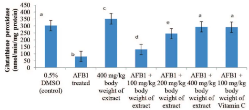
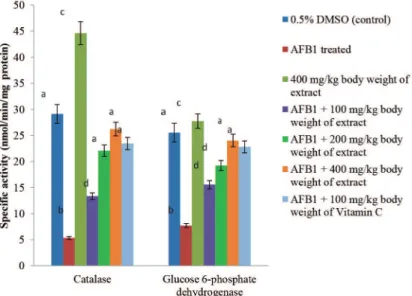
AFB1 administration significantly (p < 0.05) decrease the activities of SOD, catalase, GSH-Px, GSH-red and Glc 6-PD by 69.16, 81.74, 73.48, 63.64 and 69.74%, respectively, when compared to the control. These enzymes were significantly (p < 0.05) induced by P. biglobosa pulp extract in a dose dependent manner. The inductions significantly (p < 0.05) attenuated AFB1-mediated decrease in these activities (Figs. 1-3); with the highest dose of the extract, 400 mg/kg bodyweight, producing 67.11, 79.70, 72.77, 59.58 and 67.89%
reversal of AFB1-mediated decrease in SOD, catalase, GSH-Px, GSH-red and Glc 6-PD respectively.
Non-enzymatic antioxidant
The level of GSH reduced by 54.61% in the liver of AFB1 -intoxicated rats when compared to controls (p < 0.05). Conversely, the level of GSSG increased significantly (p < 0.05) when compared to the control. This trend was significantly reversed in rats pre-treated with phenolic extracts of
P. biglobosa pulp in a dose-dependent manner. The highest dose used in this study produced a 53.35% reversal of AFB1– mediated reduction in GSH. All the extract treated groups compared favourably (p < 0.05) with vitamin C (Table 2).
Lipid peroxidation products, protein carbonyl and DNA fragmentation
AFB1-mediated elevation in the concentrations of oxidative stress biomarkers; malondialdehyde, conjugated dienes, lipid hydroperoxides, protein carbonyl and percentage DNA fragmentation were significantly (p < 0.05) lowered by phenolic extracts of P. biglobosa pulp (Tables 3 and 4).
Figure 1 – Specific activity of glutathione peroxidase following six weeks of administration of phenolic extract of Parkia bliglobosa
Treatments Reduced glutathione (nmol/mgprotein)
Peroxidized glutathione
(nmol/mgprotein) GSH: GSSG ratio
0.5% DMSO (control) 62.52 ± 0.15a 6.54 ± 0.11a 9.56 ± 0.18a
AFB1 treated 28.38 ± 0.32b 22.62 ± 1.10b 1.25 ± 0.18b
400 mg/kg body weight of extract 91.31 ± 0.43c 5.50 ± 0.11a 16.60 ± 0.11c
AFB1 + 100 mg/kg body weight of extract 37.06 ± 1.24d 16.48 ± 0.27c 2.25 ± 0.01d
AFB1 + 200 mg/kg body weight of extract 56.66 ± 3.62a 10.89 ± 0.32d 5.20 ± 0.15e
AFB1 + 400 mg/kg body weight of extract 60.83 ± 2.25e 6.96 ± 0.57a 8.74 ± 0.09a
AFB1 + 100 mg/kg body weight of Vitamin C 64.23 ± 1.18a 6.40 ± 0.30e 10.04 ± 0.12a
Data are expressed as mean of five determinations ± SD. Specific enzymatic activities are expressed as nmol/min/mgprotein. Values with superscripts different for the liver and serum of each enzyme are significantly different (p < 0.05).
Table 2
Levels of non-enzymatic antioxidants following six weeks of administration of phenolic extract of Parkia bliglobosa pulp to aflatoxin B1-treated rats.
Figure 2 – Specific activities of SOD and Glutathione reductase following six weeks administration of phenolic extract of Parkia
bliglobosa pulp to aflatoxin B1-treated rats. Values are mean ± standard deviation (SD) of five determinations. abcdeBars with dif-ferent superscripts for each parameter are significantly difdif-ferent (p < 0.05).
Discussion
Investigations into the phytochemical constituents of fruits, medicinal plants and vegetables are important for nutraceutics and phytomedicine development (Oloyede et al., 2013). This study investigated the capability of the phenolic extract of
Parkia biglobosa (Jacq.) R. Br. ex G. Don, Fabaceae, pulp to stall oxidative rout mediated by AFB1.
Hepatocellular marker enzymes
Alterations in the activities of cellular enzymes in both tissue and serum provide a valuable tool for evaluating cellular toxicity. The significant decrease in specific activity of ALP in the liver of AFB1-treated rats with a corresponding increase in the serum indicates loss of plasma membrane integrity. Studies have reported disruption of ordered bilayer of membrane in AFB1 toxicity (Ajiboye et al., 2013; Adeleye et al., 2014), which could be due to peroxidation o f p o ly u n s a t u ra t e d f a t t y a c i d c o m p o n e n t s o f t h e membranes by ROS (O2•-, •OH and H
2O2) generated during AFB1 metabolism (Towner et al., 2003). The prevention
of AFB1-mediated alteration in ALP could be attributed to the inherent antioxidants present in the extract such as phenolics and flavonoids, capable of scavenging free radicals and ROS (Compaoré et al., 2011).
The activity of ALT (cytosolic) and AST (cytosolic and mitochondrial) is useful to confirm the integrity of the plasma membrane. The reduction in specific activities of ALT and AST in the liver of AFB1-treated rats corroborates the indication of compromised plasma membrane as revealed by the alteration in ALP. This is so because damage to plasma membrane results to leakage of cytosolic content of cell to the external milieu. The reversal of ALT and AST in both liver and serum by P. biglobosa pulp extract further substantiate protection of the membrane by the phenolic and flavonoid constituent of the extract.
Reactive oxygen species detoxifying enzymes
The decrease in specific activities of antioxidant enzymes (SOD, catalase, GSH-Px, GSH-red and Glc 6-PD) could be due to excessive mobilization of antioxidant enzymes towards detoxification of ROS (O2•-, •OH, RO
2• and H2O2) during AFB1 metabolism (Towner
Treatments Conjugated dienes
(nmol/mgprotein)
Lipid hydroperoxide (nmol/mgprotein)
Malondialdehyde (nmol/mgprotein)
0.5% DMSO (control) 35.23 ± 0.20a 23.35 ± 0.91a 6.26 ± 0.23a
AFB1 treated 92.03 ± 1.53b 117.26 ± 0.27b 20.13 ± 0.62b
400 mg/kg body weight of extract 25.12 ± 0.73a 17.84 ± 0.18a 5.01 ± 0.17a
AFB1 + 100 mg/kg body weight of extract 71.12 ± 1.21c 84.71 ± 0.58c 15.23 ± 0.31c
AFB1 + 200 mg/kg body weight of extract 57.31 ± 0.44d 55.70 ± 1.72c 10.07 ± 0.36d
AFB1 + 400 mg/kg body weight of extract 40.92 ± 4.96e 38.18 ± 0.82a 6.43 ± 0.36e
AFB1 + 100 mg/kg body weight of Vitamin C 43.62 ± 1.08e 33.10 ± 0.14d 6.22 ± 0.46a
Data are expressed as mean of five determinations ± SD. Specific enzymatic activities are expressed as nmol/min/mgprotein. Values with superscripts different for the liver and serum of each enzyme are significantly different (p < 0.05).
Treatments Protein carbonyl
(nmol/mgprotein) Fragmented DNA (%)
0.5% DMSO (control) 3.04 ± 0.02a 4.93 ± 0.31a
AFB1 treated 11.84 ± 0.13b 90.34 ± 0.35b
400 mg/kg body weight of extract 2.02 ± 0.01c 5.04 ± 0.02a
AFB1 + 100 mg/kg body weight of extract 8.14 ± 0.25b 55.72 ± 1.44c
AFB1 + 200 mg/kg body weight of extract 5.02 ± 0.24c 34.62 ± 0.23d
AFB1 + 400 mg/kg body weight of extract 3.72 ± 0.20a 10.49 ± 0.16a
AFB1 + 100 mg/kg body weight of Vitamin C 3.55 ± 0.15a 13.81 ± 0.32e
Data are expressed as mean of five determinations ± SD. Specific enzymatic activities are expressed as nmol/min/mgprotein. Values with superscripts different for the liver and serum of each enzyme are significantly different (p < 0.05).
Table 3
Levels of lipid peroxidised products following six weeks of administration of phenolic extract of Parkia bliglobosa pulp to aflatoxin B1-treated rats.
Table 4
et al., 2003). This can lead to uncontrolled oxidative attack on cellular macromolecules (lipid, protein, DNA etc.) and eventually cell death (Ajiboye et al., 2010). Similar reduction in activity of SOD, CAT, GSH-Px and GSH-Red (ROS detoxifying enzymes) were reported to be due to excessive generation of ROS during AFB1 hepatocarcinogenesis (Ramakrishnan et al., 2006; Yadav and Bhatnagar, 2007; Sivaramakrishnan et al., 2008; Ajiboye et al., 2013). Thus, significant attenuation of AFB1-mediated reduction in specific activities of ROS detoxifying enzymes by P. biglobosa pulp extract indicates a protective activity. This could be explained by the capability of the extract to scavenge ROS or enhance ROS detoxifying enzymes.
Non-enzymatic antioxidants
The significant (p < 0.05) reduction in GSH levels, a non-enzymatic antioxidant playing a complementary role in preventing ROS-induced oxidative damage, might have resulted from the depletion of GSH-Px and GSH-Red, as they have direct relationship with GSH (Kozer et al., 2003). Conversely, AFB1-mediated increase in the level of GSSG might have resulted from the oxidation of GSH or mobilization of GSH towards production of GSH-Px. The reduction in GSH:GSSG following administration of AFB1 suggests oxidative attack on liver cells. Thus, the preservation of GSH, high GSH:GSSG and low GSSG levels in the liver of AFB1-treated rats following administration of
P. biglobosa pulp extract indicates possible antioxidant activity.
Lipid peroxidation
AFB1 has been reported to induce lipid peroxidation both
in vitro and in vivo models of AFB1-induced hepatotoxicity (Shen et al., 1994; Towner et al., 2003; Theumer et al., 2010; Ravinayagam et al., 2012; Ajiboye et al., 2013). Thus, the significant increase in the levels of lipid peroxidation products (conjugated dienes, lipid hydroperoxides and malondialdehydes) shows indiscriminate oxidative assaults on cellular lipids. This can lead to disturbances of the membrane organization, functional loss and modification of proteins and DNA bases (Niki, 2009). The increase in conjugated dienes could result in mutation (Das et al., 2010). The reduction of AFB1-mediated increases in conjugated dienes, lipid hydroperoxide and malondialdehyde by phenolic extract of P. biglobosa shows protection of the membrane lipids. This might have also resulted from the capability of the extract to promote detoxification (through the induction of antioxidant enzymes) of ROS, which could cause the peroxidation of polyunsaturated fatty acids of the plasma membrane.
Protein oxidation
The significant increase in protein carbonyl, a marker of protein oxidation in the AFB1-treated rat could have resulted from the protein oxidation by the free radicals and generated ROS during AFB1 metabolism. This increase indicates irreversible oxidative damage to cellular proteins
(Dalle-Donne et al., 2003). The attenuation of AFB1-mediated increase in the levels of protein carbonyl by phenolic extract of P. biglobosa pulp further shows possible capability to promote the detoxification of ROS via the induction of antioxidant enzymes. Similar attenuation of AFB1-mediated increase in protein carbonyl level following the administration of Tridham
has been reported (Ravinayagam et al., 2012).
DNA fragmentation
Oxidative stress and accumulation of calcium ions have been reported to mediate DNA fragmentation (Amin and Hamza, 2005). This damage, which usually results from OH- can lead to either the arrest or induction of transcription, induction of signal transduction pathways, replication errors and genomic instability, all of which are associated with carcinogenesis (Cooke et al., 2006). Thus, the significant increase in the levels of fragmented DNA in the liver of AFB1-treated rats proves genotoxic activity. It also denotes possible initiation of carcinogenesis. Golli-Bennour et al. (2010) reported a similar increase in fragmented DNA in AFB1-treated rats. The reduction in the levels of fragmented DNA in the liver of AFB1-treated rat by phenolic extract of P. biglobosa pulp shows antioxidant and antigenotoxic activities. The extract could have acted as an antigenotoxic complex, which enhances the DNA repair system or DNA synthesis (Brahmi et al., 2011).
Conclusion
It is evident from this study that the phenolic extract of P. biglobosa pulp enhanced the detoxification of AFB1, possibly by enhancing the activities of reactive oxygen species detoxifying enzymes, thus preventing the oxidation and fragmentation of cellular macromolecules such as DNA, lipids and proteins as well as AFB1 induced redox imbalance. Hence, the use of the phenolic extract of P. biglobosa pulp shows promising potentials as a dietary supplement/ functional food due to its prophylactic role.
Funding
The authors received no financial support for the research, authorship, and/or publication of this article.
Authors’ contributions
Conflicts Interests
The authors declare no potential conflicts of interest with respect to the research, authorship, and/or publication of this article.
R E F E R E N C E S
Adeleye, A.O., Ajiboye, T.O., Iliasu, G.A., Abdussalam, F.A., Balogun, A., Ojewuyi, O.B., Yakubu, M.T., 2014. Phenolic extract of Dialium guineense pulp enhances reactive oxygen species detoxification in aflatoxin B1 hepatocarcinogenesis. J. Med. Food 17, 875-885.
Aebi, H., 1984. Catalase in vitro. Methods Enzymol. 105, 121-126. Ajiboye, T.O., Raji, H.O., Muritala, H.F., Ojewuyi, O.B., Yakubu,
M.T., 2013. Anthocyanin extract of Lannea microcarpa fruits stall oxidative rout associated with aflatoxin B1 hepatocarcinogenesis. Food Biosci. 4, 58-67.
Ajiboye, T.O., Salau, A.K., Yakubu, M.T., Oladiji, A.T., Akanji, M. a., Okogun, J.I., 2010. Acetaminophen perturbed redox homeostasis in Wistar rat liver: protective role of aqueous Pterocarpus osun leaf extract. Drug Chem. Toxicol. 33, 77-87. Ajiboye, T.O., Yakubu, M.T., Oladiji, A.T., 2014. Electrophilic and
reactive oxygen species detoxification potentials of chalcone dimers is mediated by redox transcription factor Nrf-2. J. Biochem. Mol. Toxicol. 28, 11-22.
Amin, A., Hamza, A.A, 2005. Oxidative stress mediates drug-induced hepatotoxicity in rats: a possible role of DNA fragmentation. Toxicology 208, 367-375.
Bankole, S., Schollenberger, M., Drochner, W., 2006. Mycotoxins in food systems in Sub Saharan Africa: A review. Mycotoxin Res. 22, 163-169.
Bergmeyer, H.U., Hørder, M., Rej, R., 1986a. International Federation of Clinical Chemistry (IFCC) Scientific
Committee, Analytical Section: approved recommendation (1985) on IFCC methods for the measurement of catalytic concentration of enzymes. Part 2. IFCC method for aspartate aminotransferase. (L-aspartate:2- oxoglutarate aminotransferase, EC 2.6.1.1) J. Clin. Chem. Clin. Biochem. 24, 497-510.
Bergmeyer, H.U., Hørder, M., Rej, R., 1986b. International
Federation of Clinical Chemistry (IFCC) Scientific Committee, Analytical Section: approved recommendation (1985) on IFCC methods for the measurement of catalytic concentration of enzymes. Part 3. IFCC method for alanine aminotransferase (L-alanine: 2-oxoglutarate aminotransferase, EC 2.6.1.2). J. Clin. Chem. Clin. Biochem. 24, 481-495.
Brahmi, D., Bouaziz, C., Ayed, Y., Ben Mansour, H., Zourgui, L., Bacha, H., 2011. Chemopreventive effect of cactus Opuntia ficus indica on oxidative stress and genotoxicity of aflatoxin B1. Nutr. Metab. (Lond). 8, 73, doi: 10.1186/1743-7075-8-73.
Burton, K., 1956. A study of the conditions and mechanism of the diphenylamine reaction for the colorimetric estimation of deoxyribonucleic acid. Biochem. J. 62, 315-323.
Campbell‐Platt, G., 1980. African locust bean (Parkia species) and its west african fermented food product, dawadawa. Ecol. Food Nutr. 9, 123-132.
Choi, C.Y., Park, S.H., Park, E.H., Cha, Y.N., 1991. Alteration of aflatoxin B1 metabolic profiles and reduction of aflatoxin B1 mutagenicity by hepatic microsomes of rats fed butylated hydroxyanisole. J. Toxicol. Sci. 16 Suppl 1, 119-132.
Compaoré, W.R., Nikièma, P.A., Bassolé, H.I.N., Savadog, A., Mouecoucou, J., 2011. Chemical composition and antioxidative properties of seeds of Moringa oleifera and pulps of Parkia biglobosa and Adansonia digitata commonly used in food fortification in Burkina Faso. Curr. Res. J. Biol. Sci. 3, 64-72. Cooke, M.S., Olinski, R., Evans, M.D., 2006. Does measurement of
oxidative damage to DNA have clinical significance ? Clin. Chim. Acta 365, 30-49.
Dalle-Donne, I., Rossi, R., Giustarini, D., Milzani, A., Colombo, R., 2003. Protein carbonyl groups as biomarkers of oxidative stress. Clin. Chim. Acta. 329, 23-38.
Das, A.K., Bag, S., Sahu, R., Dua, T.K., Sinha, M.K., Gangopadhyay, M., Zaman, K., Dewanjee, S., 2010. Protective effect of Corchorus olitorius leaves on sodium arsenite-induced toxicity in experimental rats. Food Chem. Toxicol. 48, 326-335. Djakpo, O., 2005. Fermentation contrôlée des graines de néré
(Parkia biglobosa) pour la production d’un condiment béninois de type afitin: effets del’utilisation des souches sélectionnées de Bacillus subtilis sur la qualité du produit. Thèse d’ingénieur Agronome Université d’Abomey-Calavi, Faculté des Sciences Agronomiques; 2005.
Ellman, G.L., 1959. Tissue sulfhydryl groups. Arch. Biochem. Biophys. 82, 70-77.
Golli-Bennour, E. El, Kouidhi, B., Bouslimi, A., Abid-Essefi, S., Hassen, W., Bacha, H., 2010. Cytotoxicity and genotoxicity induced by aflatoxin B1, ochratoxin A, and their combination in cultured Vero cells. J. Biochem. Mol. Toxicol. 24, 42-50. Hissin, P.J., Hilf, R., 1976. A fluorometric method for determination
of oxidized and reduced glutathione in tissues. Anal. Biochem. 74, 214-226.
Kensler, T.W., Egner, P.A., Davidson, N.E., Roebuck, B.D., Pikul, A., Groopman, J.D., 1986. Modulation of aflatoxin metabolism, aflatoxin-N7-guanine formation, and hepatic tumorigenesis in rats fed ethoxyquin: role of induction of glutathione S-transferases. Cancer Res. 46, 3924-3931.
Kozer, E., Evans, S., Barr, J., Greenberg, R., Soriano, I., Bulkowstein, M., Petrov, I., Chen-Levi, Z., Barzilay, B., Berkovitch, M., 2003. Glutathione, glutathione-dependent enzymes and antioxidant status in erythrocytes from children treated with high-dose paracetamol. Br. J. Clin. Pharmacol. 55, 234-240. Lee, J., Hye, E., Lee, K., Sook, H., 2005. Alleviation of aflatoxin B 1
-induced oxidative stress in HepG2 cells by volatile extract from Allii Fistulosi Bulbus Life Sci. 77, 2896-2910.
Levine, R.L., Garland, D., Oliver, C.N., Amici, A., Clement, I., 1990. Determination of carbonyl content in oxidatively modified proteins. Methods Enzymol. 186, 464-478.
Misra, H.P., Fridovich, I., 1972. The role of superoxide anion in the autoxidation of epinephrine and a simple assay for superoxide dismutase. J. Biol. Chem. 247, 3170-3175. Muto, S., Fujita, K., Yamazaki, Y., Kamataki, T., 2001. Inhibition by
green tea catechins of metabolic activation of procarcinogens by human cytochrome P450. Mutat. Res. 479, 197-206. Niki, E., 2009. Lipid peroxidation: physiological levels and dual
biological effects. Free Radic. Biol. Med. 47, 469-484. NRC, 2011. Guide for the Care and Use of Laboratory Animals:
8th Ed., in: Guide for the Care and Use of Laboratory Animals. National Research Council, p. 118.
Oloyede, H.O.B., Ajiboye, T.O., Komolafe, Y.O., Salau, A.K., 2013. Polyphenolic extract of Blighia sapida arilli prevents N-nitrosodiethylamine-mediated oxidative onslaught on microsomal protein, lipid and DNA. Food Biosci. 1, 48-56. Qin, G., Gopalan-kriczky, P., Su, J., Ning, Y., Lotlikar, P.D.,
1997. Inhibition of aflatoxin B1-induced initiation of
Ramakrishnan, G., Raghavendran, H.R.B., Vinodhkumar, R., Devaki, T., 2006. Suppression of N-nitrosodiethylamine induced hepatocarcinogenesis by silymarin in rats. Chem. Biol. Interact. 161, 104-114.
Rastogi, R., Srivastava, A.K., Rastogi, A.K., 2001. Long term effect of aflatoxin B(1) on lipid peroxidation in rat liver and kidney: effect of picroliv and silymarin. Phytother. Res. 15, 307-310. Rastogi, R., Srivastava, A.K., Srivastava, M., Rastogi, A.K., 2000.
Hepatocurative effect of picroliv and silymarin against aflatoxin B1 induced hepatotoxicity in rats. Planta Med. 66, 709-713.
Ravinayagam, V., Jaganathan, R., Panchanadham, S., Palanivelu, S., 2012. Potential antioxidant role of tridham in
managing oxidative stress against aflatoxin-B 1-induced experimental hepatocellular carcinoma. Int. J. Hepatol. DOI: 10.1155/2012/428373.
Reilly, C.A., Aust, S.D., 2001. Measurement of lipid peroxidation. Curr. Protoc. Toxicol. Chapter 2, Unit 2.4.
Scholl, P.F., Musser, S.M., Groopman, J.D., 1997. Synthesis and characterization of aflatoxin B1 mercapturic acids and their identification in rat urine. Chem. Res. Toxicol. 10, 1144-1151. Shen, H.M., Ong, C.N., Shi, C.Y., 1995. Involvement of reactive
oxygen species in aflatoxin B1-induced cell injury in cultured rat hepatocytes. Toxicology 99, 115-123.
Shen, H.M., Shi, C.Y., Lee, H.P., Ong, C.N., 1994. Aflatoxin B1 -induced lipid peroxidation in rat liver. Toxicol. Appl. Pharmacol. 127, 145-150.
Shen, H.M., Shi, C.Y., Shen, Y., Ong, C.N., 1996. Detection of elevated reactive oxygen species level in cultured rat hepatocytes treated with aflatoxin B1. Free Radic. Biol. Med. 21, 139-146.
Sivaramakrishnan, V., Shilpa, P.N.M., Praveen Kumar, V.R., Niranjali Devaraj, S., 2008. Attenuation of N-nitrosodiethylamine-induced hepatocellular carcinogenesis by a novel flavonol-Morin. Chem. Biol. Interact. 171, 79-88.
Sosulski, F., Krygier, K., Hogge, L., 1982. Free, esterified, and insoluble-bound phenolic acids. 3. Composition of phenolic acids in cereal and potato flours. J. Agric. Food Chem. 30, 337-340.
Spanos, G.A., Wrolstad, R.E., Heatherbell, D.A., 1990. Influence of processing and storage on the phenolic composition of apple juice. J. Agric. Food Chem. 38, 1572-1579.
Theumer, M.G., Cánepa, M.C., López, A.G., Mary, V.S., Dambolena, J.S., Rubinstein, H.R., 2010. Subchronic mycotoxicoses in Wistar rats : Assessment of the in vivo and in vitro genotoxicity induced by fumonisins and aflatoxin B 1, and oxidative stress biomarkers status. Toxicology 268, 104-110. Towner, R.A., Qian, S.Y., Kadiiska, M.B., Mason, R.P., 2003. In vivo identification of aflatoxin-induced free radicals in rat bile. Free Radic. Biol. Med. 35, 1330-1340.
Wang, C.J., Hsu, J.D., Lin, J.K., 1991. Suppression of aflatoxin B1-induced hepatotoxic lesions by crocetin (a natural carotenoid). Carcinogenesis 12, 1807-1810.
Wang, J.S., Groopman, J.D., 1999. DNA damage by mycotoxins. Mutat. Res. 424, 167-181.
World Health Organization, 1998. Basic OECD principles of GLP. Geneva, Switzerland: World Health Organization. Online at: http://www.who/int/tdr/publications 02-01-2008.
Wright, P.J., Leathwood, P.D., Plummer, D.T., 1972. Enzymes in rat urine: alkaline phosphatase. Enzymologia 42, 317-327. Yadav, A.S., Bhatnagar, D., 2007. Modulatory effect of spice
extracts on iron-induced lipid peroxidation in rat liver. Biofactors 29, 147-157.
Yakubu, M.T., Oladiji, A.T., Akanji, M.A., 2009. Mode of cellular toxicity of aqueous extract of Fadogia agrestis (Schweinf. Ex Hiern) stem in male rat liver and kidney. Hum. Exp. Toxicol. 28, 469-478.
Yates, M.S., Kwak, M.-K., Egner, P. a, Groopman, J.D., Bodreddigari, S., Sutter, T.R., Baumgartner, K.J., Roebuck, B.D., Liby, K.T., Yore, M.M., Honda, T., Gribble, G.W., Sporn, M.B., Kensler, T.W., 2006. Potent protection against aflatoxin-induced tumorigenesis through induction of Nrf2-regulated pathways by the triterpenoid 1-[2-cyano-3-,12-dioxooleana-1,9(11)-dien-28-oyl]imidazole. Cancer Res. 66, 2488-2494.