Licenciatura em Química Aplicada
Structural and functional studies of
PpcA: a key protein in the electron
transfer pathways of
Geobacter sulfurreducens
Dissertação para obtenção do Grau de Doutor em Bioquímica, ramo de Biotecnologia
Orientador: Prof. Carlos A. Salgueiro, Professor Auxiliar,
Faculdade de Ciências e Tecnologia, Universidade Nova de Lisboa
Júri:
Presidente: Prof. Doutor Fernando Jorge da Silva Pina Arguentes: Prof. Doutor Brian James Goodfellow
Doutora Inês Antunes Cardoso Pereira Vogais: Prof. Doutora Marta Bruix Bayés
Prof. Doutor Carlos Alberto Gomes Salgueiro
Maria Leonor Carvalho Morgado
Licenciatura em Química Aplicada
Structural and functional studies of PpcA: a key
protein in the electron transfer pathways of
Geobacter sulfurreducens
Dissertação para obtenção do Grau de
Doutor em Bioquímica, ramo de Biotecnologia
Orientador: Prof. Carlos A. Salgueiro, Professor Auxiliar,
Faculdade de Ciências e Tecnologia, Universidade Nova de Lisboa
iii
Structural and functional studies of PpcA: a key protein in the
electron transfer pathways of Geobacter sulfurreducens
“Copyright”
Maria Leonor Carvalho Morgado Faculdade de Ciências e Tecnologia Universidade Nova de Lisboa
Todos os capítulos foram parcialmente reproduzidos de artigos publicados sob permissão dos editores originais e sujeitos às restrições de cópia impostos pelos mesmos.
A Faculdade de Ciências e Tecnologia e a Universidade Nova de Lisboa têm o direito, perpétuo e sem limites geográficos, de arquivar e publicar esta dissertação através de exemplares impressos reproduzidos em papel ou de forma digital, ou por qualquer outro meio conhecido ou que venha a ser inventado, e de a divulgar atraves de repositórios científicos e de admitir a sua cópia e distribuição com objectivos educacionais ou de investigação, não comerciais, desde que seja dado crédito ao autor e editor.
v
A
GRADECIMENTOS
Apesar de uma tese ser apresentada em nome individual, esta resulta de um trabalho em equipa, e só foi possível devido ao trabalho e dedicação de diversas pessoas, às quais não posso deixar de agradecer.
Em primeiro lugar, ao meu orientador Prof. Doutor Carlos Salgueiro. Agradeço por ter acreditado nas minhas capacidades desde o início, por me ter iniciado no mundo da ciência e ter orientado ao longo deste trabalho. Agradeço a sua dedicação, disponibilidade, amizade, as inúmeras e infindáveis discussões, e por no final nunca desistir. Aprendi muito ao longo destes anos, especialmente a fazer um bom trabalho. E aprendi que quando se trabalha em equipa, trabalha-se melhor.
Devo um agradecimento especial à Prof. Doutora Marta Bruix, por me ter recebido por diversas vezes no seu laboratório no Instituto de Química-Física ‘Rocasolano’, Consejo Superior de Investigaciones Científicas em Madrid, e por me ter orientado e iniciado no mundo dos espectros de RMN tri-dimensionais. A sua confiança e entusiasmo são contagiantes e muito motivadores.
Ao Doutor Vítor Paixão agradeço toda a ajuda durante os cálculos para a determinação da estrutura do citocromo PpcA. Sem a sua ajuda, seria uma história sem fim.
Agradeço ao Prof. Doutor Derek R. Lovley a oportunidade de desenvolver parte deste projecto no seu laboratório na University of Massachusetts, integrado no Geobacter Project. Ao Doutor Muktak Aklukjar agradeço por me ter orientado e pela sua persistência. Agradeço ao Ashley Franks, Manju Sharma, Joy Ward, Ching Leang, Sarah, Roberto, Joanne e Zara pela sua disponibilidade e amizade durante a minha estadia em Amherst.
Agradeço à Prof. Doutora Marianne Schiffer, ao Doutor P. Raj. Pokkuluri e ao Doutor. Yuri Y. Londer do Argonne National Laboratory pela disponibilização das proteínas e dos plasmídeos, e as inúmeras discussões científicas.
Ao Prof. Doutor David L. Turner agradeço a disponibilização dos programas de cálculo para determinação dos parâmetros termodinâmicos de proteínas multihémicas e da estrutura de proteínas. Agradeço também a sua ajuda nos cálculos preliminares da estrutura do PpcA no estado paramagnético oxidado.
Agradeço ao Doutor Ricardo O. Louro e à Prof. Doutora Teresa Catarino as diversas sugestões e discussões quer de carácter experimental, quer de carácter teórico, e a disponibilidade na logística da utilização da câmara anaeróbica no ITQB.
Agradeço à Prof. Doutora Isabel Couto pelo trabalho no desenvolvimento do protocolo de expressão para marcação isotópica de proteínas e pela ajuda e disponibilidade durante a implementação das ferramentas de Biologia Molecular no laboratório.
Agradeço ao Laboratório de RMN da FCT-UNL, integrado na Rede Nacional de RMN e financiado pela Fundação para a Ciência e Tecnologia através Projecto de Re-Equipamento
vi
Científico (REDE/1517/RMN/2005), pelo acesso aos espectrómetros de RMN. Agradeço a disponibilidade do Aldino Viegas, Daniel Jana, Prof. Doutor Eurico Cabrita e Doutor Ângelo Figueiredo, durante a utilização dos espectrómetros. Ao Daniel e ao Ângelo agradeço também toda a ajuda a nível informático na instalação dos programas de cálculo.
A todos os que passaram pelo Lab. 611 agradeço a ajuda, a disponibilidade, a amizade, os inóculos matinais, por aturarem os meus dias de mau feitio, e todos os cafés, bolachas e palhaçadas. Ana, André, Sílvia, Joana, Marta, Marta, Eduardo e Verónica: obrigada. Agradeço em especial à Ana pelo trabalho no desenvolvimento do protocolo de expressão para marcação isotópica de proteínas e pelas inúmeras ajudas na expressão das diferentes proteínas; à Sílvia e à Joana que trabalharam na caracterização de alguns dos mutantes; à Marta pela ajuda durante os crescimentos, e à Verónica por se ter aventurado comigo na primeira clonagem feita neste laboratório.
Agradeço aos vários colegas do DQ (não nomeio para que não falhe ninguém) que estiveram presentes ao longo destes anos, e que me ajudaram de alguma forma, quer na utilização de um equipamento, quer no empréstimo de um reagente, ou apenas pelas conversas de corredor. Estes anos também não teriam sido os mesmos sem os almoços com o Gang do Tupperware. Agradeço a companhia e a animação durantes os nossos almoços.
Não posso deixar de agradecer à minha família, por serem uma parte fundamental da minha vida. À minha Mãe e ao meu Pai agradeço todo o apoio e por quererem sempre saber como se estavam a portar as proteínas e as bactérias, e como o trabalho estava a correr. Agradeço à minha irmã Ana Luísa e ao João por estarem sempre por perto, e por me terem “dado” dois sobrinhos lindos durante estes anos. É por estas coisas que a vida vale a pena. Agradeço aos meus Avós por, mesmo não percebendo bem o que faço, saber que os faço sentir orgulhosos. De todos senti o apoio e a força, principalmente quando estava longe ou quando as coisas não corriam como esperado.
Aos meus amigos, e em especial às minhas grandes amigas, que sabem bem que não tenho jeito para estas coisas, penso que um obrigado diz tudo. Estiveram sempre comigo, e sei que vão continuar a estar.
Ao Filipe, obrigada por estares sempre ao meu lado, em tudo.
Por último, gostaria de agradecer o financiamento à Fundação para a Ciência e a Tecnologia através da Bolsa de Doutoramento SFRH/BD/37415/2007 e do Projecto PTDC/QUI/70182/2006 atribuído ao Prof. Carlos Salgueiro, e à Fundação das Universidades Portuguesas através da Acção Integrada E-69/07.
vii
A
BSTRACT
Geobacter species show an impressive respiratory versatility and can sustain their
growth by using insoluble extracellular compounds as terminal electron acceptors. The genome of Geobacter sulfurreducens has been completely sequenced and it revealed an unprecedented number of putative c-type cytochromes, 73 of which with more than one heme binding site. Although numerous electron transfer proteins have been identified, the electron transfer pathways that allow G. sulfurreducens to obtain energy are still far from being understood.
Five homologous triheme cytochromes (PpcA-E) were identified in G. sulfurreducens periplasm and gene knockout studies revealed their involvement in Fe(III) and U(VI) extracellular reduction. This thesis focuses on the characterization of these proteins, with special emphasis on PpcA, the most abundant in G. sulfurreducens’ periplasm.
PpcA, PpcB, PpcD and PpcE were thermodynamically characterized in detail using Nuclear Magnetic Resonance and ultraviolet-visible spectroscopy. The results obtained showed that PpcA and PpcD were able to perform e-/H+ energy transduction in addition to
their role in the electron transfer pathways. No evidence for coupling of e-/H+ transfer was
observed for PpcB and PpcE. The functional implications of these results are discussed. PpcA solution structure in the fully reduced state was determined using NMR spectroscopy and the redox-Bohr center responsible for controlling the e-/H+ transfer was
identified, as well as the putative interacting regions between PpcA and its redox partners. In order to elucidate the physiologic function of PpcA individual key residues and understand its functional mechanism, a family of mutants covering the entire protein was prepared using site-directed mutagenesis. The results obtained revealed how proper tuning of the reduction potentials of the heme groups is fundamental to achieve concerted e-/H+
transfer.
Keywords: Geobacter, electron transfer, multiheme cytochromes, NMR, protein
ix
R
ESUMO
As bactérias do género Geobacter apresentam uma versatilidade respiratória impressionante. Estas conseguem crescer usando compostos extracelulares insolúveis como aceitadores finais de electrões. O genoma de Geobacter sulfurreducens foi sequenciado e possui um número nunca antes observado de possíveis citocromos do tipo c, 73 dos quais contendo mais do que um local de ligação para grupos hemo. Apesar de inúmeras proteínas de transferência electrónica terem sido identificadas, pouco se sabe sobre os caminhos de transferência electrónica que permitem a esta bactéria obter energia para o seu crescimento.
Cinco citocromos trihémicos homólogos (PpcA-E) foram identificados no periplasma de
G. sulfurreducens e estudos de crescimento com deleção dos genes que os codificam
revelaram o seu envolvimento na redução extracelular de Fe(III) e U(VI). Esta tese tem como objectivo a caracterização destas proteínas, com especial relevo no citocromo PpcA, o mais abundante no periplasma de G. sulfurreducens.
Os citocromos PpcA, PpcB, PpcD and PpcE foram caracterizados termodinamicamente pela utilização de espectroscopias de Ressonância Magnética Nuclear e ultravioleta-visível. Os resultados obtidos mostram que os citocromos PpcA e PpcD têm as propriedades necessárias para efectuar transdução de energia e-/H+ para além do seu papel nos caminhos
de transferência electrónica. No entanto, não foram encontradas evidências para a transferência acoplada e-/H+ para os citocromos PpcB e PpcE. As implicações funcionais
destes resultados são discutidas.
A estrutura do citocromo PpcA em solução no estado reduzido foi determinada por espectroscopia de RMN e o centro redox-Bohr responsável pelo controlo da transferência acoplada e-/H+ foi identificado, assim como as possíveis regiões de interacção entre o PpcA e
os seus parceiros redox.
Com o objectivo de elucidar a função fisiológica de resíduos individuais no citocromo PpcA e compreender o seu mecanismo funcional, uma familía de proteínas mutadas em diversos resíduos em toda a proteína foi preparada por mutagénese dirigida. Os resultados obtidos revelaram que a regulação precisa dos potenciais de redução dos grupos hemo é essencial para que ocorra transferência acoplada de e-/H+.
Palavras chave: Geobacter, transferência de electrões, citocromos multihémicos,
xi
T
ABLE OF CONTENTS
1. INTRODUCTION ... 3
1.1 The bacterium Geobacter sulfurreducens ... 3
1.2 Extracellular electron transfer ... 4
1.3 Gene knockout and proteomic studies in G. sulfurreducens ... 4
1.4 Periplasmic cytochromes ... 7
1.5 Solution structures of multiheme proteins ... 11
1.6 Thesis outline ... 13
1.7 References ... 14
2. EXPERIMENTAL PROCEDURES ... 21
2.1 Protein expression and purification ... 21
2.1.1 Plasmids ... 21 2.1.2 Heterologous expression... 21 2.1.2.1 Unlabeled proteins ... 21 2.1.2.2 15N labeled proteins ... 21 2.1.3 Protein purification... 22 2.2 NMR spectroscopy ... 23 2.2.1 Sample preparation ... 23 2.2.1.1 Oxidized samples ... 23 2.2.1.2 Reduced samples ... 23
2.2.1.3 Samples for redox titrations ... 23
2.2.2 NMR Experiments ... 23
2.2.2.1 Heme core structure in the reduced state ... 23
2.2.2.2 Redox titrations ... 24
2.2.2.3 Structural studies ... 24
2.2.3 Assignment strategy and methodology ... 24
2.2.3.1 Assignment of heme signals in the reduced state ... 24
2.2.3.2 Ring-current shifts calculation ... 25
2.2.3.3 Identification of heme oxidation profiles ... 26
2.2.3.4 Assignment of protein backbone and side chain signals ... 27
2.2.4 15N relaxation measurements ... 28
2.2.5 pH titrations ... 28
2.3 Redox titrations followed by UV-visible spectroscopy ... 29
2.4 Thermodynamic model ... 29
2.5 NMR structure determination ... 31
2.5.1 Determination of restraints ... 31
2.5.2 Structure calculation and analysis ... 32
2.6 References ... 33
3. THERMODYNAMIC CHARACTERIZATION OF A TRIHEME CYTOCHROME FAMILY FROM GEOBACTER SULFURREDUCENS ... 39
3.1 Results ... 40
3.1.1 Assignment of the heme signals in the reduced form ... 40
3.1.2 Thermodynamic characterization ... 41
xii
3.2.1 Structural characterization of the heme core architecture in solution ... 51
3.2.2 Thermodynamic characterization ... 52
3.2.3 Structural map of the redox-Bohr center ... 53
3.2.4 Order of oxidation of the heme groups ... 55
3.2.5 Relevant microstates in solution ... 56
3.3 Functional implications ... 58
3.4 References ... 60
4. STRUCTURAL STUDIES ON PPCA ...65
4.1 Results ... 66
4.1.1 Backbone, side chain and heme resonance assignments in the reduced state ... 66
4.1.2 Structure calculations ... 68
4.1.3 Quality and analysis of the structures ... 70
4.1.4 15N relaxation measurements ... 72
4.1.5 pH titration ... 74
4.2 Discussion ... 75
4.2.1 Comparison of PpcA solution structure with Gsc7 crystal structures ... 75
4.2.2 Backbone dynamics ... 77
4.2.3 Heme reduction potentials and redox interactions ... 78
4.2.4 Structural close-up on PpcA redox-Bohr center: pH-linked conformational changes ... 78
4.3 Conclusions ... 80
4.4 References ... 81
5. FUNCTIONAL ROLE OF PPCA KEY AMINO ACIDS ...85
5.1 Results and discussion ... 86
5.1.1 Conserved Phe15 residue ... 87
5.1.1.1 Heme core architecture and overall structure in the reduced form ... 89
5.1.1.2 Preliminary screening of the heme oxidation profiles ... 91
5.1.1.3 Thermodynamic characterizationof PpcAF15L ... 92
5.1.1.4 Role of Phe15 in the functional mechanism of PpcA ... 94
5.1.1.5 Comparison with tetraheme cytochromes c3 ... 96
5.1.2 Met58 protects heme III from solvent exposure ... 97
5.1.2.1 Heme core architecture in the reduced form ... 98
5.1.2.2 Preliminary screening of the heme oxidation profiles ... 99
5.1.2.3 Thermodynamic characterization of PpcAM58 mutants ... 101
5.1.2.4 Role of Met58 in the functional mechanism of PpcA ... 104
5.1.3 Lysine residues near the heme groups ... 106
5.1.3.1 Heme core architecture in the reduced form ... 106
5.1.3.2 Preliminary screening of the heme oxidation profiles ... 108
5.1.3.3 Thermodynamic characterization of PpcAK52 and PpcAK60 mutants ... 110
5.1.3.4 Role of Lys52 and Lys60 in the functional mechanism of PpcA ... 114
5.2 Conclusions ... 116
5.3 References ... 117
6. ONGOINGSTUDIES ... 121
6.1 Studies on PpcA in the paramagnetic oxidized state ... 121
6.1.1 Assignment of the heme resonances ... 121
6.1.2 Solution structure determination ... 123
xiii
6.3 Interaction studies with PpcA putative redox partners ...125
6.4 References ... 26
7. FINALCONSIDERATIONS ... 131
7.1 References ...132
A. APPENDIX ... 135
A.1 Assignment of the heme resonances of PpcA, PpcB, PpcD and PpcE in the reduced form ...135
A.2 Backbone, side chain and heme resonance assignments of PpcA in the reduced state ...136
A.3 Assignment of the heme signals of PpcA mutants in the reduced form ...143
A.4 Ongoing and future studies experimental procedures ...150
A.4.1 Studies on PpcA in the paramagnetic oxidized state ... 150
A.4.1.1 Protein expression and purification ... 150
A.4.1.2 NMR spectroscopy... 150
A.4.1.2.1 Sample preparation ... 150
A.4.1.2.2 NMR experiments ... 150
A.4.1.3 Assignment strategy and methodology ... 150
A.4.1.3.1 Assignment of heme signals in the oxidized state ... 150
A.4.1.3.2 Assignments of protein backbone and side chain signals ... 151
A.4.1.4 NMR structure determination ... 152
A.4.2 In vivo studies of mutated forms of PpcA ... 152
A.4.2.1 DNA manipulations ... 153
A.4.2.2 Cloning of ppcA gene in pRG5 complementation plasmid ... 153
A.4.2.3 Site directed mutagenesis of pMA59 ... 153
A.4.2.4 Preparation of G. sulfurreducens DL3 complementation mutants ... 153
A.4.2.4.1 Culturing conditions and growth media ... 153
A.4.2.4.2 Electroporation and plating ... 154
A.4.3 Interaction studies with PpcA putative redox partners ... 154
A.4.3.1 DNA manipulations ... 154
A.4.3.2 Cloning of macA gene in vector pVA203 ... 154
A.4.3.3 MacA heterologous expression ... 155
A.5 Assignment of the heme resonances of PpcA in the oxidized form ...156
xv
F
IGURES
I
NDEX
1.INTRODUCTION
Figure 1.1 – Mechanism for extracellular electron transfer by G. sulfurreducens. ... 6
Figure 1.2 – Alignment of amino acid sequences of cytochromes c7. ... 9
Figure 1.3 – Crystal structures of the five c7 cytochromes from G. sulfurreducens. ... 10
Figure 1.4 – 1D-1H NMR spectra of reduced and oxidized triheme cytochrome PpcA obtained at 25 ºC. ... 12
2.EXPERIMENTAL PROCEDURES Figure 2.1 – Diagram of heme c numbered according to the IUPAC-IUB nomenclature. ... 25
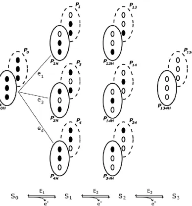
Figure 2.2 – Electronic distribution scheme for a triheme cytochrome with a proton-linked equilibrium, showing the 16 possible microstates. ... 26
Figure 2.3 – Connectivities observed in tridimensional spectra for 15N labeled proteins. ... 27
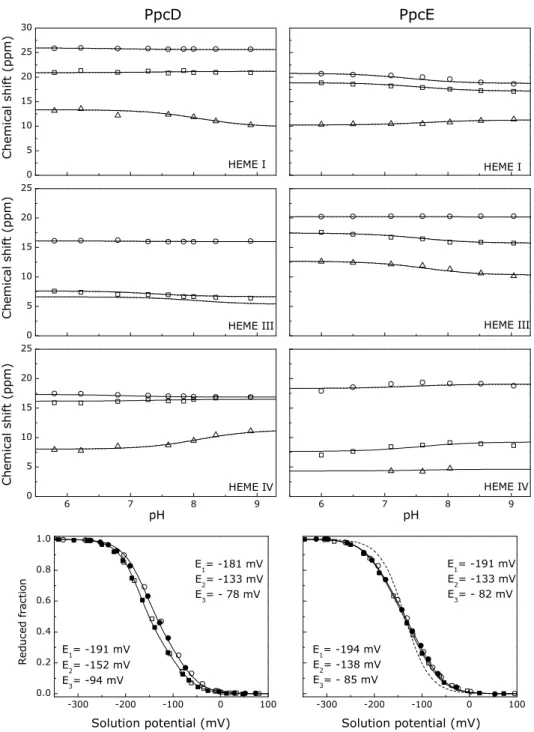
3. THERMODYNAMIC CHARACTERIZATION OF A TRIHEME CYTOCHROME FAMILY FROM GEOBACTER SULFURREDUCENS Figure 3.1 – Comparison between calculated and observed chemical shifts for the heme substituents in the reduced forms of PpcA, PpcB, PpcD and PpcE. ... 40
Figure 3.2 – Comparison between the observed heme proton chemical shifts of reduced PpcB, PpcD and PpcE with those of PpcA. ... 41
Figure 3.3 – Expansions of 2D-EXSY NMR spectra obtained at 15 °C and pH 8 for PpcA, PpcB, PpcE and PpcD at different degrees of oxidation. ... 42
Figure 3.4 – UV–visible spectra of PpcA in the fully oxidized and fully reduced states. ... 46
Figure 3.5 – Fitting of the thermodynamic model to the experimental data for PpcA, PpcB, PpcD, and PpcE. ... 47
Figure 3.6 – Heme cores of PpcB and PpcA as observed in their crystal structures. ... 51
Figure 3.7 – pH dependence for PpcB and PpcA heme IV methyl group paramagnetic shifts. ... 53
Figure 3.8 – Individual heme oxidation fractions for PpcA, PpcB, PpcD, and PpcE. ... 55
Figure 3.9 – Molar fractions of the 16 individual microstates of PpcA, PpcB, PpcD, and PpcE at pH 7.5. ... 55
Figure 3.10 – Thermodynamic and mechanistic bases for energy transduction by PpcA and PpcD. ... 58
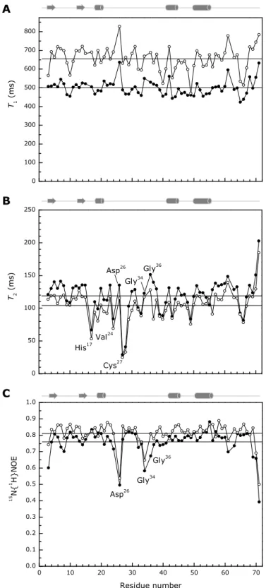
4.STRUCTURAL STUDIES ON PPCA Figure 4.1 – 1H-15N HSQC spectrum of fully reduced PpcA (1 mM protein in 45 mM sodium phosphate buffer pH 7.1, 25 °C). ... 67
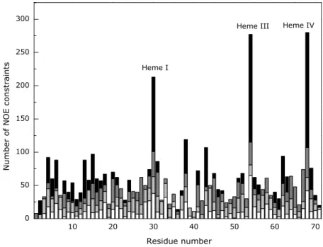
Figure 4.2 – Number of constraints per residue used for the calculation of the structure of PpcA. ... 69
Figure 4.3 – PpcA solution structure. ... 70
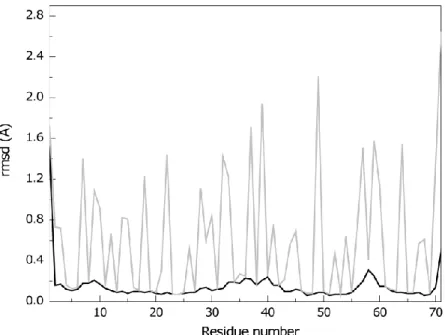
Figure 4.4 – Average pairwise backbone and heavy atom rmsd values per residue of the family of 20 conformers obtained for PpcA solution structure. ... 71
Figure 4.5 – 15N relaxation parameters for PpcA backbone in the reduced state. ... 72
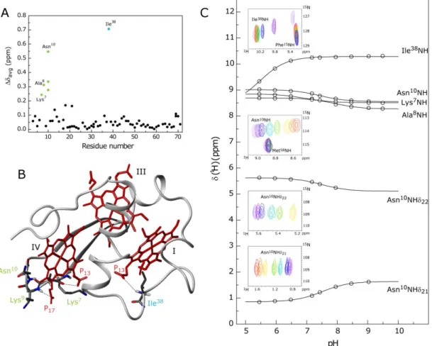
Figure 4.6 – pH-linked conformational changes in PpcA. ... 74
Figure 4.7 – Comparison of PpcA lowest energy solution structure with PpcA and PpcB crystal structures. ... 76
xvi
5.FUNCTIONAL ROLE OF PPCA KEY AMINO ACIDS
Figure 5.1 – Spatial localization of the mutated residues in PpcA solution structure. ... 86 Figure 5.2 – Spatial localization of residue Phe15 in PpcA solution structure. ... 87
Figure 5.3 – Expansions of the 1D-1H-NMR low-field regions obtained for PpcA and PpcA mutants. .... 88
Figure 5.4 – Expansion of 2D 1H-15N HSQC NMR spectra of PpcAF15L and PpcA in the reduced form... 89
Figure 5.5 – Comparison of the observed heme proton chemical shifts of reduced PpcAF15L and those
of PpcA at pH 8 and 15 °C. ... 90
Figure 5.6 – Oxidation fraction of PpcAF15L and PpcA at pH 6 and pH 8. ... 91 Figure 5.7 – Fitting of the thermodynamic model to the experimental data for PpcAF15L. ... 92 Figure 5.8 – Molar fractions of the 16 individual microstates and oxidation fractions of individual hemes
in PpcAF15L and wild-type cytochromes. ... 95
Figure 5.9 – Spatial localization of residue Met58 in PpcA solution structure. ... 97
Figure 5.10 – Comparison of the observed heme proton chemical shifts of reduced PpcAM58 mutants
and those of PpcA at pH 8 and 15 °C. ... 98
Figure 5.11 – Oxidation fraction of PpcAM58 mutants and PpcA at pH 6 and pH 8. ... 99 Figure 5.12 – Fitting of the thermodynamic model to the experimental data for PpcAM58 mutants. . 101 Figure 5.13 – Molar fractions of the 16 individual microstates and oxidation fractions of individual
hemes in PpcAM58 mutants and wild-type cytochromes. ... 104
Figure 5.14 – Spatial localization of lysine residues in PpcA solution structure. ... 106 Figure 5.15 – Comparison of the observed heme proton chemical shifts of reduced PpcA lysine mutants
and those of PpcA at pH 8 and 15 °C. ... 107
Figure 5.16 – Oxidation fraction of PpcAK9, PpcAK18 and PpcAK22 mutants and PpcA at pH 6 and pH 8.
... 108
Figure 5.17 – Oxidation fraction of PpcAK52 and PpcAK60 mutants and PpcA at pH 6 and pH 8. ... 109 Figure 5.18 – Fitting of the thermodynamic model to the experimental data for PpcAK52Q and
PpcAK52E. ... 111
Figure 5.19 – Fitting of the thermodynamic model to the experimental data for PpcAK60Q and
PpcAK60E. ... 112
Figure 5.20 – Molar fractions of the 16 individual microstates and oxidation fractions of individual
hemes in PpcAK52Q and wild-type cytochromes. ... 114
Figure 5.21 – Molar fractions of the 16 individual microstates and oxidation fractions of individual
hemes in PpcAK60Q and wild-type cytochromes. ... 115 6.ONGOING STUDIES
Figure 6.1 – 2D-1H-13C HSQC NMR spectra of the unlabeled and labeled PpcA (1.2 mM) obtained at 25 °C, with 640 and 80 scans, respectively.. ... 122 7.FINAL CONSIDERATIONS
Figure 7.1 – Figurative representation of the work developed. ... 131
APPENDIX
Figure A.1 – Connectivities observed in tridimensional spectra for 13C/15N labeled proteins. ... 151
xvii
T
ABLES
I
NDEX
1.INTRODUCTION
Table 1.1 – Summary of gene knockout and proteomic studies on G. sulfurreducens c-type
cytochromes. ... 4
3. THERMODYNAMIC CHARACTERIZATION OF A TRIHEME CYTOCHROME FAMILY FROM GEOBACTER SULFURREDUCENS Table 3.1 – Redox-dependent heme methyl chemical shifts of PpcA, PpcB, PpcD and PpcE at pH 8. .... 45
Table 3.2 – Energy parameters for PpcA, PpcB, PpcD, and PpcE. ... 49
Table 3.3 – Macroscopic pKa values of the redox-Bohr center for PpcA, PpcB, PpcD, and PpcE at each stage of oxidation. ... 49
4.STRUCTURAL STUDIES ON PPCA Table 4.1 – Summary of restraint violations and quality analysis for the final families of structures for PpcA. ... 68
Table 4.2 – Heme geometry for G. sulfurreducens cytochromes c7. ... 75
Table 4.3 – Heme reduction potentials and pairwise interactions of the fully reduced and protonated forms of PpcA. ... 78
5.FUNCTIONAL ROLE OF PPCA KEY AMINO ACIDS Table 5.1 – Calculated oxidation fractions 𝒙𝒊 for each heme group in each oxidation stage at pH 8.0 for PpcAF15L and wild-type cytochromes. ... 93
Table 5.2 – Energy parameters of PpcAF15L and PpcA. ... 93
Table 5.3 – Macroscopic pKa values of the redox-Bohr center for PpcAF15L and wild-type cytochromes. ... 94
Table 5.4 – Energy parameters for PpcAM58 mutants. ... 103
Table 5.5 – Energy parameters for PpcAK52 mutants... 113
Table 5.6 – Energy parameters for PpcAK60 mutants... 113
Table 5.7 – Summary of the results obtained from the thermodynamic characterization of PpcA mutated forms. ... 116
APPENDIX Table A.1 – Chemical shifts of the heme protons of PpcA, PpcB, PpcD and PpcE in the reduced state at pH 8.0 and 15 °C. ... 135
Table A.2 – Chemical shifts of PpcA in the reduced state at pH 7.1 and 25 °C. ... 136
Table A.3 – Chemical shifts of the heme protons of PpcAF15L in the reduced state at pH 8.0 and 15 °C. ... 143
Table A.4 – Chemical shifts of the heme protons of PpcAM58 mutants in the reduced state at pH 8.0 and 15 °C. ... 144
Table A.5 – Chemical shifts of the heme protons of PpcAK9 mutants in the reduced state at pH 8.0 and 15 °C. ... 145
Table A.6 – Chemical shifts of the heme protons of PpcAK18 mutants in the reduced state at pH 8.0 and 15 °C. ... 146
xviii
Table A.7 – Chemical shifts of the heme protons of PpcAK22 mutants in the reduced state at pH 8.0
and 15 °C. ... 147
Table A.8 – Chemical shifts of the heme protons of PpcAK52 mutants in the reduced state at pH 8.0
and 15 °C. ... 148
Table A.9 – Chemical shifts of the heme protons of PpcAK60 mutants in the reduced state at pH 8.0
and 15 °C. ... 149
Table A.10 – Primers used for site-directed mutagenesis of pMA59. ... 153 Table A.11 – 1H and 13C chemical shifts of the heme substituents in the oxidized triheme PpcA... 156
xix
A
BBREVIATIONS
,
SYMBOLS AND CONSTANTS
1D – one dimensional 2D – two dimensional 3D – three dimensional
AQDS – anthraquinone-2,6-disulfonate AMP – ampicillin
ATP – adenosine triphosphate
BLAST – Basic Local Alignment Search Tool BMRB – Biological Magnetic Resonance Data Bank CLO – chloramphenicol
COSY – COrrelation SpectroscopY
D. acetoxidans – Desulfuromonas acetoxidans Dac7 – Desulfuromonas acetoxidans c7 cytochrome
DL1 – G. sulfurreducens wild-type strain
DL3 – G. sulfurreducens strain with ppcA gene knocked-out DNA – deoxyribonucleic acid
DvHc3 – Desulfovibrio vulgaris strain Hildenborough tetraheme cytochrome c3
DvMc3 – Desulfovibrio vulgaris strain Miyazaki tetraheme cytochrome c3
E. coli – Escherichia coli
eapp – apparent midpoint reduction potential
EDTA – ethylenediamine tetraacetic acid EXSY – EXchange SpectroscopY
G. sulfurreducens – Geobacter sulfurreducens Gsc7 – Geobacter sulfurreducens cytochromes c7
HSQC - Heteronuclear Single-Quantum Correlation IPTG – isopropyl β-D-1-thiogalactopyranoside
IUPAC-IUB – International Union of Pure and Applied Chemistry and International Union of Biochemistry
lov – lower limit volume MQH2 – menaquinol
MQ – menaquinone
NMR – Nuclear Magnetic Resonance NOE – Nuclear Overhauser Effect
NOESY – Nuclear Overhauser Enhancement SpectroscopY OD600 – optical density at 600 nm
PDB – Protein Data Bank pI – isoelectric point
PpcA – G. sulfurreducens c7 cytochrome (GSU0612)
PpcB – G. sulfurreducens c7 cytochrome (GSU0364)
xx
PpcD – G. sulfurreducens c7 cytochrome (GSU1024)
PpcE – G. sulfurreducens c7 cytochrome (GSU1760)
ppm – parts per million
rmsd – root mean square deviation
SDS-PAGE – sodium dodecyl sulfate polyacrylamide gel electrophoresis SHE – Standard Hydrogen Electrode
TOCSY – TOtal Correlation SpectroscopY Tris – tris(hydroxymethyl)aminomethane upl – upper distance limit
upv – upper limit volume UV-visible – ultraviolet-visible
δ - chemical shift
F – Faraday constant (96485 Cmol-1)
R – molar gas constant (8.314 JK-1mol-1)
Amino acid abbreviations
Alanine Ala A Arginine Arg R Asparagine Asn N Aspartate Asp D Cysteine Cys C Glutamate Glu E Glutamine Gln Q Glycine Gly G Histidine His H Isoleucine Ile I Leucine Leu L Lysine Lys K Methionine Met M Phenylalanine Phe F Proline Pro P Serine Ser S Threonine Thr T Tryptophan Trp W Tyrosine Tyr Y Valine Val V1
Introduction
2
1. INTRODUCTION ... 3 1.1 The bacterium Geobacter sulfurreducens ... 3 1.2 Extracellular electron transfer ... 4 1.3 Gene knockout and proteomic studies in G. sulfurreducens ... 4 1.4 Periplasmic cytochromes ... 7 1.5 Solution structures of multiheme proteins ... 11 1.6 Thesis outline ... 13 1.7 References ... 14
3
1. I
NTRODUCTION
Dissimilatory metal reduction is the process by which microorganisms couple the oxidation of organic matter with the reduction of metal ions for other purposes than its assimilation into cellular components. These processes play important roles in the biogeochemical cycles of metals as iron, manganese, uranium and chromium [1].
Geobacter species show an impressive respiratory versatility. These bacteria are able to
sustain their growth by using extracellular compounds, as Fe(III), U(VI) or Mn(IV) oxides, as terminal electron acceptors, in addition to the more frequent respiratory processes, which use both soluble electron donors (e.g. acetate) and acceptors (e.g. fumarate) [1,2]. Some of these compounds are toxic or radioactive, making this organism a potential target for bioremediation and biotechnological applications.
Based on these skills and the fact that these bacteria are highly enriched in subsurface environments, several bioremediation applications towards the decontamination of different environments have been developed [2-4]. This includes the reduction of soluble U(VI) to insoluble U(IV) for the immobilization of uranium in contaminated ground waters [5,6] and the anaerobic benzene degradation in petroleum-contaminated aquifers [7]. These strategies rely on the stimulation of Geobacter growth by addition of an electron donor to the groundwater [4].
Geobacter species are also being used to harvest electricity from aquatic sediments and
waste organic matter by growing with electrodes as electron acceptors in microbial fuel cells [8-11]. This characteristic of Geobacter as “The microbe electric” was highlighted as one of the 50 best inventions of 2009 by the Time Magazine.
1.1 The bacterium Geobacter sulfurreducens
The bacterium Geobacter sulfurreducens (G. sulfurreducens) PCA was described for the first time in 1994 after being isolated from surface sediments of hydrocarbon-contaminated ditch near Norman, Oklahoma, USA [12]. This was the first bacterium described that couples the oxidation of acetate or hydrogen to the reduction of Fe(III). Phylogenetic analysis of its 16S rRNA placed G. sulfurreducens in the subgroup of -proteobacteria [12].
The genome of G. sulfurreducens has been sequenced and it revealed an unprecedented number of 111 putative c-type cytochromes, 73 of which with more than one heme binding site [13]. G. sulfurreducens is the microorganism with the largest number of c-type cytochrome genes [14]. The co-existence of such a large number of multiheme cytochromes might help the organism to efficiently use a diverse range of respiratory pathways and highlights their involvement in a broad range of essential cellular functions.
In the last years, G. sulfurreducens DL1 (wild-type strain maintained for many transfers in laboratory) has been used as a model for the study of Geobacteraceae because a genetic system was developed for this bacterium [15]. This system allows the mutagenesis of the chromosome by gene replacement, to introduce foreign DNA by electroporation and to
4
express proteins from extrachromossomal elements. These advances enabled to study the effect of the deletion of some genes, as well as their expression in trans.
1.2 Extracellular electron transfer
Many of the terminal electron acceptors that can be used by G. sulfurreducens are insoluble and, thus, unable to diffuse inside the cells. Therefore, reduction of these acceptors cannot occur in the periplasm as for soluble acceptors, and requires electron transfer across the outer membrane [16,17].
To assist electron transfer to the cell exterior, the spatial disposition of the redox components in G. sulfurreducens cells differs from that of the majority of other microorganisms. In fact, in addition to the usual location in the inner membrane and periplasmic space, multiheme cytochromes have been identified in G. sulfurreducens’ outer membrane [18].
Although numerous electron transfer proteins have been identified in G. sulfurreducens, the electron transfer pathways that allow this microorganism to obtain energy are still far from being understood. It is essential to obtain a detailed characterization of G.
sulfurreducens electron transfer proteins before understanding these electron transfer
mechanisms and delineate their physiological roles. Insights into electricity production mechanisms can arise from the elucidation of the cellular strategies that allow energy production from the reduction of natural extracellular terminal acceptors [9]. This is of major interest for the design of improved biotechnological applications.
1.3 Gene knockout and proteomic studies in G. sulfurreducens
Several studies have been made in order to identify the proteins involved in the different reduction pathways used by G. sulfurreducens. Proteomic analysis under different growth conditions and gene deletion studies were performed, with special focus on c-type cytochromes since these proteins may play an important role in electron transfer to extracellular electron acceptors. The results of these studies are summarized in Table 1.1.
Table 1.1 – Summary of gene knockout and proteomic studies on G. sulfurreducens c-type cytochromes.
Protein (GSU number) Predicted number of heme groups Predicted cellular
localization Gene knockout and proteomic studies PpcB
(GSU0364) 3 Periplasm Detected in both Fe(III) citrate and Fe(III) oxide cultures, but more abundant in Fe(III) citrate [19]. Double deletion mutant with PpcC has U(VI) reduction affected [20].
PpcC
5
Table 1.1 (cont.) – Summary of gene knockout and proteomic studies on G. sulfurreducens c-type cytochromes.
Protein (GSU number) Predicted number of heme groups Predicted cellular
localization Gene knockout and proteomic studies MacA
(GSU0466) 2 Periplasm Deletion mutant reduction of U(VI) is affected [20]. Deletion mutant growth on Fe(III) citrate is impaired,
by affecting OmcB expression [21].
More abundant during growth with Fe(III) oxides vs. Fe(III) citrate as electron acceptor [19].
PpcA
(GSU0612) 3 Periplasm Deletion mutant U(VI) and AQDS reduction is affected when acetate is electron donor [22]. Deletion mutant Fe(III) oxides reduction is affected [20].
Detected in both Fe(III) citrate and Fe(III) oxide cultures [19].
GSU0616 8 Periplasm Deletion mutant has increased Fe(III) and U(VI) reducing activity [20].
OmcE
(GSU0618) 4 Outer membrane Deletion mutant growth in Fe(III) and Mn(IV) oxides is affected [18]; power production is affected but adapts with time [23].
More abundant during growth with Fe(III) citrate vs. fumarate as electron acceptor [24].
Deletion mutant reduction of U(VI) is affected [20]. No impact on current production [25].
PpcD
(GSU1024) 3 Periplasm More abundant during growth with Fe(III) oxides vs. Fe(III) citrate as electron acceptor [19].
GSU1334 7 Outer
membrane Reduction of U(VI) and Fe(III) oxides is affected [20]. More abundant during growth with Fe(III) oxides vs. Fe(III) citrate as electron acceptor [19].
PpcE
(GSU1760) 3 Periplasm Found only in cultures with Fe(III) citrate [19]. PgcA
(GSU1761)
3 Periplasm More abundant during growth with Fe(III) oxides vs. Fe(III) citrate as electron acceptor [19].
Increased expression in strains adapted to growth in Fe(III) oxides [26].
OmcZ
(GSU2076) 7 Outer membrane Severely inhibited current production and biofilm formation [25].
OmcF
(GSU2432) 1 Outer membrane Deletion mutant growth on Fe(III) citrate is impaired, by affecting OmcB expression [27]. Deletion mutant reduction of U(VI) is affected [20]. Deletion mutant decreased current production [28].
OmcS
(GSU2504) 6 Outer membrane Deletion mutant growth in Fe(III) and Mn(IV) oxides is affected [18]. Deletion mutant has no impact on current production [25].
Localized along the pili [29].
OmcC
(GSU2731) 12 Outer membrane Deletion mutant has no effect on Fe(III) [30] and U(VI) reduction [20].
OmcB
(GSU2737) 12 Outer membrane Deletion mutant grows poorly in Fe(III) citrate and doesn’t grow in Fe(III) oxide [30] but there is no effect on reduction of electrodes and current production [23,25] or on U(VI) reduction [20].
More abundant during growth with Fe(III) citrate vs. fumarate as electron acceptor [24].
Affected by deletion of omcF [27], omcG and omcH genes [31].
GSU3332 2 Outer
6
From the results presented in Table 1.1 is possible to verify that c-type cytochromes can be involved not only in electron transfer but also in transcriptional and post-transcriptional regulation or processing in G. sulfurreducens [27,31]. It was also shown that deletion of individual genes for outer surface c-type cytochromes only partially inhibited humic substance or anthraquinone-2,6-disulfonate (AQDS, model compound for humics) reduction and suggested that there are multiple routes for transfer of electrons to these acceptors [32].
Based on all these studies, a model for electron transfer to Fe(III) oxides in G.
sulfurreducens has been proposed (Figure 1.1) [9,17,29].
Figure 1.1 – Mechanism for extracellular electron transfer by G. sulfurreducens. Potential route for electron transfer to Fe(III) oxides based on previous
models [9,17] and subsequent findings [26,29,33,34] (Table 1.1). The small white hexagons represent heme groups. MQH2 is menaquinol and MQ is menaquinone.
Besides, the several cytochromes associated with the inner membrane, the periplasm and the outer membrane, it was shown that G. sulfurreducens forms pili when growing in Fe(III) oxides, and that these are essential for the contact with the oxides and for electron transfer [16]. Initial studies showed that pili were highly conductive and could serve as biological nanowires, working as the electrical connection between the cell and the surface of Fe(III) oxides [16]. However, more recent studies revealed that the hexaheme cytochrome OmcS was localized along the pili when G. sulfurreducens grows in Fe(III) oxides [29]. This suggests that pili may play a structural role by supporting c-type cytochromes along the way
7 to the surface of iron oxides [33,34]. It was proposed that cytochromes can also play a role in electron storage when an electron acceptor is not available [35]. However, the precise functions of these proteins are not yet elucidated and only a few components of the respiratory chain have been individually characterized so far.
The cytochrome OmcS has a molecular weight of 47 kDa, six heme groups and was purified from a strain that overproduces this protein [36]. Nuclear Magnetic Resonance (NMR) together with UV-visible spectroscopic studies allowed determining that all the heme groups are bis-histidinyl hexacoordinated and low spin in both fully oxidized and reduced states. The redox behavior of OmcS was studied by redox titrations followed by UV-visible [36]. It was observed that the six redox centers are not equivalent and the redox curve spans over a large range of reduction potentials from -360 mV to -40 mV. The midpoint reduction potential at pH 7 was -212 mV. Reduced OmcS was able to transfer electrons in
vitro to different substrates as Fe(III) and Mn(IV) oxides and humic substances [36].
More related to electron transfer to electrodes, the cytochromes OmcF and OmcZ have also been characterized.
OmcF is a monoheme cytochrome with sequence similarity to soluble c6 cytochromes of
photosynthetic algae and cyanobacteria [37]. OmcF crystal structure was determined and was superimposable with a root mean square deviation (rmsd) of 1.1 Å to the structure of the cytochrome c6 from the green alga Monoraphidium braunii. However the function of
these two proteins is probably different, since their biochemical properties are very distinct. OmcF has an isoelectric point (pI) of 7.8 while M. braunii cytochrome has a pI of 4.2, and their reduction potentials at pH 7 are +180 mV and +357 mV, respectively [37].
OmcZ was shown be present in two forms in G. sulfurreducens: a large one (OmcZL)
and a small one (OmcZS) that is a cleaved product from the first. OmcZS is most probably
the extracellular and active form [38]. OmcZS has eight heme groups, seven bound to the
typical heme binding motif CXXCH (where X represents any amino acid) and one with the unusual binding motif CX14CH. A similar motif (CX15CH) was identified in an octaheme protein
from Wolinella succinogenes [39]. Redox titrations revealed that OmcZS functional working
potential range is between -420 mV and -60 mV with a midpoint reduction potential of -220 mV. In vitro, OmcZS was able to transfer electrons to Fe(III) citrate, U(VI), Cr(VI), Au(III),
Mn(IV) oxides, and AQDS, but not Fe(III) oxide [38].
1.4 Periplasmic cytochromes
In addition to the outer membrane cytochromes, an unusual periplasmic pool of five homologous triheme cytochromes (also known as c7 cytochromes) was identified in G.
sulfurreducens [40]. This family is considered essential in the bacterium electron transfer
pathways since soluble periplasmic cytochromes are crucial for shuttling electrons from the cytoplasmic compartment to the outer membrane [9].
8
The five cytochromes are small proteins with approximately 10 kDa and a pI 9, due to the high content in lysine residues. The cytochromes are designated PpcA, PpcB, PpcC, PpcD, and PpcE (Gsc7) and share a 77%, 62%, 57%, and 65% amino acid sequence identity with
PpcA, respectively [40].
Using the PpcA amino acid sequence, the non-redundant amino acid database of NCBI using the Basic Local Alignment Search Tool (BLAST) [41] was searched and in addition to these five cytochromes c7, 18 other cytochromes were found: five in Geobacter
metallireducens, four in Geobacter uraniireducens, three in Geobacter bemidjiensis, two in Anaeromyxobacter dehalogenans and Geobacter lovleyi, and one in Desulfuromonas acetoxidans strain 5071 (D. acetoxidans) and in Pelobacter propionicus. A sequence
alignment of these proteins is depicted in Figure 1.2 and shows that of the 21 highly conserved residues, only nine are not cysteine or histidine residues directly involved in heme binding.
9 F ig u re 1 .2 – Ali g n m en t o f am in o acid sequ enc es o f cyt o ch ro m es c7 . PpcA , PpcB , PpcC , PpcD, PpcE , cy to ch ro me s c7 fro m Ge o b act er su lf u rre d u cen s; D ac7 , cy to ch ro me c7 fro m D . acet o xi d an s; Gmet , Geo b ac te r me tal lire d u cen s; Gu ra , Geo b act er u ran iire d u cen s; Gbe m , Geo b act er b emi d ji en si s; Gl o v, Geo b act er lo vl ey i; Ppro , Pe lo b act er p ro p io n icu s; A d eh , An ae ro my xo b act er d eh al og en an s. Th e n u mb ers re fe r to th e g en e th at en co d es each cy to ch ro me . Th e co n served re si d u es in t h e p ro te in s are b o xe d : g ray h eme -at tach ed re si d u es an d b lack n o n -h eme -at tach ed re si d u es. Th e spe ci fi c h eme an d t h e re sp ect iv e at tach ed r esi d u es are i n d ica te d a t th e b ot to m o f th e las t cy to ch ro m e c7 a mi n o aci d seq u en ce. Th e se q u en ce id en ti ty f or each cy to ch ro me i n re lat io n t o PpcA i s al so i n d icat ed .
10
The crystal structures of the five G. sulfurreducens c7 cytochromes have been
determined, showing that they have a high level of structural homology as depicted in Figure 1.3 [40,42,43].
Figure 1.3 – Crystal structures of the five c7 cytochromes from G. sulfurreducens. PpcA is represented in gray with the deoxycholate acid molecule used for
crystallization in black (PDB 1OS6 [40]), PpcB in green (PDB 3BXU [43]), PpcC in blue (PDB 3H33), PpcD in orange (PDB 3H4N) and PpcE in cyan (PDB 3H34) [42]. PpcB and PpcD displayed two molecules in the crystal asymmetric unit (monomers A and B) and monomer A is represented. The molecules are all in the same orientation.
The spatial arrangements of the hemes in cytochromes c7 are superimposable with
those of the structurally homologous tetraheme cytochromes c3, with the sole difference
being lack of heme II and the corresponding polypeptide segment. For this reason, the three heme groups in cytochromes c7 have been numbered as I, III, and IV [44].
The three heme groups form the protein core and are covalently linked to the cysteine residues of the CXXCH binding motifs (Figure 1.2). All the hemes are axially coordinated by two histidine residues and are low-spin both in the reduced (Fe(II), S = 0) and in the oxidized (Fe(III), S = ½) forms. The heme core structures are similar, with hemes I and III roughly parallel to each other and both nearly perpendicular to heme IV [40,42,43].
A two-strand -sheet at the N-terminal is conserved in all the structures, and is followed by different helical regions in the different proteins [40,42,43]. The most conserved region is the positively charged surface around heme IV and the lowest similarity is found near heme I [42].
The only other c7 cytochrome with structural information available is the one isolated
from D. acetoxidans (Dac7). The structure of this cytochrome has been studied by X-ray in
its oxidized form [45], and by NMR in both redox states [46,47]. The overall fold of the polypeptide is similar to the G. sulfurreducens cytochromes; however, the heme core
11 rearrangement is somehow different, especially in the iron to iron distance between hemes I and IV (19.3 Å in Dac7 compared to an average of 18.3 Å in the other proteins) [42].
The best studied G. sulfurreducens c7 cytochrome is PpcA. In addition to the gene
knockout and proteomic studies already described, the thermodynamic properties of the heme groups have been determined [48]. The results obtained showed that the heme groups of PpcA have negative reduction potentials that are modulated by heme-heme interactions and interactions with protonated groups (redox-Bohr effect). Taken together, the thermodynamic parameters obtained for PpcA showed that this protein is designed to present a preferential electron transfer pathway at physiologic pH coupled with proton transfer [48].
It had been previously showed that for growth in the presence of extracellular Fe(III) oxides, G. sulfurreducens needs additional e-/H+ coupling mechanisms in comparison to
those used in fumarate respiration [49]. The authors suggested that the most likely mechanism for additional membrane potential generation is the coupling of electron transfer to the periplasmic cytochromes involved in the Fe(III) reduction with proton translocation, so that additional membrane potential can be generated for ATP production.
Since PpcA is known to be involved in Fe(III) reduction pathways, it was proposed that in the presence of extracellular electron acceptors PpcA might contribute to the H+
electrochemical potential gradient across the periplasmic membrane that drives ATP synthesis [48]. These results contrast with the ones obtained for the triheme cytochrome
Dac7, which seems to be designed to work as simple electron transfer protein in the
physiologic pH range [50].
1.5 Solution structures of multiheme proteins
Despite the cellular localization and phenotype associated to some G. sulfurreducens multiheme cytochromes are already known, no structural information in solution is available for any of these proteins. This relates with the traditional difficulties in obtaining isotopic labeled recombinant multiheme cytochromes with the correct fold and post-translational modification of the heme groups in a cost-effective manner [51-53].
Important contributions for the solution structure determination of multiheme cytochromes have been made by the research groups of Prof. António Xavier [54-58], Prof. Ivano Bertini [46,47] and Prof. Hideo Akutsu [59]. Although the proteins’ molecular weights are appropriate for structural studies in solution, only a few structures have been determined for multiheme proteins. Indeed, reports of solution structures are still limited to five tetraheme cytochromes c3 [54-59] and to one triheme cytochrome [46,47]. In total, ten
solution structures have been reported, five for fully reduced proteins [46,54,56-58] and three for fully oxidized [47,54,55,58]. This clearly contrasts with the large number of structures determined by X-ray crystallography [60].
The smaller number of solution structures obtained in the oxidized form is undoubtedly associated with the inherent complexity of their NMR spectra (Figure 1.4).
12
Figure 1.4 – 1D-1H NMR spectra of reduced and oxidized triheme cytochrome
PpcA obtained at 25 ºC. The typical regions of the heme substituents are indicated.
Indeed, in the oxidized form, the paramagnetic effect of the iron unpaired electrons, causes the spread of the signals of the heme cofactors (Figure 1.4), as well as those of the amino acid residues located in their neighborhoods, all over large NMR spectral widths [61]. Additionally, these resonances are generally broader, which makes the complete assignment of the heme and polypeptide resonances a laborious and very time consuming task.
On the other hand, for diamagnetic multiheme proteins, as it is the case of the reduced PpcA, the assignment of the heme substituents is facilitated since they are dominated by the porphyrin ring-current shifts and, therefore, appear in well defined regions of the 1H NMR
spectra (Figure 1.4). The only exception is observed for the heme propionate protons as they are structurally more variable.
An efficient expression system to produce multiheme c-type cytochromes, using
Escherichia coli (E. coli) as host, was recently described and successfully applied to the
expression of multiheme cytochromes containing up to 12 heme groups [62,63]. This was overcome by using a lac promoter in the expression plasmid and co-expressing the cytochrome c maturation gene cluster on a separate plasmid [63].
This system was also used to achieve cost-effective labeling of multiheme cytochromes using an experimental labeling methodology that is based on two major aspects: (i) use of a two-step culture growth procedure, where cell growth in rich media was followed by transfer
13 to minimal media containing 15N-labeled ammonium chloride, and (ii) incorporation of the
heme precursor -aminolevulinic acid in minimal culture media [64].
1.6 Thesis outline
In order to contribute to the studies of the electron transfer chains of G. sulfurreducens, the work developed on this thesis focused on the study of the periplasmic triheme cytochrome family, with special emphasis on PpcA, the most abundant in G. sulfurreducens.
The thermodynamic characterization of cytochromes PpcA, PpcB, PpcD and PpcE by NMR and UV-visible spectroscopy is described on Chapter 3. Chapter 4 is dedicated to structural studies of PpcA in solution. The assignment of the resonances of the protein and its co-factors is described, and the solution structure in the reduced state is presented. The physiological role of key amino acids in PpcA is discussed in Chapter 5. Finally, on Chapter 6, ongoing studies are presented.
14
1.7 References
[1] DR Lovley (1993) Dissimilatory metal reduction, Annu Rev Microbiol 47, 263-290.
[2] JR Lloyd, DR Lovley (2001) Microbial detoxification of metals and radionuclides, Curr Opin Biotechnol 12, 248-253.
[3] DR Lovley (2003) Cleaning up with genomics: applying molecular biology to bioremediation, Nat Rev Microbiol 1, 35-44.
[4] DE Holmes, KT Finneran, RA O'Neil, DR Lovley (2002) Enrichment of members of the family Geobacteraceae associated with stimulation of dissimilatory metal reduction in uranium-contaminated aquifer sediments, Appl Environ Microbiol 68, 2300-2306.
[5] RT Anderson, HA Vrionis, I Ortiz-Bernad, CT Resch, PE Long, R Dayvault, K Karp, S Marutzky, DR Metzler, A Peacock, DC White, M Lowe, DR Lovley (2003) Stimulating the
in situ activity of Geobacter species to remove uranium from the groundwater of a
uranium-contaminated aquifer, Appl Environ Microbiol 69, 5884-5891.
[6] KB Gregory, DR Lovley (2005) Remediation and recovery of uranium from contaminated subsurface environments with electrodes, Environ Sci Technol 39, 8943-8947.
[7] JN Rooney-Varga, RT Anderson, JL Fraga, D Ringelberg, DR Lovley (1999) Microbial communities associated with anaerobic benzene degradation in a petroleum-contaminated aquifer, Appl Environ Microbiol 65, 3056-3063.
[8] DR Bond, DR Lovley (2003) Electricity production by Geobacter sulfurreducens attached to electrodes, Appl Environ Microbiol 69, 1548-1555.
[9] DR Lovley (2006) Bug juice: harvesting electricity with microorganisms, Nat Rev Microbiol 4, 497-508.
[10] AE Franks, KP Nevin (2010) Microbial Fuel Cells, A Current Review, Energies 3, 899-919.
[11] DE Holmes, DR Bond, RA O'Neil, CE Reimers, LR Tender, DR Lovley (2004) Microbial communities associated with electrodes harvesting electricity from a variety of aquatic sediments, Microb Ecol 48, 178-190.
[12] F Caccavo, Jr., DJ Lonergan, DR Lovley, M Davis, JF Stolz, MJ McInerney (1994)
Geobacter sulfurreducens sp. nov., a hydrogen- and acetate-oxidizing dissimilatory
metal-reducing microorganism, Appl Environ Microbiol 60, 3752-3759.
[13] BA Methé, KE Nelson, JA Eisen, IT Paulsen, W Nelson, JF Heidelberg, D Wu, M Wu, N Ward, MJ Beanan, RJ Dodson, R Madupu, LM Brinkac, SC Daugherty, RT DeBoy, AS Durkin, M Gwinn, JF Kolonay, SA Sullivan, DH Haft, J Selengut, TM Davidsen, N Zafar, O White, B Tran, C Romero, HA Forberger, J Weidman, H Khouri, TV Feldblyum, TR Utterback, SE Van Aken, DR Lovley, CM Fraser (2003) Genome of Geobacter
sulfurreducens: metal reduction in subsurface environments, Science 302, 1967-1969.
[14] SH Thomas, RD Wagner, AK Arakaki, J Skolnick, JR Kirby, LJ Shimkets, RA Sanford, FE Loffler (2008) The mosaic genome of Anaeromyxobacter dehalogenans strain 2CP-C suggests an aerobic common ancestor to the delta-proteobacteria, PLoS One 3, e2103. [15] MV Coppi, C Leang, SJ Sandler, DR Lovley (2001) Development of a genetic system for
Geobacter sulfurreducens, Appl Environ Microbiol 67, 3180-3187.
[16] G Reguera, KD McCarthy, T Mehta, JS Nicoll, MT Tuominen, DR Lovley (2005) Extracellular electron transfer via microbial nanowires, Nature 435, 1098-1101.
15 [17] KA Weber, LA Achenbach, JD Coates (2006) Microorganisms pumping iron: anaerobic
microbial iron oxidation and reduction, Nat Rev Microbiol 4, 752-764.
[18] T Mehta, MV Coppi, SE Childers, DR Lovley (2005) Outer membrane c-type cytochromes required for Fe(III) and Mn(IV) oxide reduction in Geobacter sulfurreducens, Appl Environ Microbiol 71, 8634-8641.
[19] YH Ding, KK Hixson, MA Aklujkar, MS Lipton, RD Smith, DR Lovley, T Mester (2008) Proteome of Geobacter sulfurreducens grown with Fe(III) oxide or Fe(III) citrate as the electron acceptor, Biochim Biophys Acta 1784, 1935-1941.
[20] ES Shelobolina, MV Coppi, AA Korenevsky, LN Didonato, SA Sullivan, H Konishi, H Xu, C Leang, JE Butler, BC Kim, DR Lovley (2007) Importance of c-type cytochromes for U(VI) reduction by Geobacter sulfurreducens, BMC Microbiol 7, 16.
[21] BC Kim, DR Lovley (2008) Investigation of direct vs. indirect involvement of the c-type cytochrome MacA in Fe(III) reduction by Geobacter sulfurreducens, FEMS Microbiol Lett. [22] JR Lloyd, C Leang, AL Hodges Myerson, MV Coppi, S Cuifo, B Methe, SJ Sandler, DR Lovley (2003) Biochemical and genetic characterization of PpcA, a periplasmic c-type cytochrome in Geobacter sulfurreducens, Biochem J 369, 153-161.
[23] DE Holmes, SK Chaudhuri, KP Nevin, T Mehta, BA Methe, A Liu, JE Ward, TL Woodard, J Webster, DR Lovley (2006) Microarray and genetic analysis of electron transfer to electrodes in Geobacter sulfurreducens, Environ Microbiol 8, 1805-1815.
[24] YH Ding, KK Hixson, CS Giometti, A Stanley, A Esteve-Núñez, T Khare, SL Tollaksen, W Zhu, JN Adkins, MS Lipton, RD Smith, T Mester, DR Lovley (2006) The proteome of dissimilatory metal-reducing microorganism Geobacter sulfurreducens under various growth conditions, Biochim Biophys Acta 1764, 1198-1206.
[25] KP Nevin, BC Kim, RH Glaven, JP Johnson, TL Woodard, BA Methe, RJ Didonato, SF Covalla, AE Franks, A Liu, DR Lovley (2009) Anode biofilm transcriptomics reveals outer surface components essential for high density current production in Geobacter
sulfurreducens fuel cells, PLoS One 4, e5628.
[26] PL Tremblay, ZM Summers, RH Glaven, KP Nevin, K Zengler, CL Barrett, Y Qiu, BO Palsson, DR Lovley (2011) A c-type cytochrome and a transcriptional regulator responsible for enhanced extracellular electron transfer in Geobacter sulfurreducens revealed by adaptive evolution, Environ Microbiol 13, 13-23.
[27] BC Kim, C Leang, YH Ding, RH Glaven, MV Coppi, DR Lovley (2005) OmcF, a putative c-type monoheme outer membrane cytochrome required for the expression of other outer membrane cytochromes in Geobacter sulfurreducens, J Bacteriol 187, 4505-4513. [28] BC Kim, BL Postier, RJ Didonato, SK Chaudhuri, KP Nevin, DR Lovley (2008) Insights
into genes involved in electricity generation in Geobacter sulfurreducens via whole genome microarray analysis of the OmcF-deficient mutant, Bioelectrochemistry 73, 70-75.
[29] C Leang, X Qian, T Mester, DR Lovley (2010) Alignment of the c-type cytochrome OmcS along pili of Geobacter sulfurreducens, Appl Environ Microbiol 76, 4080-4084.
[30] C Leang, MV Coppi, DR Lovley (2003) OmcB, a c-type polyheme cytochrome, involved in Fe(III) reduction in Geobacter sulfurreducens, J Bacteriol 185, 2096-2103.
[31] BC Kim, X Qian, C Leang, MV Coppi, DR Lovley (2006) Two putative c-type multiheme cytochromes required for the expression of OmcB, an outer membrane protein essential for optimal Fe(III) reduction in Geobacter sulfurreducens, J Bacteriol 188, 3138-3142.
16
[32] JW Voordeckers, BC Kim, M Izallalen, DR Lovley (2010) Role of Geobacter
sulfurreducens outer surface c-type cytochromes in reduction of soil humic acid and
anthraquinone-2,6-disulfonate, Appl Environ Microbiol 76, 2371-2375.
[33] DR Lovley (2008) Extracellular electron transfer: wires, capacitors, iron lungs, and more, Geobiology 6, 225-231.
[34] G Reguera, RB Pollina, JS Nicoll, DR Lovley (2007) Possible nonconductive role of
Geobacter sulfurreducens pilus nanowires in biofilm formation, J Bacteriol 189,
2125-2127.
[35] A Esteve-Núñez, J Sosnik, P Visconti, DR Lovley (2008) Fluorescent properties of c-type cytochromes reveal their potential role as an extracytoplasmic electron sink in
Geobacter sulfurreducens, Environ Microbiol 10, 497-505.
[36] X Qian, T Mester, L Morgado, T Arakawa, ML Sharma, K Inoue, C Joseph, CA Salgueiro, MJ Maroney, DR Lovley (2011) Biochemical characterization of purified OmcS, a c-type cytochrome required for insoluble Fe(III) reduction in Geobacter sulfurreducens, Biochim Biophys Acta 1807, 404-412.
[37] PR Pokkuluri, YY Londer, SJ Wood, NE Duke, L Morgado, CA Salgueiro, M Schiffer (2009) Outer membrane cytochrome c, OmcF, from Geobacter sulfurreducens: high structural similarity to an algal cytochrome c6, Proteins 74, 266-270.
[38] K Inoue, X Qian, L Morgado, BC Kim, T Mester, M Izallalen, CA Salgueiro, DR Lovley (2010) Purification and characterization of OmcZ, an outer-surface, octaheme c-type cytochrome essential for optimal current production by Geobacter sulfurreducens, Appl Environ Microbiol 76, 3999-4007.
[39] RS Hartshorne, M Kern, B Meyer, TA Clarke, M Karas, DJ Richardson, J Simon (2007) A dedicated haem lyase is required for the maturation of a novel bacterial cytochrome c with unconventional covalent haem binding, Mol Microbiol 64, 1049-1060.
[40] PR Pokkuluri, YY Londer, NE Duke, WC Long, M Schiffer (2004) Family of cytochrome c7
-type proteins from Geobacter sulfurreducens: structure of one cytochrome c7 at 1.45 Å
resolution, Biochemistry 43, 849-859.
[41] SF Altschul, TL Madden, AA Schaffer, J Zhang, Z Zhang, W Miller, DJ Lipman (1997) Gapped BLAST and PSI-BLAST: a new generation of protein database search programs, Nucleic Acids Res 25, 3389-3402.
[42] PR Pokkuluri, YY Londer, X Yang, NE Duke, J Erickson, V Orshonsky, G Johnson, M Schiffer (2010) Structural characterization of a family of cytochromes c7 involved in
Fe(III) respiration by Geobacter sulfurreducens, Biochim Biophys Acta 1797, 222-232. [43] L Morgado, M Bruix, V Orshonsky, YY Londer, NE Duke, X Yang, PR Pokkuluri, M
Schiffer, CA Salgueiro (2008) Structural insights into the modulation of the redox properties of two Geobacter sulfurreducens homologous triheme cytochromes, Biochim Biophys Acta 1777, 1157-1165.
[44] IB Coutinho, DL Turner, MY Liu, J LeGall, AV Xavier (1996) Structure of the three-haem core of cytochrome c551.5 determined by 1H NMR, J Biol Inorg Chem 1, 305-311.
[45] M Czjzek, P Arnoux, R Haser, W Shepard (2001) Structure of cytochrome c7 from
Desulfuromonas acetoxidans at 1.9 Å resolution, Acta Crystallogr D Biol Crystallogr 57,
670-678.
[46] M Assfalg, L Banci, I Bertini, M Bruschi, MT Giudici-Orticoni, P Turano (1999) A proton-NMR investigation of the fully reduced cytochrome c7 from Desulfuromonas acetoxidans.