THE ROLE OF MITOCHONDRIA IN DROSOPHILA WOUND HEALING
SUSANA ISABEL PEREIRA DA PONTE
Tese para obtenção do grau de Doutor em
Envelhecimento e Doenças Crónicas
Doutoramento em associação entre:
Universidade NOVA de Lisboa (Faculdade de Ciências Médicas | NOVA Medical School -
FCM|NMS/UNL)
Universidade de Coimbra (Faculdade de Medicina - FM/UC)
Universidade do Minho (Escola de Medicina - EMed/UM)
THE ROLE OF MITOCHONDRIA IN DROSOPHILA WOUND HEALING
Susana Isabel Pereira da Ponte
Orientadores:
António Jacinto, Investigador Principal e Professor da Faculdade de Ciências Médicas | NOVA
Medical School, Universidade NOVA de Lisboa
Paulo J. Oliveira, Investigador Principal do Centro de Neurociências e Biologia Celular (CNC) e
Professor assistente convidado da Universidade de Coimbra
Tese para obtenção do grau de Doutor em Envelhecimento e Doenças Crónicas
VERSÃO PROVISÓRIA
Doutoramento em associação entre:
Universidade NOVA de Lisboa (Faculdade de Ciências Médicas | NOVA Medical School -
FCM|NMS/UNL)
Universidade de Coimbra (Faculdade de Medicina - FM/UC)
Universidade do Minho (Escola de Medicina - EMed/UM)
The research described here was performed at the Chronic Diseases Research Center (CEDOC), NOVA Medical School - Faculdade de Ciências Médicas, between 2015 and 2019. Its execution was supported by a PhD fellowship from the Portuguese Foundation for Science and Technology (PD/BD/106058/2015).
Este trabalho de investigação foi realizado no Centro de Estudos de Doenças Crónicas (CEDOC) da NOVA Medical School - Faculdade de Ciências Médicas, entre 2015 e 2019, ao abrigo de uma bolsa de Doutoramento, financiada pela Fundação para a Ciência e a Tecnologia (PD/BD/106058/2015).
Trabalho realizado ao abrigo da Bolsa (Ref. PD/00291/2012), com o apoio da FCT (Fundação para a Ciência e a Tecnologia), do FSE (Fundo Social Europeu) e do POCH (Programa Operacional Capital Humano)
“One, remember to look up at the stars and not down at your feet.
Two, never give up work. Work gives you meaning and purpose and life is empty without it.
Three, if you are lucky enough to find love, remember it is there and don't throw it away.”
xi
TABLE OF CONTENTS
TABLE OF CONTENTS ... xi
THESIS PUBLICATIONS ... xiii
THESIS CONTRIBUTIONS ... xv
ACKNOWLEDGEMENTS... xvii
ABSTRACT ... xix
RESUMO ... xxi
LIST OF ABBREVIATIONS ... xxiii
LIST OF FIGURES ... xxix
LIST OF TABLES ... xxxi
CHAPTER 1. INTRODUCTION... 33
1. EPITHELIAL WOUND HEALING ... 35
1.1. Embryonic and simple epithelia ... 35
1.2. Repair in complex and adult epithelia ... 43
1.3. Repair of single cell wounds ... 45
1.4. Comparison between wound healing mechanisms ... 48
2. MITOCHONDRIAL BIOLOGY ... 49 2.1. Origin ... 49 2.2. Structure ... 49 2.3. Mitochondrial DNA ... 51 2.4. Inheritance ... 51 2.5. Functions ... 52 2.6. Mitochondrial dynamics ... 59
3. THE ROLE OF MITOCHONDRIA AND EPITHELIAL WOUND HEALING ... 70
4. AIMS ... 71
CHAPTER 2. MATERIALS & METHODS ... 73
1. Drosophila strains and handling ... 75
2. Generation of recombinant fly lines ... 75
3. Reagents ... 76
4. Wounding assay ... 76
5. Embryo permeabilization ... 76
6. Embryo microinjection ... 77
7. Immunohistochemistry and imaging of fixed samples ... 77
xii
7.2. TUNEL assay... 78
7.3. Mounting and imaging ... 78
8. Live imaging ... 79
9. Image analysis and quantifications ... 79
9.1. Mitochondrial morphology ... 79
9.2. Wound area ... 80
9.3. Fluorescence intensity measurements ... 80
9.4. Statistics ... 81
CHAPTER 3. RESULTS ... 87
1. Mitochondrial dynamics proteins are required for wound healing ... 89
2. Drp1 is present in the embryonic epidermis ... 92
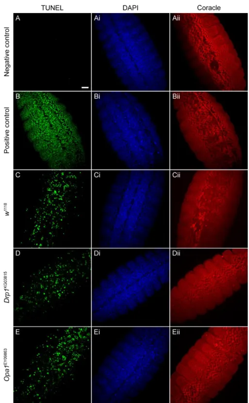
3. Drp1 and Opa1 mutations do not compromise cell viability in Drosophila embryos ... 93
4. Analysis of mitochondrial morphology and localization in the embryonic epidermis ... 95
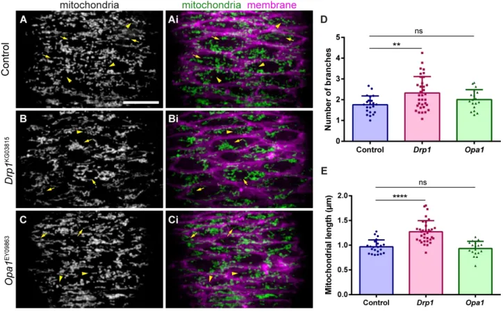
4.2. Drp1 mutants have altered mitochondrial morphology ... 95
4.3. Epithelial wounding leads to changes in mitochondrial morphology ... 98
4.4. Mitochondrial localization is unaffected during wound closure ... 102
5. Measurement of the mitochondrial membrane potential in the embryonic epidermis ... 103
6. Assessment of mitophagy during wound healing ... 107
7. Characterization of the wound healing phenotype of Drp1 and Opa1 mutants... 108
7.1. Drp1 mutants show delayed wound healing ... 108
7.2. Drp1 and Opa1 mutants have F-actin defects during wound closure ... 111
7.3. Rok localization at the wound edge is affected in Opa1 mutants ... 115
7.4. E-cadherin remodelling during wound repair is unaffected in Drp1 and Opa1 mutants ... 117
7.5. Drp1 and Opa1 mutants have altered cytosolic calcium dynamics upon wounding ... 117
7.6. Drp1 mutants have altered mitochondrial calcium dynamics upon wounding ... 121
7.7. Measurement of Reactive Oxygen Species in the embryonic epidermis ... 123
CHAPTER 4. DISCUSSION ... 125
1. Mitochondrial dynamics proteins as novel embryonic wound healing regulators ... 127
2. Epithelial wounding induces changes in mitochondrial morphology ... 129
3. Mitochondrial fusion and fission proteins regulate wound healing events ... 131
3.1. The mitochondrial fusion protein Opa1 is required for calcium and F-actin dynamics during wound closure ... 132
3.2. The mitochondrial fission protein Drp1 is essential for wound healing ... 133
4. Conclusions and future perspectives... 137
xiii
THESIS PUBLICATIONS
Part of the work shown in this thesis is included in the following publication:
Susana Ponte, Lara Carvalho, Maria Gagliardi, Isabel Campos, Paulo J. Oliveira, António Jacinto. Drp1-mediated mitochondrial fission regulates calcium and F-actin dynamics during wound healing.
In submission
The work performed during one of the lab rotations of this PhD is included in the following publication:
Susana Ponte, António Jacinto, Lara Carvalho. The occluding junction protein Neurexin-IV is required for tissue integrity in the Drosophila wing disc epithelium. Matters (ISSN: 2297-8240) 2019. DOI: 10.19185/matters.201903000014
During the course of this PhD the author has contributed to the following publications:
Lara Carvalho, Pedro Patricio, Susana Ponte, Carl-Philipp Heisenberg, Luis Almeida, André S. Nunes, Nuno A.M. Araújo, Antonio Jacinto. Occluding junctions as novel regulators of tissue mechanics during wound repair. J Cell Biol. 2018 Dec 3;217(12):4267-4283. doi: 10.1083/jcb.201804048.
Inês Cristo, Lara Carvalho, Susana Ponte, António Jacinto, Novel role for Grainy head in the regulation of cytoskeletal and junctional dynamics during epithelial repair. J Cell Sci. 2018 Sep 3;131(17). pii: jcs213595. doi: 10.1242/jcs.213595.
xv
THESIS CONTRIBUTIONS
The author designed and performed the great majority of experimental procedures, analysis of the results and assembly of the figures in this thesis. The author wrote all the Chapters of this thesis.
Lara Carvalho designed, performed the experiments, analysed the results shown in Figure 28. Lara Carvalho also helped to perform the experiments shown in Figure 27.
Telmo Pereira and Carolina Crespo helped with feedback on image analysis and quantifications in Figures 14, 15, 29 and 30.
xvii
ACKNOWLEDGEMENTS
“You cannot teach a man anything; you can only help him discover it in himself.”
- Galileo
I would like to thank the people who helped me accomplish this PhD.
To my supervisor, António Jacinto, for giving me the opportunity to work in his lab for the past 8 years. Thank you for making me and independent scientist and always trusting my ideas. Thank you for recruiting the best lab mates one could wish for. Thank you for all the parties and lab retreats, it sure is fun to work in your lab. To my co-supervisor, Paulo J. Oliveira, for always being available to discuss my work and for reviewing this thesis.
To the PHDOC Board, for giving me the opportunity to do a PhD. It was not always pleasant, but it was definitely lifechanging and I will never regret it.
To my favourite post-doc of all times, Lara Carvalho, for letting me grow as a scientist whilst supporting me all the way through this PhD. I once told you that 99% of what I know about being a good scientist I have learnt from you. This is still true. Thank you for all your help during this PhD and for reviewing this thesis. Thank you for the yoga classes. Namaste!
To Telmo, for all his help with image acquisition, image analysis and quantifications. Sorry for all the times I complained about the Spinning Disk!
To Carolina Crespo, for always trying to help me get the best images and the best graphs. Thank you for all your motivational speeches and for reviewing this thesis.
To Ana Sofia Brandão, for being a role model of what a good PhD student should be, for all the times you changed the laying pots, for reviewing this thesis, for always being there for me.
To all my lab colleagues, past and present, for all the laughs, for all the fun, for always supporting me. It is definitely easier to a PhD with your company. All of you helped me somehow along this way and I will be forever grateful to have met you.
To Inês Cristo, for all the motivation from far far away.
To Maria Gagliardi and Isabel Campos, for providing the preliminary results that led to this project and for always encouraging me.
xviii
To Marta Santos and Teresa Gomes for making life easier for me and for the flies. To the CEDOC fly community, for all the insightful discussion about my work.
To Rita Teodoro, for providing the anti-HA antibody and for all the feedback regarding my work.
To the IGC, specially to Nuno Pimpão Martins and Gabriel Martins from the imaging facility and to Élio Sucena for hosting me for a couple of months to do the calcium experiments.
To Petra Pintado and Fábio Valério for helping me with the microinjection and to the Catarina Craveiro (from the CCU) for the microinjection needles.
To Ana Roberto, Fernanda Baptista, Ângela Dias and Ana Sofia Almeida, for making my life easier when it comes to bureaucracy.
To Cláudia Pereira and Ana Soares, for sharing this journey with me and helping me endure the difficult times and celebrate the good ones.
To all my fellow PHDOC colleagues, for all the fun and good food in Braga and Coimbra (and a bit of science as well).
To my friends and family, for always supporting me even though they have no idea what I do for a living. To Eduardo, for all your help and understanding, for believing in me and always motivate me to do better. Love is not in grand gestures; love is in the little things.
xix
ABSTRACT
Epithelia form the barrier that protects our body against the external environment. An injury represents a challenge to barrier homeostasis and must be repaired efficiently to maintain epithelial integrity and function. Wound healing in different types of epithelia varies in complexity but encompasses a series of conserved responses. The wound triggers the release of molecules that signal to the surviving tissue, leading to a cascade of events that include an immune response, and coordinated changes in the cellular cytoskeleton and adhesion machineries to close the wound and restore epithelial integrity. Understanding the molecular mechanisms involved in these different steps is highly relevant from the biological and biomedical perspective. Embryonic tissues have a remarkable ability to deal with injury by closing the wounds in a quick and scarless manner. The lessons learned from this model system should be useful to improve the current therapeutics for wound healing complications in humans, such as chronic wounds. Embryonic wound healing relies on the formation of a contractile cable at the wound leading edge, formed by the actin and myosin cytoskeleton, that coordinates the collective tissue movement that brings the wound edges together and closes the gap. This process is coordinated with cell crawling, cellular rearrangements and shape changes, in order to close the wound without the involvement of cell proliferation.
Mitochondria are pivotal organelles for cell survival. Known as the powerhouse of the cell due to their energy production capability, they also perform other critical cellular functions, such as the regulation of calcium and redox homeostasis and apoptosis. Mitochondria are dynamic organelles, being able to change their shape, number and localization to adapt to the cellular needs. These events are collectively termed mitochondrial dynamics and play an important role in modulating mitochondrial functions. Mitochondrial dynamics includes fusion and fission events, which modulate morphology; mitochondrial biogenesis and mitophagy, which regulate mitochondrial number and quality control; and mitochondrial trafficking, which controls the subcellular distribution of mitochondria in response to the cellular needs. Dysfunction in the molecular machinery that governs mitochondrial dynamics is associated with a plethora of human pathologies, such as cancer, and neurodegenerative and metabolic diseases. However, the role of mitochondria and mitochondrial dynamics in epithelial repair in vivo has so far not been investigated.
In this work, we took advantage of genetically-encoded fluorescent markers, high-resolution imaging and advanced laser ablation techniques to understand the contribution of mitochondrial dynamics to epithelial repair in vivo, in the Drosophila embryonic epidermis. Using a genetic screen assay, we identified proteins involved in mitochondrial fission, fusion and trafficking as novel wound healing regulators. In vivo live imaging of the wound closure process revealed that Dynamin related protein 1 (Drp1) and Optic Atrophy 1 (Opa1) proteins, that are central players in mitochondrial fission and fusion, respectively, regulate calcium
xx
and actin cytoskeleton dynamics during wound healing. Moreover, we showed that wounding induces changes in mitochondrial morphology in the cells facing the wound, suggesting that the injury induces mitochondrial fission. We found that Drp1 loss of function leads to defects in both cytosolic and mitochondrial calcium dynamics upon wounding. Calcium ions are important second messengers in a myriad of signalling pathways and key players in the most important wound healing events. Wounding induces a quick and striking increase in intracellular calcium in the cells closer to the wound, which triggers a cascade of events that leads to the formation of the contractile cable and consequent wound closure. We showed that, besides this rise in cytosolic calcium, wounding also prompts an increase in mitochondrial calcium upon wounding. The uptake of calcium by mitochondria is known as an essential mechanism in controlling cytosolic calcium levels. Given the pleiotropic effects of calcium ions in the cell, its concentration needs to be tightly regulated in a spatial and temporal manner. Based on our results, we propose that Drp1 regulates the mitochondrial calcium buffering capacity, which then controls cytosolic calcium levels. Consistent with the described role of calcium in coordinating F-actin dynamics during wound healing, we also show that Drp1 mutants display significant defects in F-actin accumulation at the wound edge and in wound closure kinetics. Altogether, our results lead us to propose a model where mitochondrial fission is induced upon injury; this leads to a controlled increase in intracellular calcium, which then activates the main cytoskeleton changes needed to promote efficient wound healing. This work places Drp1 and mitochondrial fission as upstream players in the cascade leading to the main events of wound healing. As calcium and F-actin are crucial elements of the wound healing response across different types of epithelia, our work has relevant implications in the understanding of epithelial repair in other systems. Finally, our results also expand the knowledge about mitochondria biology and their relevance for different cellular processes.
xxi
RESUMO
Os epitélios formam uma barreira que protege o nosso corpo do ambiente externo. Uma lesão constitui um desafio a esta função de barreira e deve ser resolvida de forma eficiente para manter a integridade e função dos epitélios. A cicatrização de feridas varia no grau de complexidade depedendo do tipo de epitélio, mas envolve uma série de respostas conservadas. A ferida promove a libertação de moléculas que servem como sinais para o tecido circundante coordenar uma cascata de eventos, que inclui uma resposta imunitária e alterações coordenadas no citoesqueleto e adesões celulares, de forma a fechar a ferida e restaurar a integridade epitelial. Compreender os mecanismos moleculares envolvidos nestes diferentes passos é extremamente relevante do ponto de vista biológico e biomédico. Os tecidos embrionários têm uma capacidade notável de resolver lesões, fechando as feridas de forma rápida e sem deixar cicatriz. O conhecimento adquirido através destes modelos poderá ser útil para melhorar as terapêuticas atuais para complicações inerentes à cicatrização de feridas em humanos, como é o caso das feridas crónicas. A cicatrização de feridas no estádio embrionário envolve a formação de um cabo contrátil de actina e miosina na margem da ferida que coordena o movimento coletivo do tecido de forma a aproximar os limites da ferida e fechar a brecha. Este processo acontece em paralelo com migração celular e rearranjos na forma e posição das células, de forma a fechar a ferida sem o envolvimento de proliferação celular.
As mitocôndrias são organelos cruciais para a sobrevivência das células. São organelos conhecidos principalmente pela sua capacidade de produção de energia, mas desempenham outras funções críticas nas células, tais como a regulação do cálcio e do estado redox, e da morte celular. As mitocôndrias são organelos dinâmicos, com a capacidade de mudar a sua forma, número e localização como forma de adaptação às necessidades celulares. Estes processos são denominados de dinâmica mitocondrial e possuem um papel importante no controlo das funções mitocondriais. A dinâmica mitocondrial inclui eventos de fusão e fissão que modulam a morfologia das mitocôndrias, processos de biogénese e mitofagia que regulam o número de mitocôndrias e o seu controlo de qualidade, e mecanismos de tráfego mitocondrial que controlam a localização subcelular das mitocôndrias em resposta às necessidades da célula. A disfunção na maquinaria molecular que controla a dinâmica mitocondrial está associada a várias patologias humanas, tais como cancro e doenças neurodegenerativas e metabólicas. Contudo, o papel das mitocôndrias e dinâmica mitocondrial na reparação de tecidos in vivo não foi até agora investigado.
Neste trabalho tirámos partido de marcadores fluorescentes geneticamente codificados, imagiologia de alta resolução e técnicas de ablação avançadas para compreender a contribuição da dinâmica mitocondrial na reparação epitelial in vivo, usando a epiderme do embrião da mosca-da-fruta (Drosophila melanogaster) como modelo. Este estudo permitiu-nos identificar proteínas envolvidas na fissão, fusão e tráfego mitocondrial como novos reguladores da cicatrização de feridas. A observação do processo de fecho de ferida
xxii
respetivamente, regulam o cálcio e o citoesqueleto de actina durante a cicatrização da ferida. Adicionalmente, descobrimos que a ferida promove alterações na morfologia mitocondrial que sugerem uma indução de fissão mitocondrial. A perda de função de Drp1 conduz a defeitos na dinâmica do cálcio citosólico e mitocondrial em resposta à ferida. Os iões de cálcio são importantes segundos-mensageiros em várias vias de sinalização e são reguladores fundamentais no processo de cicatrização de feridas. A ferida induz um aumento rápido e dramático no aumento dos níveis intracelulares de cálcio, que despoleta a cascata de eventos que culmina na formação do cabo de actina e miosina e consequente fecho da ferida. Neste trabalho mostrámos que, para além do aumento de cálcio no citosol, a ferida também induz um aumento no cálcio mitocondrial. Sabe-se que o influxo de cálcio para o interior das mitocôndrias é um mecanismo essencial para a regulação dos níveis de cálcio no citosol. Dado a variedade de funções desempenhadas pelo cálcio, a sua concentração celular deve ser regulada no tempo e no espaço. Com base nos nossos resultados, propomos que Drp1 regula a capacidade de captação de cálcio pelas mitocôndrias, o que por sua vez controla os níveis de cálcio no citosol. Em concordância com o papel descrito do cálcio na regulação do citoesqueleto de actina durante a cicatrização de feridas, mostrámos ainda que mutantes para Drp1 possuem defeitos significativos na acumulação de actina nas margens da ferida e na dinâmica de fecho da ferida. Em suma, os nossos resultados levam-nos a propor um modelo onde a fissão mitocondrial é induzida pela ferida, levando a um aumento controlado dos níveis intracelulares de cálcio, o que então regula as alterações no citoesqueleto necessárias para promover a cicatrização de forma eficiente. Este trabalho coloca a proteína Drp1 e a fissão mitocondrial no topo da cascata de sinalização que leva aos principais eventos na cicatrização de feridas. Dado que o cálcio e a actina são elementos cruciais na resposta à ferida em diferentes tipos de tecidos epiteliais, o nosso trabalho tem implicações relevantes na compreensão da reparação de tecidos noutros sistemas. Por fim, os nossos resultados aumentam o conhecimento da biologia mitocondrial e da sua relevância para diferentes processos celulares.
xxiii
LIST OF ABBREVIATIONS
Abbreviations – Full form
% - percent, percentage (-) – minus (+) – plus [O2]•− - superoxide ’ – minutes µg - microgram µL – microliter µm – micrometer µM – micromolar 1O2 - singlet oxygenADP - Adenosine diphosphate AJs - Adherens Junctions
AMP - Adenosine monophosphate AMPK - AMP-activated protein kinase Arp2/3 - actin-related proteins 2/3 Atg - autophagy-related protein ATP - Adenosine triphosphate Bcl-2 - B-cell lymphoma 2
Bnip3 - Bcl2/adenovirus E1B 19-kDa interacting protein 3 BSA - Bovine Serum Albumin
bw – before wounding
C. elegans - Caernorhabditis elegans Ca2+ - calcium ions
Caf4 – CCR4-associated factor 4
CaMKIV - calcium/calmodulin-dependent protein kinase IV CaMKIα - calcium/calmodulin-dependent protein kinase Iα cAMP – cyclic adenosine monophosphate
caspase - cysteine protease cleaving after Asp CCR4 - C-C chemokine receptor type 4
Cdc42 - Cell division control protein 42 homolog ChFP- Cherry fluorescent protein
xxiv
CoA - coenzyme A
CREB - cAMP-response-element-binding protein Cu - copper
cyt c - cytochrome c
DABCO - 1,4-diazabicyclo[2.2.2]octane DAPI - 4', 6-diamidino-2-phenylindole Dia – Diaphanous
DIABLO - direct IAP-binding protein with low pI DMSO - Dimethyl sulfoxide
DN - dominant-negative DNA - deoxyribonucleic acid Dnm1 – Dynamin-related protein 1 Drosophila – Drosophila melanogaster Drp1 - Dynamin-related protein 1 dUTP - Deoxyuridine Triphosphate EAT-3 – C. elegans homolog of Opa1 E-cad – E-cadherin
ECM – extracellular matrix EM – electron microscopy
EMRE - essential MCU regulatory element ER – endoplasmic reticulum
ETC - electron transport chain
EYFP – enhanced yellow fluorescent protein F-actin – filamentous actin
FAs – Focal adhesions
Fis1 - Mitochondrial fission 1 protein FUNDC1 - FUN14 Domain Containing 1 Fzo – fuzzy onions
g – gram
G-actin – globular actin
Gdap1 - ganglioside-induced differentiation-associated protein 1 GDP - Guanosine diphosphate
GED - GTPase effector domain GFP – greeh fluorescent protein GSH – reduced Glutathione
xxv GSSG – oxidized Glutathione GTP – Guanosine triphosphate GTPase – GTP hydrolase h – hour H+ - proton H2O – water
H2O2 – hydrogen peroxide
HCX - Na+ independent (H+/Ca2+) exchanger HOCl - hypochlorous acid
HTRA2 - high temperature requirement protein A2 IAP - inhibitor of apoptosis protein
IBM - inner boundary membrane IFN2 - Inverted Formin 2
IMM – inner mitochondrial membrane IMS - intermembrane space
IP3R - inositol triphosphate receptor L – liter
LC3 - Microtubule-associated protein 1A/1B-light chain 3 LC3-II - lipidated form of LC3
LETM1 - leucine zipper EF-hand- containing transmembrane protein 1 LIR - LC3 interacting region
L-Opa1 – large isoform of Opa1 M – molar
MAMs - mitochondria-associated ER membranes Marf - Mitochondrial assembly regulatory factor mCherry – mCherry fluorescent protein
MCU – mitochondrial calcium uniporter MCUb - mitochondrial calcium uniporter b MDCK - Madin-Darby Canine Kidney
Mdm36 – Mitochondrial distribution and morphology protein 36 Mdv1 – Mitochondrial division protein 1
MELC – myosin essential light chain Mff - mitochondrial fission factor Mfn1 – Mitofusin 1
xxvi
Mgm1 – mitochondrial genome maintenance 1 MHC – myosin heavy chain
MICU1 - mitochondrial calcium uptake 1 MICU2 - mitochondrial calcium uptake 2
MiD49 - mitochondrial dynamics proteins of 49 kDa MiD51 - mitochondrial dynamics proteins of 51 kDa Min – minutes
MiNA - Mitochondrial Network Analysis Miro - Mitochondrial Rho
mL – milliliter mm – millimeter mM – millimolar
MOMP - mitochondrial outer membrane permeabilization MPT - mitochondrial permeability transition
mpw - minutes post-wounding MRLC – myosin regulatory light chain mRYR - mitochondrial ryanodine receptor mtDNA – mitochondrial DNA
MTP18 - mitochondrial protein of 18 kDa
Mtpα - Mitochondrial trifunctional protein α subunit Myo19 - Myosin XIX
NA – numerical aperture
NAD+ – Nicotinamide adenine dinucleotide NADH – reduced form of NAD+
NCX - Na+ dependent (Na+/Ca2+) exchanger nm – nanometers
nM – nanomolar NO - nitric oxide
NRF-1 - nuclear respiratory factor 1 NRF-2 - nuclear respiratory factor 2 NSF - N-ethylmaleimide-sensitive factor Num1 – Nuclear migration protein 1 ºC – degrees Celsius
OJs – Occluding Junctions
xxvii OMM – outer mitochondrial membrane
Opa1 - Optic atrophy 1
Orp1 - oxidant receptor peroxidase 1 OXPHOS - oxidative phosphorylation PBS - phosphate-buffered saline PGC-1α - PPAR-γ coactivator-1α PH - pleckstrin homology
PI3K - phosphatidylinositol (3)-phosphate kinase PINK1 - PTEN-induced kinase 1
Pkn – Protein kinase N PLCγ - Phospholipases Cγ
PPAR - peroxisome proliferator-activated receptor
PTEN - Phosphatase and Tensin Homolog deleted on Chromosome 10 Rho – Ras homologous
RhoGAPs - Rho GTPase-activating proteins
RhoGEFs – Rho guanine nucleotide exchange factors RNA - ribonucleic acid
Rok – Rho kinase
ROS - reactive oxygen species RT – room temperature
SAM - sorting and assembly machinery SJs – Septate Junctions
SNARE - soluble NSF attachment receptor Sod1 - superoxide dismutase 1
S-Opa1 – small isoform of Opa1 Spire1C - Protein spire homolog 1 spw - seconds post-wounding Sqh - spaghetti squash
Src – pronounced sarc, as it comes from the word “sarcoma” SUMO - small ubiquitin-like modifier
TCA - Tricarboxylic Acid
TdT - terminal deoxynucleotidyl transferase Tfam - mitochondrial transcription factor A TJs – Tight Junctions
xxviii
TNTs - intercellular tunneling nanotubes
TOM - translocator of the outer mitochondrial membrane TORC - transducer of regulated CREB-binding protein TRPC3 - short transient receptor potential channel 3
TRPM - transient receptor potential channel, melastatin family TUNEL - TdT-mediated dUTP-X nick end labelling
UCP1 - mitochondrial uncoupling protein 1 UCP2 - mitochondrial uncoupling protein 2 VDAC - voltage-dependent anion channel WASp - Wiskott–Aldrich syndrome protein
WAVE - WASp-family verprolin homologous protein X. laevis – Xenopus laevis
Zip – Zipper Zn – zinc
xxix
LIST OF FIGURES
Figure 1. Wound healing in embryonic tissues. ... 36 Figure 2. Wound healing of complex epithelia. ... 43 Figure 3. Cell wound repair. ... 46 Figure 4. Models of mitochondrial membrane structure. ... 49 Figure 5. Mitochondrial bioenergetics. ... 53 Figure 6. Mitochondrial morphology in different cell types. ... 59 Figure 7. Mitochondrial fission. ... 61 Figure 8. Mechanism of mitochondrial fusion... 63 Figure 9. Mitochondrial trafficking. ... 64 Figure 10. Mitophagy... 66 Figure 11. Mitochondrial dynamics proteins are required for wound healing. ... 92 Figure 12. Drp1 localization in the embryonic epidermis. ... 92 Figure 13. Drp1 and Opa1 mutations do not seem to affect apoptosis. ... 94 Figure 14. Drp1 mutant embryos have altered mitochondrial morphology. ... 96 Figure 15. Different quantification methods applied to the analysis of mitochondrial morphology in the Drosophila embryonic epidermis. ... 97 Figure 16. Mitochondrial morphology before and during wound healing. ... 99 Figure 17. Mitochondrial morphology changes upon wounding. ... 101 Figure 18. Mitochondrial localization upon wounding... 102 Figure 19. Mitochondrial membrane potential assessment with Mitotracker after embryo permeabilization. ... 104 Figure 20. Mitochondrial membrane potential assessment by microinjection of TMRM. ... 107 Figure 21. Wounding does not seem to induce autophagy. ... 107 Figure 22. Drp1 mutant embryos show impaired wound closure dynamics. ... 109 Figure 23. Opa1 mutant embryos show no wound closure dynamics defects. ... 110 Figure 24. Drp1 mutants show F-actin defects during wound closure. ... 112 Figure 25 Opa1 mutants show F-actin defects during wound closure. ... 113 Figure 26. Myosin accumulation at the wound edge is unaffected in Drp1 and Opa1 mutants. ... 114 Figure 27. Rok localization in control, Drp1 and Opa1 mutants during the wound healing response. ... 116 Figure 28. E-cadherin localization in control, Drp1 and Opa1 mutants after wounding. ... 119 Figure 29. Drp1 and Opa1 mutant embryos show altered cytosolic calcium dynamics. ... 120 Figure 30. Drp1 mutants show altered mitochondrial calcium dynamics. ... 122 Figure 31. Changes in roGFP emission spectrum upon oxidation after injection of Diamide. ... 123
xxxi
LIST OF TABLES
Table 1. Fly lines used in this study ... 82 Table 2. Reagents used in this study ... 84
33
Chapter 1. Introduction
34
“What we know is a drop, what we don’t know is an ocean.”
Chapter 1. Introduction
35
1. EPITHELIAL WOUND HEALING
For Metazoans, the maintenance of epithelial integrity is critical to sustain life. Epithelial tissues cover all the exposed surfaces of our body, inside and out, and form the functional units of secretory glands. Epithelia are cohesive sheets of specialized cells for absorption, secretion and to act as a barrier against abrasion, radiation, chemical stress and invasion by pathogens. Epithelia are classified in terms of the number of cell layers and the shape of the epithelial cells. They can be simple, when composed of just one cell layer, or stratified, containing several layers of epithelial cells. Epithelia are classified as squamous (flat cells), cuboidal (similar cellular width and height) or columnar (tall cells). The cellular shape and the tissue stratification are related to the epithelial function. For example, the stratified nature of our skin is ideal for its barrier function. In contrast, the squamous shape of the alveolar cells from our lungs facilitates gas exchanges (Lowe and Anderson, 2015). Given all the critical functions of epithelial tissues, it is of the upmost importance to deal with injury in a quick, efficient way. Epithelial cells have developed mechanisms to cope with wounds, either at the single cell level, or at the tissue scale.
This thesis has focused on wound healing of the Drosophila melanogaster (hereafter Drosophila) embryonic epidermis, a simple epithelium. A detailed review of embryonic and other simple epithelia repair mechanisms will be provided in this section, along with brief mentions to single cell repair and wound healing of more complex epithelia.
1.1.
Embryonic and simple epithelia
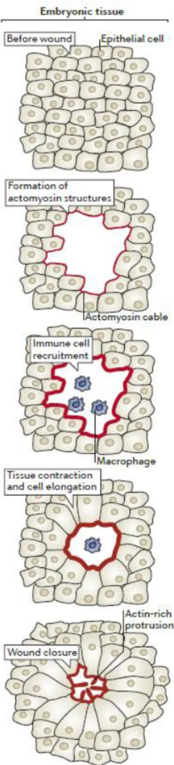
Embryonic tissues have been used as models to understand epithelial repair because of their remarkable ability to efficiently deal with injury (Garcia-Fernandez et al., 2009). Early studies, relying on electron microscopy, have highlighted that the embryonic wound closure process is very fast, ranging from minutes to less than a day (Smedley and Stanisstreet, 1984; Stanisstreet et al., 1980). The current view of the phases of this process are depicted in Figure 1. Damage signals released by the wounded tissue trigger a response by the surviving cells. This response leads to the accumulation of cytoskeleton components, namely actin filaments and non-muscle myosin II (hereafter called myosin) molecular motors at the wound edge, forming a ring-like contractile structure called the actomyosin cable (Rothenberg and Fernandez-Gonzalez, 2019). As the actomyosin structures form, immune cells from the leukocytic lineage, such as macrophages and neutrophils, are recruited to the wound in order to clear cell debris and fight the entry of pathogens. Sliding of myosin motors along actin filaments at the front-edge of wound-facing cells leads to tissue movement, contraction and concomitant reduction of the wound area over time. In addition, and possibly as
Chapter 1. Introduction
36
a consequence, wound edge cells elongate to further reduce the wound size. In addition to the actomyosin cable, leading edge cells form actin-rich protrusions that promote cell crawling and mediate contacts between opposing sides of the wound (Cordeiro and Jacinto, 2013).
We will now address these steps in more detail, highlighting the known and unknown molecular mechanisms involved in the repair of embryonic and other simple epithelia.
1.1.1.
Actomyosin cable
Wound healing of embryonic epithelia is characterized by accumulation of F-actin and Myosin at the cell membranes that face the wound, forming an actomyosin cable. Actin is the most abundant intracellular protein and exists in two conformations: as a globular monomer called G-actin and as a filamentous polymer called F-actin. Actin filaments are formed by polymerization of G-actin subunits, with consequent expenditure of adenosine triphosphate (ATP). F-actin organization in complex networks forms a cytoskeleton that modulates cellular shape. The polymerization, depolymerization and organization of F-actin into different types of networks are regulated by actin-binding proteins. Actin is also involved in the formation of cellular protrusions, like microvilli, which increase the apical membrane surface area, or filopodia and lamellipodia, which establish contacts with the underlying cell substrate and are involved in cell migration (Lodish et al., 2000). Myosins are molecular motors that move along actin filaments by conformational changes induced by hydrolysis of ATP. Myosin is a multimeric protein, composed of two heavy chains (MHC), two essential light chains (MELC) and two regulatory light chains (MRLC) (Betapudi, 2014). The movement of myosin across F-actin networks generates contractile forces. The actin-myosin contractile machinery spans the apical cell surface of the epithelial cells and is anchored to cell-cell junctions, namely the Adherens
Figure 1. Wound healing in embryonic tissues.
The wound healing process includes the formation of actomyosin structures that promote tissue contraction and cell elongation to bring the wound edge cells together. Immune cells are recruited to the wounded region to clear cell debris and prevent infection. Actin protrusions mediate the final adhesion of the wound edge cells. Adapted from (Cordeiro and Jacinto, 2013).
Chapter 1. Introduction
37 Junctions (AJs). This localization allows contraction that lead to changes in cell shape and are coupled with tissue movements (Kasza and Zallen, 2011).
In the early 90s, Martin and Lewis characterized the closure of the chick embryo epidermis and observed that the epidermal tissue moves inwards over time. The fact that lamellipodia were not detected in the leading-edge cells suggested that this movement was independent of cell migration. Combined with the observation that the tissue was under tension, it led them to propose that closure must rely on circumferential contractile forces at the wound margin, that bring the wound edges together, closing the hole in a purse string manner. Consistent with this hypothesis, they observed accumulation of F-actin at the wound edge minutes after wounding and remaining there until the wound was closed (Martin and Lewis, 1992). The purse string hypothesis (Martin and Lewis, 1992) was compatible with previous observations that the shape of the epidermal cells changes upon wounding, suggesting that the tissue is under tension, and that the leading-edge cells elongate during wound closure, indicating that they are being pulled by contractile forces (Smedley and Stanisstreet, 1984; Stanisstreet et al., 1980). A few years later, the same purse string wound closure mechanism was observed in the mouse embryo and disruption of the actin cable by cytochalasin D treatment impaired re-epithelialization, confirming its requirement for proper wound closure (McCluskey and Martin, 1995). It was also found that myosin was part of the cable, accumulating at the wound edge together with F-actin, further highlighting the contractile nature of this ring-like structure (Bement et al., 1993; Brock, 1996).
Over the past years, many efforts have been made to understand how epithelia sense the wound and form the actomyosin cable. Actomyosin cables are not exclusive of wound healing. They have been observed during morphogenesis in embryonic development (Jacinto et al., 2002; Wood et al., 2002; Young et al., 1993), in extrusion of apoptotic cells (Ninov et al., 2007; Rosenblatt et al., 2001), and during the separation of dividing cells in the process of cytokinesis (Pollard, 2010). Studies on these processes have also helped to understand how actomyosin cables work in wound healing. Advances in microscopy techniques, allowing the live imaging of the wound closure process have improved our understanding of how the actomyosin cable is formed and how it drives wound closure.
1.1.2. Cell migration
Studies in epithelial monolayers in vitro have shown that wounds can close either by actomyosin cable-mediated contraction or by cell migration/crawling to cover the wound or a combination of the two processes (Altan and Fenteany, 2004; Begnaud et al., 2016; Bement et al., 1993; Fenteany et al., 2000; Tamada et al.,
Chapter 1. Introduction
38
2007). Cell migration is not exclusive of wound healing in vitro. Wounds in the Drosophila abdomen epidermis also close through cell shape changes and lamellipodia formation (Rämet et al., 2002) and in embryonic wound healing the actin protrusions work together with the actomyosin cable to drive the collective movement of the epithelial tissue.
Cell migration-mediated wound closure involves actomyosin cytoskeleton remodelling to form protrusive structures and to create the intracellular forces required for cell movement. Cells adjacent to the wound repolarize and become migratory and lead the other epithelial cells, in a process referred to as collective cell migration (Begnaud et al., 2016). Polarized leader cells extend protrusions in the direction of movement. Cell migration is associated with two types of F-actin protrusions: lamellipodia, which look like large sheets, and contain highly branched and cross-linked actin filaments (Ballestrem et al., 2000); and filopodia, thin finger-like structures, with parallel bundles of F-actin, that often project beyond the edge of the lamellipodium (Mattila and Lappalainen, 2008). The generation of the intracellular forces occurs through attachment sites, called focal adhesions (FAs). FAs link the intracellular actin cytoskeleton with the extracellular matrix (ECM) and this interaction is mediated by integrins. New FAs form behind the leading edge of the cell and pull the cell forward. Release of attachment sites at the rear of the cell allows the rear end to move in the direction of movement (Lambrechts et al., 2004). The regulators of the actomyosin cable and F-actin protrusions are conserved and are discussed below (1.1.3 Rho GTPases and their effectors and 1.1.4 Calcium).
1.1.3. Rho GTPases and effectors
The regulation of actomyosin contractile structures relies on the action of Ras homologous (Rho) GTPase protein family. Rho GTPases are cytoskeletal regulators that alternate between an inactive (GDP-bound) and an active (GTP-bound) form. This switch is regulated by Rho guanine nucleotide exchange factors (RhoGEFs), which catalyse the phosphorylation of GDP to GTP, and by Rho GTPase-activating proteins (RhoGAPs), which hydrolyse the GTP to GDP. When active, Rho GTPases are able to activate effector proteins that regulate F-actin polymerization and myosin contraction. In mammals, this protein family is composed of 20 proteins, but the most well studied and conserved members are Rho, Rac and the Cell division control protein 42 homolog (Cdc42) (Heasman and Ridley, 2008; Sit and Manser, 2011). Rho GTPases are known regulators of the actin cytoskeleton in mammalian cultured cells (Hall, 1994) and are also important during wound healing
Chapter 1. Introduction
39 In the wound healing of the chick wing bud during development, treatment with C3 transferase, a bacterial enzyme that inactivates Rho, prevents the assembly of the actomyosin cable, leading to failure of wound closure. The Rho protein was found to be indispensable for actomyosin cable formation (Brock, 1996). In Drosophila, Rho1 mutants or embryos expressing dominant-negative (DN) versions of Rho1 fail to form the actomyosin cable during dorsal closure, a morphogenetic movement during Drosophila embryonic development that resembles wound closure (Lu and Settleman, 1999; Magie et al., 1999). The family of Rho GTPases cooperates during wound healing: Rho1 regulates the formation of the actomyosin cable and Cdc42 mediates filopodia and lamellipodia formation. F-actin protrusions are important to mediate cell crawling during the contraction phase and to mediate the final approximation and adhesion of the wound-edge cells in the final stages of wound closure (Abreu-Blanco et al., 2012b; Verboon and Parkhurst, 2015; Wood et al., 2002). In vitro, Rac is the Rho GTPase responsible for lamellipodia formation (Das et al., 2015; Fenteany et al., 2000; Yamaguchi et al., 2015). The role of Rac in wound closure in vivo is less clear. Expression of DN-Rac in both the chick wing bud (Brock, 1996) and mutations in Rac genes in the Drosophila embryo (Wood et al., 2002) do not lead to detectable wound healing defects. However, a more recent study has identified a significant wound healing delay in Rac mutants, although no visible impairment in either the actomyosin cable or the actin protrusions was detected (Verboon and Parkhurst, 2015).
Active Rho GTPases exert their function by activating effector proteins that regulate F-actin and myosin. Rho1 effectors include the formin Diaphanous (Dia) and the Rho kinase (Rok). Dia promotes the polymerization of unbranched F-actin (Narumiya et al., 1997). Rok acts on myosin by activating MRLC, either directly, by phosphorylation, or indirectly, by inactivation of myosin phosphatases, leading to actomyosin contractility (Amano et al., 1996; Kimura et al., 1996; Ueda et al., 2002). Knockdown of both Rok and Dia impairs actomyosin dynamics in the Drosophila pupa wound healing (Antunes et al., 2013). Rok2 mutant embryos show delayed wound healing (Verboon and Parkhurst, 2015) and dia5 mutants have defects in the formation of the actomyosin cable and actin protrusions (Matsubayashi et al., 2015). The Cdc42 effector protein Wiskott-Aldrich Syndrome protein (WASp) and the Rac effector WASp-family verprolin homologous protein (WAVE), both implicated in the nucleation of branched actin filaments (Miki and Takenawa, 2003), have also been linked to Drosophila embryonic wound healing, but their roles are still not well understood (Matsubayashi et al., 2015).
The actomyosin cable and the actin protrusions collaborate to drive wound closure (Abreu-Blanco et al., 2012b; Ducuing and Vincent, 2016). Disruption of either of the actin-based structures leads to delayed wound healing, but the wounds eventually close. Rho1 and zip1 (zipper, the Drosophila MHC gene) mutants, in which the actomyosin cable does not fully form, are able close their wounds by increased formation of actin protrusions. Conversely, the wounds in cdc42 mutants, are able to contract through the action of the
Chapter 1. Introduction
40
actomyosin cable and defects are only observed in the final adhesion stage (Abreu-Blanco et al., 2012b; Wood et al., 2002). The absence of either mechanism of wound closure seems to be compensated by the other. Only the simultaneous disruption of the actomyosin cable and actin protrusions leads to fully impairment of wound healing (Abreu-Blanco et al., 2012b).
1.1.4. Remodelling of cell junctions
The AJs mediate adhesion between neighbouring cells, thus being essential to maintain tissue architecture. AJs are composed of:
- calcium-dependent transmembrane proteins called cadherins. About 20 different cadherins have been described but E-cadherin is characteristic of epithelial tissues (Takeichi, 1988). Binding of calcium controls the conformation of the cadherin extracellular domain, leading to homophilic interactions between cadherins of neighbouring cells (Pokutta et al., 1994).
- cytosolic proteins called catenins, which include p120-catenin, α-catenin and β-catenin. These catenins in turn bind a variety of other molecules.
AJs are connected to the actin cytoskeleton via α-catenin, that binds both β-catenin and actin cytoskeleton regulators such as vinculin. In polarized epithelial cells, AJs are localized apically on the cell lateral membrane and an adhesion belt, called the circumferential actin belt, as they completely encircle the cells along with the F‑actin lining on the cytosolic side (Hartsock and Nelson, 2008; Meng and Takeichi, 2009).
During wound healing, at the same time of actomyosin cable formation, E-cadherin has been found to localize in clusters at the wound margin, presumably representing the sites that link the actomyosin cable in adjacent cells (Brock, 1996). The AJ components E-cadherin (Abreu-Blanco et al., 2012b; Brock, 1996), α-catenin (Wood et al., 2002) and β-α-catenin (Zulueta-Coarasa et al., 2014) are removed from the cell cortex that face the wound and remain only at the cell-cell junctions linking adjacent cells. E-cadherin exclusion from the wound edge is mediated through remodelling by endocytosis (Hunter et al., 2015; Matsubayashi et al., 2015) and by transcriptional regulation by the NFB-pathway (Carvalho et al., 2014). The dynamics of E-cadherin localization is critical for the formation of the actomyosin cable, as both E-E-cadherin mutations and overexpression impair the formation of the actomyosin cable (Abreu-Blanco et al., 2012b; Hunter et al., 2015; Matsubayashi et al., 2015).
Besides AJs, other types of cell junctions have been implicated in wound healing. Occluding Junctions (OJs) localize close to the AJs. OJs are known for their permeability barrier function, controlling the transepithelial passage of molecules, and for maintenance of cell polarity, constituting a “fence” that
Chapter 1. Introduction
41 separates the apical and basolateral membrane compartments. OJs are termed Tight Junctions (TJs) in vertebrates and Septate Junctions (SJs) in invertebrates (Jonusaite et al., 2016; Shen, 2012). Recently it was reported that several mutants for SJ components fail to close epithelial wounds. A functional analysis of the mutant kune-kune (kune), a transmembrane SJ component of the Claudin family, revealed that SJ loss of function severely impairs the wound closure process and actomyosin cable formation. As seen for AJs, SJ proteins are also removed from the wound edge, but the mechanisms are still unknown. Interestingly, SJ loss of function affects the mechanical properties of the epithelial tissue and the cell shape changes and rearrangements that occur during wound healing (Carvalho et al., 2018). However, the molecular mechanisms involved remain completely unknown.
1.1.5. Calcium
An increase in cytoplasmic calcium in cells adjacent to the wound is the first response signal to be detected upon injury. This has been observed both in in vitro (Hinman et al., 1997; Leiper et al., 2006; Shabir and Southgate, 2008; Sung et al., 2003), and in in vivo models (Antunes et al., 2013; Razzell et al., 2013; Xu and Chisholm, 2011).
It is still unclear which are the calcium sources contributing to the increase of cytoplasmic calcium in the different wound healing models. In fact, there is evidence supporting both calcium influx from the extracellular environment and calcium release from internal stores. In the epidermis of the nematode
Caenorhabditis elegans (C. elegans), the rise in intracellular calcium is mediated by the transient receptor
potential calciumchannels of the melastatin subfamily (TRPM) at the plasma membrane. Additionally, there is calcium release from the endoplasmic reticulum (ER) via the Inositol 1,4,5-trisphosphate (IP3) receptor (IP3R), that seems to be mediated by G-protein coupled receptor (GPCR) signalling (Xu and Chisholm, 2011). Knockdown of TRPM also reduces the wound-induced intracellular calcium levels in the Drosophila pupal epithelium (Antunes et al., 2013). In both C. elegans and Drosophila models, impairment of the intracellular calcium rise leads to actomyosin cable defects (Antunes et al., 2013; Hunter et al., 2018a; Xu and Chisholm, 2011; Xu and Chisholm, 2014). Depletion of ER calcium stores or extracellular calcium also promote a reduction in Reactive Oxygen Species (ROS) production (Hunter et al., 2018a; Razzell et al., 2013; Xu and Chisholm, 2014) and immune cell recruitment (Razzell et al., 2013).
High-speed imaging of the wound-induced calcium rise has shown that calcium increases in the leading cells and then spreads a few cell rows away from the wound, propagating in an intercellular wave manner. After this initial dispersion, the calcium levels decrease from the periphery towards the wound edge (Antunes
Chapter 1. Introduction
42
et al., 2013; Narciso et al., 2015; Razzell et al., 2013; Restrepo and Basler, 2016; Shannon et al., 2017). The propagation of the intercellular calcium wave depends on IP3-mediated calcium release from internal stores and calcium transport across cells via Gap Junctions (Narciso et al., 2015; Razzell et al., 2013; Restrepo and Basler, 2016).
The exact mechanisms through which calcium mediates the wound healing response are still not fully understood. Data suggests that calcium regulates the actomyosin cable by activating actomyosin regulators. Knockdown of the calcium-dependent actin filament–severing protein Gelsolin (Sun et al., 1999) in the
Drosophila pupa impairs the actomyosin flow towards the wound and consequent cable formation (Antunes
et al., 2013). There is also evidence that the activation of Rho and Cdc42 GTPases upon wounding is calcium-dependent (Benink and Bement, 2005). Recent work has also shown that the regulation of the actomyosin cable occurs through the calcium-mediated production of ROS (Hunter et al., 2018a; Xu and Chisholm, 2014). ROS can regulate Rho GTPases and Rok by acting on redox-sensitive motifs in these proteins (Muliyil and Narasimha, 2014; Xu and Chisholm, 2014) and control E-cadherin remodelling by oxidizing the Src kinase Src42A (Hunter et al., 2018a).
1.1.6. Immune cell recruitment
Although the immune system differs between embryos and adults, wounding of embryonic tissues also triggers an inflammatory response, that involves the recruitment of immune cells to the wound site (Babcock et al., 2008; Moreira et al., 2010; Niethammer et al., 2009b; Razzell et al., 2013; Stramer et al., 2005; Wood et al., 2006). Upon wounding, immune cells such as macrophages and neutrophils are attracted to and migrate towards the wound site, to phagocytose pathogens and cellular debris resulting from the death of wounded cells (Babcock et al., 2008; Stramer et al., 2005). Wound-induced hydrogen peroxide production, which is downstream of calcium signalling triggered by the injury (Razzell et al., 2013), seems to contribute to immune cell recruitment (Moreira et al., 2010; Niethammer et al., 2009b; Razzell et al., 2013). Interestingly, it was shown that hemocytes, the Drosophila equivalent to macrophages, are not required for reepithelialisation (Stramer et al., 2005). This suggests that wound closure is not mediated by signals coming from these immune cells, neither the cell debris removal is critical to achieve wound healing. Nevertheless, although they do not affect the wound healing process, it is possible that hemocytes are important to prevent infection and ensure embryo survival, but this is yet to be addressed.
Chapter 1. Introduction
43
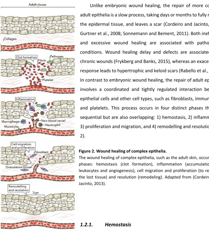
1.2. Repair in complex and adult epithelia
Unlike embryonic wound healing, the repair of more complex adult epithelia is a slow process, taking days or months to fully restore the epidermal tissue, and leaves a scar (Cordeiro and Jacinto, 2013; Gurtner et al., 2008; Sonnemann and Bement, 2011). Both inefficient and excessive wound healing are associated with pathological conditions. Wound healing delay and defects are associated with chronic wounds (Frykberg and Banks, 2015), whereas an exacerbated response leads to hypertrophic and keloid scars (Rabello et al., 2014). In contrast to embryonic wound healing, the repair of adult epithelia involves a coordinated and tightly regulated interaction between epithelial cells and other cell types, such as fibroblasts, immune cells and platelets. This process occurs in four distinct phases that are sequential but are also overlapping: 1) hemostasis, 2) inflammation, 3) proliferation and migration, and 4) remodelling and resolution (Fig. 2).
1.2.1.
Hemostasis
This initial phase is characterized by the formation of a clot that serves as a shield against the physical and chemical extracellular environment, preventing tissue/fluid leakage and pathogen entry. During this phase, blood vessels constrict, mediated by the vascular smooth muscle cells, to limit blood loss. Platelets leak from damaged blood vessels and aggregate to form a plug at the lesioned area (Palta et al., 2014). The subsequent release of secreted factors such as platelet-derived growth factor (PDGF), epidermal growth factor (EGF) and transforming growth factor-β (TGF-β) lead to the formation of a fibrin clot that plugs the wound hole. The factors released by the platelets also serve as
Figure 2. Wound healing of complex epithelia.
The wound healing of complex epithelia, such as the adult skin, occurs in 4 phases: hemostasis (clot formation), inflammation (accumulation of leukocytes and angiogenesis), cell migration and proliferation (to replace the lost tissue) and resolution (remodeling). Adapted from (Cordeiro and Jacinto, 2013).
Chapter 1. Introduction
44
chemoattractants to the immune cells that are recruited to mediate the next phase of wound healing (Pakyari et al., 2013; Pierce et al., 1991; Schultz et al., 1991).
1.2.2. Inflammation
Immune cells infiltrate the wound. The first cells to arrive are neutrophils, whose main function is to kill pathogens, through the release of proteases and ROS (Wilgus et al., 2013). Later on, monocytes arrive at the wound site, where they differentiate into macrophages and remove debris and apoptotic neutrophils by phagocytosis (Zaja-Milatovic and Richmond, 2008). Macrophages also secrete cytokines and growth factors to recruit other immune cells, such as lymphocytes, fibroblasts and endothelial cells (Park and Barbul, 2004). T-lymphocyte infiltration is also observed. CD4+ cells (T-helper cells) play a positive role in wound healing, whereas CD8+ cells (T-suppressor-cytotoxic cells) have an inhibitory effect in wound healing (Park and Barbul, 2004). There are also skin -resident T cells (γδ-T cells) that have roles in epidermal keratinocyte proliferation and survival (Havran and Jameson, 2010). Angiogenesis, the formation of new blood vessels, is also triggered at this stage. Vessels close to the wound produce branches that reach the lesioned area facilitating the migration of immune cells and providing oxygen and nutrients to the wound site (Rosenkilde and Schwartz, 2004).
1.2.3. Proliferation and migration
Re-epithelialization is achieved by proliferation of epithelial cells, specifically keratinocytes, and tissue contraction. Keratinocytes undergo a transient dedifferentiation process: they change shape to a more flattened and elongated phenotype and remodel the contacts with the ECM and the F-actin cytoskeleton to form lamellipodia. These changes allow keratinocytes to migrate into the wound area as a cohesive sheet, referred as the migrating tongue. Keratinocytes behind the migrating tongue proliferate to provide sufficient number of cells to reconstitute the lost tissue (Pastar et al., 2014).
Fibroblasts also proliferate and migrate to the wound. They secrete a large amount of ECM proteins, such as collagen, into the wound area. Some fibroblasts also differentiate into myofibroblasts, which are contractile cells. The newly formed ECM, together with the fibroblasts, myofibroblasts, and the new blood vessels form the so-called granulation tissue. Contraction of the myofibroblasts pulls the cells associated with the granulation tissue, leading to tissue contraction and alignment of the ECM collagen fibres, and contributing to the re-epithelialization (Li et al., 2007)
.
Chapter 1. Introduction
45
1.2.4. Remodelling and resolution
After re-epithelialization, the structures formed in the previous stages are removed or remodelled. Epidermal cell migration and proliferation stops and the remaining leukocytes either leave the wound site or undergo apoptosis. The blood vessel network is reorganized and the granulation tissue is removed by metalloproteinases secreted by the remaining immune cells. At the end of this phase, only the aligned ECM filaments are maintained, forming the scar tissue (Gurtner et al., 2008; Li et al., 2007).
1.3. Repair of single cell wounds
Damage to the plasma membrane poses a threat to cell survival. The cell must avoid leakage of internal contents and the entry of foreign unwanted material, and needs to maintain the electrical and chemical gradients required for normal cellular functions (Nakamura et al., 2018). How cells sense a plasma membrane breach is still not clearly understood. The first signal to be detected upon membrane injury is the influx of extracellular
calcium
. This rise in intracellular calcium has been detected in different cellular models and is required for the wound response (Bement et al., 1999; Bi et al., 1995; Heilbrunn, 1930; Miyake and McNeil, 1995; Steinhardt et al., 1994; Terasaki et al., 1997; Yumura et al., 2014).Calcium
triggers the initiation of wound repair by regulating membrane (Bi et al., 1995; Luxardi et al., 2014; Steinhardt et al., 1994) and cytoskeleton changes (Bement et al., 1999). Alternative signals that trigger wound healing include the entry of ROS (Cai et al., 2009), plasma membrane depolarization (Luxardi et al., 2014) and the decrease in membrane tension (Togo et al., 2000). Single cell wound healing occurs at two levels: plasma membrane resealing and cortical cytoskeleton remodelling.Cell repair occurs through the successive fusion of cytosolic vesicles with each other and with the plasma membrane to form an impermeant and transient patch at the site of the membrane lesion (Fig. 3) (Cooper and McNeil, 2015; Davenport and Bement, 2016; McNeil et al., 2000; Terasaki et al., 1997). Proteins necessary for vesicle exocytosis have been shown to be required for this process. These proteins include the calcium/calmodulin kinase, kinesin and soluble N-ethylmaleimide-sensitive factor (NSF) attachment receptor (SNARE) proteins (Steinhardt et al., 1994). Based on increasing evidence, the current model proposes that calcium-dependent exocytosis of lysosomal-derived vesicles is immediately followed by endocytosis, which leads to lesion internalization and restoration of plasma membrane integrity (Corrotte et al., 2013; Idone et al., 2008; Tam et al., 2010).
Chapter 1. Introduction
46
Regarding cortical cytoskeleton remodelling, an enrichment in cortical F-actin close to the lesion has been observed in different single cell repair models and is required for wound closure (Nakamura et al., 2018). Different mechanisms seem to control this localized F-actin accumulation. In Drosophila and Xenopus
laevis (X. laevis) models, the membrane lesion triggers the formation of F-actin and myosin rings, that
contract and reduce the wound area progressively until it closes. The contraction of the cortical cytoskeleton ring is accompanied by concomitant movement of the overlying membrane to fully repair the wound (Abreu-Blanco et al., 2011; Bement et al., 1999; Mandato and Bement, 2001). The assembly and contraction of the
Figure 3. Cell wound repair.
Plasma membrane disruptions result in calcium influx that activates vesicular exocytosis and fusion of cytoplasmic vesicles. Exocytic fusion reduces membrane tension, and vesicle-vesicle fusion events form a transient patch to replace the membrane barrier missing at the lesion site. The membrane patch is subsequently remodelled and removed via exocytic and/or endocytic machinery. Adapted from (Cooper and McNeil, 2015).
Chapter 1. Introduction
47 actomyosin cortical ring is mediated by members of the Rho GTPase family and their effectors, which also localize at the vicinity of the wound (Abreu-Blanco et al., 2014; Benink and Bement, 2005; Nakamura et al., 2017). When active, Rho GTPases are able to activate effector proteins that regulate F-actin polymerization and myosin contraction (Benink and Bement, 2005). In other models, in which F-actin rings are absent (Henson et al., 2002; Yumura et al., 2014), cortical F-actin polymerization is mediated by the actin-related proteins 2/3 (Arp2/3) complex (Henson et al., 2002).
Both cortical cytoskeleton changes and membrane resealing mechanisms contribute to single cell wound closure, but it is still unknown how they are coordinated. In some cell types, the cortical F-actin constitutes a barrier for the vesicle-plasma membrane fusion events. Destabilization of actin favours the membrane resealing process (Miyake et al., 2001; Togo et al., 1999; Xie and Barrett, 1991), whereas treatments to stabilize F-actin have the opposite effect (Miyake et al., 2001). An initial transient disassembly of the cortical cytoskeleton may be needed to allow the vesicle-plasma membrane fusion (Miyake et al., 2001). The fact that the cortical cytoskeleton contraction is accompanied by the plasma membrane suggests that they must be connected (Mandato and Bement, 2001). One possible link appears to be the AJ protein E- cad, that co-localizes with the F-actin ring. Mutants for E-cadherin shown wound overexpansion and F-actin ring defects. However, wounds still manage to close in these mutants, suggesting that other cellular components are needed to mediate cytoskeleton-plasma membrane tethering (Abreu-Blanco et al., 2011).
Chapter 1. Introduction
48
1.4. Comparison between wound healing mechanisms
To summarize this section about wound healing, there are obvious differences and similarities between wound repair in single cells, simple epithelia and complex epithelia.
Single cell wound repair involves resealing of the plasma membrane and cytoskeletal rearrangements (Nakamura et al., 2018). When we go from a single cell to an epithelial tissue, the actomyosin cable still seems to be the most accepted main driving force for wound healing, and the actomyosin regulators, Rho GTPases and their effectors, are conserved. However, there is another layer of complexity: epithelial cells in a tissue are closely linked to each other and the integrity of the tissue must be maintained during the wound repair. For this purpose, epithelial cells rely on cellular junctions, which are also required for wound healing (Rothenberg and Fernandez-Gonzalez, 2019).
In complex epithelia, different cell types have to mount a coordinated wound healing response, so the process takes longer. Although with different degrees of complexity, an immune response is common to embryonic and adult (complex epithelia) wound healing. The mechanisms involved in cell migration in both simple and complex epithelia share similarities. Unlike most embryonic wound healing models, healing of adult complex epithelia also requires cell proliferation (Thiruvoth et al., 2015).
Despite all the mentioned differences, understanding how wound healing occurs in different cells and tissues is a fascinating subject from the cell biology perspective. Importantly, the fundamental knowledge gathered in simple epithelia wound closure models may be useful to improve current therapeutics for wound healing-related disease in humans.
Chapter 1. Introduction
49
2. MITOCHONDRIAL BIOLOGY
2.1. Origin
The origin and evolution of mitochondria has long fascinated biologists. Mitochondria seem to be as old as the first Eukaryote, since all known eukaryotic lineages possess mitochondria or mitochondrion-related organelles (Van Der Giezen, 2009) or at least have contained them at some point (Karnkowska et al., 2016). The endosymbiont hypothesis (Sagan, 1967) is the most widely accepted theory of mitochondrial origin. Although many questions in the evolution of mitochondria and eukaryotes remain unanswered, mitochondria are thought to be derived from an α-proteobacterial endosymbiont that integrated into an archaebacteria host (Cox et al., 2008; Gray, 2012; Gray, 2017; Lane and Martin, 2010).
2.2. Structure
Mitochondria are double-membrane organelles, composed of an outer mitochondrial membrane (OMM), an inner mitochondrial membrane (IMM) and two aqueous compartments, the intermembrane
Figure 4. Models of mitochondrial membrane structure.
(a) Infolding or Baffle Model. (b) Crista Junction Model. Opposed to the Baffle Model, which shows large openings connecting the intercristal space to the intermembrane space, the Crista Junction Model shows that these connections are narrow tubular openings (crista junctions). Cristae can have more than one crista junction, on the same side of the mitochondrial periphery, or on opposite sides. Adapted from (Logan, 2006).