UNIVERSIDADE DE TRÁS-OS-MONTES E ALTO DOURO
Prevalence, antibiotic resistance and genetic characterization of
Enterococcus spp. strains isolated from fish species used in sushi
preparation
Dissertação de Mestrado em Biotecnologia para as Ciências da Saúde
Anicia Cardoso Gomes
Orientadora: Professora Doutora Patrícia Alexandra Curado Quintas Dinis Poeta Coorientadora: Professora Doutora Carmen Torres Manrique
II
UNIVERSIDADE DE TRÁS-OS-MONTES E ALTO DOURO
Prevalence, antibiotic resistance and genetic characterization of
Enterococcus spp. strains isolated from fish species used in sushi
preparation
Dissertação de Mestrado em Biotecnologia para as Ciências da Saúde
Anicia Cardoso Gomes
Orientadora: Professora Doutora Patrícia Alexandra Curado Quintas Dinis Poeta Coorientadora: Professora Doutora Carmen Torres Manrique
Composição do Júri:
Professora Doutora Ana Lúcia Pinto e Sintra
Professora Doutora Carmen Torres Manrique
Professor Doutor Gilberto Paulo Peixoto Igrejas
Professor Doutor Francisco José Vieira e Brito
III The ideas here exposed are the author’s full responsibility.
V “Nothing in life is to be feared, it is only to be understood. Now is the time to understand more, so that we may fear less.” Marie Curie
VII Acknowledgments
Ao Magnífico Reitor da Universidade de Trás-os-Montes e Alto Douro, Professor Doutor António Fontainhas Fernandes, pelas facilidades concedidas.
À Professora Doutora Patrícia Poeta pela orientação neste projeto, otimismo, ensinamentos transmitidos e pela disponibilidade demonstrada sempre que necessitei. Também ao Professor Doutor Gilberto Igrejas por estar sempre disponível e por se mostrar pronto a ensinar sempre que necessário. Obrigada a ambos por me integrarem neste projeto e por acreditarem nas minhas capacidades e no meu trabalho. Foi sem dúvida um privilégio trabalhar com ambos.
À Professora Doutora Carmen Torres Manrique, por me coorientar no trabalho desenvolvido e por tão bem me ter recebido no Laboratório de Biologia Molecular da Universidade de La Rioja, Logroño, Espanha. Sem dúvida que lhe fico muitíssimo grata por me ter integrado na sua equipa de trabalho, por me encorajar em todas as fases do trabalho e por não me deixar desistir quando os resultados não eram os esperados. Tenho a agradecer-lhe também por toda a disponibilidade demonstrada em ajudar-me fora do laboratório, quando Logroño se tornou temporariamente a minha casa. Muito obrigada!
À Dona Fátima Fraga do Laboratório de Microbiologia Médica da Universidade de Trás-os-Montes e Alto Douro pela prontidão em ajudar e esclarecer dúvidas quando necessário.
A todos os que ao longo deste trabalho partilharam o laboratório de Microbiologia Médica e a Unidade de Genómica Funcional e Proteómica comigo e que, de certa forma, também contribuíram para a realização deste trabalho, não só ajudando quando necessário, mas também por terem tornado o trabalho mais fácil com a boa disposição e companhia. Obrigada especialmente à Margarida, ao Miguel e à Andreia por manterem o espírito animado, por ajudarem a que o trabalho decorresse de forma mais descontraída, mas eficiente, e também por todo o convívio e bons momentos partilhados fora do laboratório.
Às “chicas del labo 229” (Sara, Laura, Ohiana, Fêmi, Rosa e Liliana) por todo o apoio e ajuda. Por todos os bons momentos passados, pelo espírito de entreajuda no laboratório, tanto nas tarefas individuais como coletivas. Obrigada por me mostrarem o melhor de Logroño e por terem sido a minha família durante a estadia em Espanha. Sem dúvida que marcaram este meu percurso e irei sempre recordar este momento com carinho.
À Vânia, por toda a amizade e companheirismo que já partilhamos desde a Licenciatura. Obrigada por toda a paciência nos momentos mais difíceis, pelo grande apoio
VIII ao longo deste trabalho e por todos os bons momentos e aventuras que já partilhamos. A tua amizade é das coisas mais bonitas que levo de Vila Real.
Obrigada à Yana, à Sofia, a todos os amigos que fiz no Mestrado de BCS e aos meus amigos de longa data. Pelos bons momentos, brincadeiras, gargalhadas, momentos mais sérios e por tudo aquilo que partilhamos. O vosso apoio foi muito importante para mim.
À Sara e ao Rafael, por serem para mim um exemplo e por me ajudarem sempre em tudo o que podem. Pelo carinho, incentivo e por me ouvirem quando necessito. Por confiarem e acreditarem no meu trabalho e no meu potencial e, acima de tudo, por nunca me terem deixado desistir. Obrigada por todos os bons e maus momentos e pela enorme amizade, tenho a certeza que as nossas aventuras não se ficarão por aqui.
Por fim, e claramente não menos importante, obrigada à minha mãe, irmã e avó. Obrigada por não duvidarem do meu potencial e por me levantarem a moral nos momentos em que mais precisei. Por todos os esforços que fizeram para que pudesse alcançar os meus sonhos e objetivos. Obrigada por apoiarem todas as minhas decisões, mesmo as menos fáceis, sem nunca deixarem de acreditar que algum dia eu iria conseguir. Muito obrigada, do fundo do coração, por tudo!
IX Resumo
O aumento da resistência antimicrobiana resulta do uso abusivo de antibióticos na saúde humana e animal e na produção animal ao longo dos anos, exercendo uma pressão seletiva sobre os microrganismos, favorecendo o surgimento de bactérias resistentes e a sua disseminação no meio ambiente. Enterococcus spp. são bactérias comensais da microbiota intestinal, extremamente versáteis e podem sobreviver numa ampla diversidade de condições, tornando-se cada vez mais relatadas como um patógeno oportunista e capaz de causar infeções graves. A resistência a determinados antibióticos é cada vez mais emergente, sendo este um problema preocupante.
O objetivo deste estudo foi avaliar a prevalência de Enterococcus spp. e a diversidade de espécies em amostras de peixes para consumo humano, avaliar o fenótipo/genótipo de resistência a antibióticos dos isolados recuperados, bem como a sua capacidade de produção de bacteriocinas. Além disso, determinou-se a taxa de enterococos resistentes à vancomicina nas amostras (inoculadas em Slanetz-Bartley suplementadas com vancomicina), os mecanismos de resistência à vancomicina, o tipo de Tn1546, a presença de genes de virulência e as linhagens genéticas de VRE.
Recolheu-se Enterococcus em 63 das 150 amostras de peixes testadas (42%), detetadas quando inoculadas em Slanetz-Bartley (sem vancomicina), e foi caracterizado um isolado/amostra. Foram detetadas as seguintes espécies: E. faecium-E. faecalis (84,7%) e E. hirae-E. gallinarum-E. mundtii (15,3%). Foi detetado um fenótipo de multirresistência em 15,2% dos enterococos. Detetaram-se as seguintes frequências de resistência: tetraciclina (40,7%, principalmente pelo gene tetM, mas também tetL/tetK), eritromicina (33,9%, principalmente pelo gene ermB, mas também ermT/msrA), canamicina (35,6%, aph(3)-III), ampicilina (25,4%), estreptomicina-ciprofloxacina (10,2-18,6%), gentamicina-cloranfenicol-quinupristina/dalfopristina (1,7-5%) e linezolide-vancomicina-teicoplanina (0%). O gene aac(6’)-Ie-aph(2”)-Ia foi detetado numa estirpe E. faecium resistente a gentamicina de alta carga, da nova linhagem ST1396. Sessenta por cento dos enterococos das amostras de peixes foram capazes de produzir substâncias antimicrobianas com atividade contra diferentes bactérias indicadoras. Os genes que codificam as bacteriocinas entA, entB, ent1071A/B e/ou entL50A/B foram detetados em metade das estirpes testadas.
Além disso, foram detetados VRE em 4 das 150 amostras de peixes (2,7%), correspondendo a 3 E. faecium e 1 E. faecalis, todos com genótipo vanA. Aos 3 E. faecium foi atribuído o ST139 (CC5) e ao E. faecalis o ST16 (CC16). Os isolados VRE eram multirresistentes e continham os genes tetM, tetL, tetK, ermB, aac(6’)-Ie-aph(2”)-Ia e/ou
X catpC223. Os genes hyl e gel não foram detetados entre os isolados VRE, mas o gene esp foi identificado no E. faecalis. As estirpes VRE mostraram a estrutura Tn1546 padrão e o gene vanA não pôde ser transferido por conjugação para as estirpes E. faecalis e E. faecium recetoras.
Estes resultados indicam que as espécies de peixe para consumo humano estão contaminadas com enterococos com resistências antimicrobianas relevantes, como é o caso de VRE com o genótipo vanA, entre outros, com potencial implicação na saúde pública. Os dados obtidos neste estudo são importantes para estabelecer políticas para o uso prudente de antibióticos, bem como para alertar a população sobre os riscos do uso inadequado de antimicrobianos e o consumo de produtos crus ou mal cozidos.
XI Abstract
The increase of antimicrobial resistance results from the abusive use of antibiotics in human and animal health and in animal production over the years, exerting selective pressure on the microorganisms, favouring the emergence of resistant bacteria and their dissemination in the environment. Enterococcus spp. are commensal bacteria of the intestinal microbiota, extremely versatile and can survive in a wide diversity of conditions, becoming increasingly reported as an opportunistic pathogen and capable of causing serious infections. Resistance to certain antibiotics is increasingly emerging, being this a worrying problem.
The aim of this study was to evaluate the prevalence of Enterococcus spp. and the diversity of species in fish samples for human consumption, to evaluate the phenotype/genotype of antibiotic resistance of the recovered isolates, as well as their capacity to produce bacteriocins. In addition, it was determined the rate of vancomycin-resistant enterococci among the samples (inoculated in Slanetz-Barley supplemented with vancomycin), the mechanisms of vancomycin resistance, the type of Tn1546, the presence of virulence genes and the genetic lineages of VRE.
Enterococcus was recovered in 63 of 150 fish samples tested (42%), detected when inoculated in Slanetz-Bartley (without vancomycin), and one isolate/sample was characterized. The following species were detected: E. faecium-E. faecalis (84.7%), and E. hirae-E. gallinarum-E. mundtii (15.3%). A multidrug resistance phenotype (MDR) was found in 15.2% of enterococci. The following frequencies of resistance were detected: tetracycline (40.7%, mostly by tetM, but also tetL/tetK genes), erythromycin (33.9%, mostly by ermB, but also ermT/msrA genes), kanamycin (35.6%, aph(3´)-III), ampicillin (25.4%), streptomycin-ciprofloxacin (10.2-18.6%), gentamicin-chloramphenicol-quinupristin/dalfopristin (1.7-5%), and linezolid-vancomycin-teicoplanin (0%). The aac(6’)-Ie-aph(2”)-Ia gene was detected in one high-level-gentamicin-resistant E. faecium strain, of the new lineage ST1396. Sixty-per-cent of enterococci of fish samples were able to produce antimicrobial substances with activity against different indicator bacteria. The entA, entB, ent1071A/B and/or entL50A/B bacteriocin encoding genes were detected in half of bacteriogenic tested strains.
Moreover, VRE carriage was detected in 4 of 150 fish samples (2.7%), corresponding to 3 E. faecium and one E. faecalis, all with vanA genotype. The 3 E. faecium were ascribed to ST139 (CC5), and the E. faecalis to ST16 (CC16). VRE isolates were MDR and carried the genes tetM, tetL, tetK, ermB, aac(6’)-Ie-aph(2”)-Ia and/or catpC223. The hyl and gel genes were not detected among VRE isolates, but the esp gene was identified in the E. faecalis. VRE
XII strains showed the standard Tn1546 structure and the vanA gene couldn’t be transferred by conjugation to E. faecalis and E. faecium receptors strains.
These results indicate that fish species for human consumption are contaminated with enterococci with relevant antimicrobial resistance, as is the case of VRE with the vanA genotype, among others, with potential implication in public health. The data obtained in this study are important to establish policies for the prudent use of antibiotics, as well as to warn the population about the risks of inappropriate use of antimicrobials and the consumption of raw or undercooked products.
XIII General Index
Acknowledgments ... VII Resumo ... IX Abstract ... XI Figure Index ... XVII Table Index ... XIX Abbreviations List ... XXI
1. Introduction ... 1
1.1 Importance of the antibiotic resistance study in fish species ... 1
1.2 Enterococcus spp. ... 1
1.2.1 Characterization of the genus Enterococcus ... 1
1.2.2 Enterococcus spp. habitats ... 3
1.3 Antibiotics ... 4
1.3.1 Evolution of the use of antibiotics as growth promoters ... 4
1.3.2 The mechanisms of action of antibiotics and microbial metabolism ... 5
1.4 Antibiotic resistance in enterococci ...11
1.4.1 Acquired resistances ...13
1.5 Vancomycin resistance mechanism and its epidemiology ...17
1.5.1 vanA genotype ...19
1.5.2 Epidemiology of vancomycin resistance ...20
1.6 Virulence in enterococci ...21 1.6.1 Esp ...21 1.6.2 Hyl ...21 1.6.3 GelE ...22 1.6.4 Cyl ...22 1.6.5 Bacteriocin Production ...23
1.7 Circulating genetic lineages. MLST typing of E. faecalis and E. faecium ...23
1.7.1 E. faecium ...24
1.7.2 E. faecalis ...25
XIV
1.8.1 Transposable elements ...26
1.8.2 Plasmids……….27
2. Objectives ...29
3. Materials and Methods ...31
3.1 Fish samples ...31
3.2 Processing of the fish samples and isolation of Enterococcus spp. ...31
3.3 Growth mediums ...31
3.4 Strain conservation ...32
3.5 Identification tests ...32
3.5.1 Gelatinase activity ...32
3.5.2 Hemolytic activity of cytolysin ...33
3.6 Antimicrobial susceptibility studies ...33
3.6.1 Antibiotics used...33
3.6.2 Determination of the sensitivity to antibiotics through the Kirby-Bauer method ...33
3.7 Species Identification using the MALDI Biotyper® (Bruker) technique ...34
3.8 Molecular characterization ...35
3.8.1 DNA extraction with InstaGene™ Purification Matrix Protocol ...35
3.8.2 DNA quantification ...35
3.8.3 Polymerase Chain Reaction ...36
3.8.4 Electrophoresis in agarose gel ...43
3.8.5 Sequencing ...44
3.9 Bacterial conjugation assay ...44
3.10 Screening of Antimicrobial Activities production ...45
4. Results ...49
4.1 Enterococcus spp. isolated from SB-medium (non-supplemented with vancomycin) ...49
4.1.1 Origin and species identification ...49
4.1.2 Gelatinase activity and hemolytic activity of cytolysin ...50
4.1.3 Phenotypic characterization of resistance ...50
4.1.4 Genotypic characterization of resistance ...52
XV 4.2 Enterococcus spp. isolated from plates containing SB-Van medium
(supplemented with vancomycin) ...53
4.2.1 Origin of the samples and species identification ...53
4.2.2 Gelatinase activity and hemolytic activity of cytolysin ...53
4.2.3 Phenotypic characterization of resistance ...54
4.2.4 Genotypic characterization of resistance ...54
4.2.5 Genetic lineages (MLST) of E. faecium and E. faecalis strains with selected genotypes ...54
4.2.6 Production of virulence factors (esp, hyl and gelE) ...55
4.2.7 Characterization of the Tn1546 transposon……….55
4.3 Conjugation assay ...56
4.4 Study of antimicrobial activities against indicator strains and detection of bacteriocin structural genes ...56
5. Discussion ...63
5.1 Prevalence of enterococcal species on fish samples ...63
5.2 Gelatinase activity and hemolytic activity of cytolysin ...64
5.3 Phenotypic and genotypic resistance ...65
5.4 Genetic lineages (MLST) of E. faecium and E. faecalis strains ...69
5.5 Production of virulence factors (esp, hyl and gelE) ...69
5.6 Structural characterization of the Tn1546 transposon ...70
5.7 Conjugation assay ...70
5.8 Study of production of antimicrobial activity and the genes implicated ...70
6. Conclusion ...73
References ...75
XVII Figure Index
Figure 1. Phylogenetic dendrogram of Gram-positive genera and Enterococcus position by
16S rRNA.. ... 2
Figure 2. The four main mechanisms through which antibiotics act on the bacterial cell. ... 5
Figure 3. β-lactams (A) and glycopeptides (B) mechanism of action. ... 7
Figure 4. Action site of the tetracyclines... 8
Figure 5. Action site of the macrolides and chloramphenicol... 8
Figure 6. Quinolone target on the bacterial cell and its cell death effect. ...10
Figure 7. Mechanisms of antibiotic resistance acquisition. ...11
Figure 8. Acquired mechanisms of enterococcal antimicrobial resistance. ...17
Figure 9. Mechanism or glycopeptide resistance. ...17
Figure 10. Comparative scheme of the structure of the different vancomycin resistance clusters, from vanA to vanM. ...18
Figure 11. Schematic illustration of the Tn1546 transposon, which includes the vanA operon ...19
Figure 12. eBURST scheme representing the most important clonal complexes (CCs) found in E. faecium. ...24
Figure 13. Template of the plate areas that must contain the strains under test for the bacteriocin production. ...45
Figure 14. Antimicrobial Activity production classification. ...46
Figure 15. Enterococcal species identified and its respective percentages ...50
Figure 16. Percentage of detected antibiotic resistance of enterococci of each fish species from which the strains were isolated. ...51
Figure 17. Number of strains showing antimicrobial activity against each of the indicator strains used in the study and their respective intensity of production. ...57
XIX Table Index
Table 1. Distribution, into 5 different groups, of the enterococcal species according to acid formation on mannitol and sorbose broths and arginine hydrolysis ... 3 Table 2. Intrinsic and acquired antibiotic resistance in enterococci ...12 Table 3. Most prevalent aminoglycoside resistance genes in enterococci and the antibiotics to which they give resistance. ...13 Table 4. Function and resulting protein of each gene included in the vanA operon ...19 Table 5. Antibiotics used in Enterococcus spp. and their respective disk contents and diameter breakpoints. ...34 Table 6. Components and its respective volumes used in the PCR reactions. ...36 Table 7. Nucleotide sequence, amplification conditions and amplicon sizes of the primers used to identify the Enterococcus spp. species. ...37 Table 8. Nucleotide sequences, amplification conditions and amplicon sizes of the primers used to detect genes related to antibiotic resistance. ...38 Table 9. Nucleotide sequence, amplification conditions and size of the resultant amplicon of the primers used in the detection of virulence genes. ...41 Table 10. Nucleotide sequence, amplification conditions and amplicon size of the primers used to characterize the Tn1546 transposon. ...42 Table 11. Nucleotide sequences of the primers used to amplify the 7 housekeeping genes used to type the MLST of E. faecium and their respective amplicon size. ...42 Table 12. Nucleotide sequences of the primers used to amplify the 7 housekeeping genes used to type the MLST of E. faecalis and their respective amplicon sizes. ...43 Table 13. Nucleotide sequence, amplification conditions and resultant amplicon size of the primers used to detect the genes associated with bacteriocin production. ...47 Table 14. Distribution, per fish species, of the number of enterococcal isolates studied ...49 Table 15. Phenotype of antibiotic resistance of the enterococcal strains studied...51 Table 16. Genes detected among the strains showing antibiotic resistance, their percentage of prevalence, the enterococcal species in which they were found and the fish species of origin of the isolates. ...52 Table 17. Allelic combinations detected on the E. faecium isolate characterized, as well as its respective ST. ...53 Table 18. Genes detected among the VRE strains and their enterococcal species in which they were found. ...54 Table 19. Allelic combinations detected on the E. faecium isolates characterized, as well as their respective ST’s and CC’s. ...55
XX Table 20. Allelic combinations detected on the E. faecalis isolate characterized, as well as the
respective ST and CC. ...55
Table 21. Genes encoding bacteriocins detected among the strains. ...58
Table 22. Results of the molecular characterization of the vancomycin-resistant strains. ...59
XXI Abbreviations List
% Percentage
(w/v) weight per volume µg SI unit for microgram µm SI unit for micrometre µM SI unit for micromolar
AAC Aminoglycoside Acetyl-transferase protein
aac(6’)-Ie-aph(2”)-Ia Gene encoding the bifunctional enzyme
ABC ATP-binding cassette transporter AbR Antibiotic Resistance
adk adenylate kinase
AMEs Aminoglycoside-Modifying Enzymes AMP Ampicillin
ANT Aminoglycoside
Nucleotidyltransferase protein
ant(6) Streptomycin nucleotidyltransferase AntAct Antimicrobial Activities
APH Aminoglycoside Phosphotransferase protein
aph(3’) Aminoglycoside 3’-phosphotransferase
aroE shikimate dehydrogenase atpA ATP synthase, alpha subunit Bac+ Positive for bacteriocin activity BHI Brain Heart Infusion
bla β-lactamase gene bp base pair
CAT Chloramphenicol acetyltransferase catA Chloramphenicol acetyltransferase gene
CC Clonal Complex
cfr Chloramphenicol-florfenicol resistance gene
CFU Colony-forming unit CHL Chloramphenicol CIP Ciprofloxacin
CLSI Clinical and Laboratory Standards Institute
CTns Conjugative Transposons cyl Cytolytic toxin gene
cylLL/S Cyl operon
D-Ala D-Alanine
ddl D-alanine:D-alanine ligase D-Lac D-Lactate
DNA Deoxyribonucleic Acid
dNTP Deoxynucleotide triphosphate D-Ser D-Serine
dso double-stranded origin
EEC European Economic Community ent Gene encoding the production of enterocins A, B, P, Q, L50A/B and AS-48
XXII erm Erythromycin resistance gene
ERM Erythromycin resistance methylase ERY Erythromycin
esp Enterococcal Surface Protein gene EtOH ethanol
EU European Union
FDA Food and Drug Administration fsr Faecal streptococci regulator locus g SI unit for gram
gdh glucose-6-phosphate dehydrogenase GEH Gentamicin in high concentration gelE Gelatinase gene
GI Gastro-intestinal gki putative glucokinase
gyd glyceraldehyde-3-phosphate dehydrogenase
h hours
HCCA α-Cyano-4-hydroxycinnamic acid HLR High-level resistance
hyl Hyaluronidase gene I Intermediate
ICEs Integrative Conjugative Elements IS Insertion Sequence
KAH Kanamycin in high concentration LAB Lactic Acid Bacteria
LGT Lateral Gene Transfer LNZ Linezolid
MDR Multi-Drug Resistance
mef macrolide efflux transporter MgCl2 Magnesium Chloride MGE Mobile Genetic Element MH Mueller-Hinton
MIC Minimum Inhibitory Concentration min minutes
mL Milliliter
MLSB Macrolide- Lincosamide- Streptogramin B antibiotics
MLST Multilocus Sequence Typing mM millimolar
mm millimetre
mRNA messenger RNA
msr methionine sulfoxide reductase NaCl Sodium Chloride
NAGA N-acetylglucosamine NAMA N-acetylmuramic acid ng/µL nanograms per microliter nm nanometers
No. Number ºC degree Celsius
orf open reading frame gene PAI Pathogenicity Island PBP Penicillin-Binding-Protein PCR Polymerase Chain Reaction PFGE Pulsed-Field Gel Electrophoresis pstS phosphate ATP binding cassette transporter
XXIII purK phosphoribosylaminoimidazol
carboxylase ATPase subunit
PYR L-pyrrolidonyl-β-naphthylamide QD Quinupristin-Dalfopristin
QRDR Quinolone Resistance Determining Regions
R Resistant
RCR Rolling Circle Replication RD Rifampicin
rep replication initiator proteins RNA Ribonucleic Acid
rpm revolutions per minute S Sensitive
SB Slanetz-Bartley
SB-Van Plates containing Slanetz-Bartley medium supplemented with vancomycin
ST Sequence Type
STH Streptomycin in high concentration Taq-polymerase Thermus aquaticus-polymerase
TBE Tris/Boric Acid/EDTA TC Transconjugant
TE Transposable Element TEC Teicoplanin
TET Tetracycline
tet Tetracycline resistance gene Tn Transposon
TnpA Transposase
TnpR Resolvase
Topo IV Topoisomerase IV tRNA transfer RNA
TSA Tryptic-Soy Agar TSB Tryptic-Soy Broth VAN Vancomycin
van Vancomycin resistance gene vat Virginiamycin B acetyltransferase vgb Virginiamycin B lysase
VRE Vancomycin- Resistant enterococci VSE Vancomycin- Sensitive enterococci xpt shikimate 5-dehydrogenase
1 1. Introduction
1.1 Importance of the antibiotic resistance study in fish species
In the past, the consumption of raw or other undercooked dishes was associated with specific cultures and geographical areas of the world. Nevertheless, over time, some consumption habits changed and with the increase of travel, the globalization of food supplies and the embracement of foreign eating habits, the diseases that were considered rare and exclusive of certain geographic areas are now becoming increasingly widespread (Broglia & Kapel, 2011).
The consumption of sushi, sashimi or nigiri, which are raw or undercooked dishes, originating from Japan, and consisting mainly of rice and raw fish became very popular around Europe in the last years. In Japanese restaurants and sushi bars, these dishes are preferably prepared from marine fish such as tuna, salmon, yellowtail and red snapper, although other fish species can be used (Nawa et al., 2005).
While fish is generally considered healthy, just like many other animal products, it is a potential carrier of public health risks, as the ingestion of the musculature can lead to the ingestion of pathogenic bacteria or parasites if not properly cooked (Atanassova et al., 2008). Beyond sushi and sashimi, other raw fish preparations have also sparked global interest. In Latin American countries, dishes such as ceviche (usually raw fish marinated in lemon), and smoked fish dishes can be sources of bacterial infections and some European specialities, such as fermented salmon, can be risks to human health (Broglia & Kapel, 2011).
As the intestinal microbiota is the natural habitat of some enterococcal species, its occurrence in the fish intestinal microbiota or fish environments has been already reported and although enterococci are usually seen as harmless commensal bacteria, these organisms are also considered as one of the causes of nosocomial infections (Hammad et al., 2014). These microorganisms are frequently present in food either because of their occurrence in the raw materials or because they were introduced during the manufacture, storage or commercialization, thus contaminating the food products. The consumption of contaminated food products, especially those that are not cooked or are undercooked, constitute an imminent risk to public health, taking into account the possibility of transmission of virulence factors or genes implicated in resistance to antibiotics (Soares-Santos et al., 2015).
1.2 Enterococcus spp.
1.2.1 Characterization of the genus Enterococcus
The name “enterococcus” was introduced at the end of the 19th century, by Thiercelin, to describe the origin of an intestinal organism, saprophytic coccus capable of causing
2 infection. The name proposed was “Enterocoque”, to emphasize the morphology and intestinal origin of the microbe. MacCallum and Hastings, in the same year, characterized a similar organism, now known as Enterococcus faecalis, from a lethal case of endocarditis, providing a detailed description of its pathogenic abilities (Lebreton et al., 2014).
Enterococci are Gram-positive organisms, non-spore-forming, catalase-negative, and facultative anaerobes. These cocci can be arranged in pairs or short chains, although some species are motile, such as Enterococcus gallinarum and Enterococcus casseliflavus (Fisher & Phillips, 2009; Vu & Carvalho, 2011). As
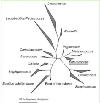
members of the lactic acid bacteria (LAB) group and bacteriocin producers, these organisms are identified by a low G+C content of ˂50mol%. In addition, they are chemoorganotrophic and produce L-lactic acid from hexoses by homofermentative lactic acid fermentation. Through DNA hybridization and 16S rRNA sequencing, it was established in 1984 that two species, Streptococcus faecium and Streptococcus faecalis, were sufficiently distinct from the other streptococci to be designated another genus, Enterococcus, meaning that the D group antigen can be found in both
streptococci and enterococci (Figure 1) (Franz et al., 2003; Fisher & Phillips 2009).
Enterococci are able to survive in a wide range of environmental conditions, such as extreme temperatures (5 to 50 ºC) and pH (4.6-9.9). They can also hydrolyze esculin in the presence of 40% (w/v) bile salts, survive at 60 ºC for 30 minutes and grow in 6.5% NaCl. Most enterococci are able to hydrolyze L-pyrrolidonyl-β-naphthylamide (PYR) (Fisher & Phillips, 2009; Lebreton et al., 2014). These characteristics allow enterococci to be distinguished from other Gram-positive, homofermentative and catalase-negative organisms.
Based on acid formation in mannitol and sorbose broth and hydrolysis of arginine, Facklam and collaborators divided the enterococcal species into 5 different groups, from Group I to Group V (Table 1). Over thirty Enterococcus species have been described so far with distinct phenotype, habitat and metabolic characteristics (Facklam et al., 2002).
Figure 1. Phylogenetic dendrogram of Gram-positive genera and Enterococcus position by 16S rRNA (retrieved from Fisher & Phillips (2009)).
3
Table 1. Distribution, into 5 different groups, of the enterococcal species according to acid formation on mannitol and sorbose broths and arginine hydrolysis (based on Facklam et al., (2002)).
Group I Group II Group III Group IV Group V
A c id F ormati on Mannitol Broth Sorbose Broth Arginine Hydrolysis Enterococcal Species • E. avium, • E. malodoratus, • E. raffinosus, • E. pseudoavium, • E.saccharolyticus, • E. pallens, • E. gilvus. • E. faecalis, • E. faecium, • E. casseliflavus, • E. mundtii, • E. gallinarum. • E. durans, • E. porcinus, • E. ratti, • E. hirae • E. dispar. • E. faecalis and E. faecium variants • E. asini, • E. sulfureus, • E. cecorum. • E. columbae, • E. faecalis, gallinarum and casseliflavus variants negative for arginine hydrolisis 1.2.2 Enterococcus spp. habitats
Enterococci are widely distributed in the environment. In addition of being ubiquitous in the gastrointestinal (GI) tract of humans and a wide range of animals, they can also be frequently found in food, vegetables, water, soil, insects and other sources (Torres et al., 2018).
As typical colonizers of the GI tract, these organisms are the most abundant Gram-positive coccus found in human faeces, being isolated at a rate of up to 105-108 CFU/g, with E. faecalis and E. faecium as the most prevalent species found. However, other species such as E. avium and E. durans can also be present. Despite being the natural colonizers of the lower GI tract, these bacteria can also appear in the vaginal tract, oral cavity and upper GI tract and from these sites, enterococci are able to colonize other tissues, such as urinary tract, wounds (commonly intra-abdominal and pelvic sites), bloodstream and endocardium, thus becoming nosocomial pathogens (Shepard & Gilmore, 2002; Martínez, 2011).
Enterococci are used as markers of faecal contamination of food products (e.g., raw meat, milk and milk products) and the acquisition of these bacteria through food may result in intestinal colonization (Tyson et al., 2017). Although these organisms are considered food contaminants, they are also used as probiotics in order to improve the microbial balance of the intestines, to treat gastroenteritis or as starter cultures to produce cheese (Eaton & Gasson 2001; Biavasco et al., 2007).
4 Although E.faecalis and E.faecium are the most prevalent species among the different environments, some enterococcal species are typically associated with a specific environment or animal species being the key to the Enterococcus spp. broad distribution in nature the capacity to survive under extreme conditions, allowing them to become pathogens with clinical relevance (Aarestrup et al., 2002; Vu & Carvalho, 2011).
1.3 Antibiotics
Antibiotics are molecules capable of stopping bacteria, by killing or preventing them from growing. Produced by fermentation or chemical synthesis, antibiotics that stop bacterial growth are bacteriostatic and the ones causing cell death are bactericidal, lowering the bacterial count (Walsh 2003a; Maddison et al., 2008). The antibacterial agents can be natural or synthetic products, both with the aim of blocking crucial processes in microbial cells. The natural antimicrobial compounds are produced by bacteria and fungi, being the major producer of these substances the actinomycetes group (Walsh, 2003a). Antibiotics are also classified according to their spectrum of activity, describing the extent of microorganisms that are sensitive to the drug. Broad spectrum antibiotics are able to target different microbes from different species whilst narrow spectrum is only effective against a few species (Kester et al., 2012).
1.3.1 Evolution of the use of antibiotics as growth promoters
In the 1940s, chlortetracycline was discovered to have a growth promoting effect when chickens were fed with fermentation offal of this compound produced by Streptomyces aureofaciens. Ever since, several antibiotics had been used as growth promoters, as they improved nutrient absorption, feed intake, fatty acid absorption and reduced mortality due to clinical diseases, allowing a growth improvement between 4 and 8% (Gaskins et al., 2002; Butaye et al., 2003). In 1951, the U.S Food & Drug Administration (FDA) approved the use of antimicrobials as additives without a veterinary prescription. In Europe, in the 1950s and 1960s, each European state approved national guidelines about the use of antibiotics in animal feed (Castanon, 2007).
When livestock producers started to routinely use antimicrobials, the scientific community questioned the possible side effects of this practice in human health, as many of the antimicrobials used as feed additives were identical or related to drugs used to treat diseases in human medicine, in addition to the fact that the resistance genes usually encode resistance to an entire class of antibiotics, with the risk of cross-resistance. In 1969, the Swann Committee was formed in the UK to discuss these concerns and it was stated that antibiotics
5 used in livestock should be divided into feed and therapeutic classes, being that the feed class shouldn’t include therapeutic agents used in medicine (Butaye et al., 2003; Tollefson & Karp, 2004). In 1970, the European Economic Community (EEC) decided that only antibiotics having a proven effect on animal growth, acting on bacteria and showing no intestinal absorption could be used as growth promoters in order to prevent the presence of residues in meat (Barros, 2010). Since 1997, avoparcin was prohibited in the European Union (EU) as a growth promoter and in 1999 tylosin and virginiamycin were also banned (Poeta et al., 2006a). As a consequence of these directives, in 2006, it was decided by the EU to eliminate antibiotics as growth promoters (European Union, 2006).
1.3.2 The mechanisms of action of antibiotics and microbial metabolism
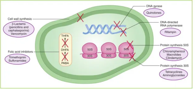
As previously referred, antibiotics are molecules capable of stopping bacterial activity, being classified according to the mechanism of action, as described in Figure 2. There are four major mechanisms through which most antibiotics act on microbial cells, although only three will be described, which are: i) Antibiotics targeting the cell wall; ii) Inhibitors of the protein biosynthesis; and iii) Inhibitors of DNA replication (Kester et al., 2012).
Figure 2. The four main mechanisms through which antibiotics act on the bacterial cell. The action of oxazolidinones and streptogramins is not represented however it is the same as that of chloramphenicol and macrolides (retrieved from Kester et al., (2012)).
1.3.2.1 Antibiotics targeting the cell wall
Bacterial cells are enveloped by an outer layer, the cell wall, which is made of peptidoglycan (long sugar polymers) (Kapoor et al., 2017). As the internal osmotic pressure of bacterial cells is high, any injury or inhibition of this layer can lead to cell lysis (Allison &
6 Lambert, 2015). The peptidoglycan is, therefore, crucial to maintaining the integrity of bacteria. It consists of N-acetylglucosamine (NAGA) and N-acetylmuramic acid (NAMA) that alternate between each other, crosslinked by short strands of peptides by transglycosidase action. Most bacteria have cell membrane-binding proteins, called penicillin-binding-protein (PBP), which consists of enzymes involved in the terminal stages of assembling the cell wall, being responsible for cross-linking the D-alanyl-D-alanine portion of the peptide chain. This process strengthens the cell wall and reshapes it during division and growth (Maddison et al., 2008; Kapoor et al., 2017).
β-lactams and glycopeptides are examples of antibiotics whose target is the cell wall synthesis, inhibiting enzymes or sequestering substrates involved in the peptidoglycan assembly and cross-linking (Walsh, 2003b).
β-lactams were firstly discovered in 1928, by Alexander Fleming, who named the compound as “penicillin” (Hauser, 2012b). Penicillin is formed by a four-member ring denominated β-lactam ring. The modifications on this ring led to the production of different antibacterial compounds, including penicillins (e.g. ampicillin), cephalosporins, carbapenems and monobactams, each with a specific spectrum of activity. β-lactams inhibit the PBPs, thus not allowing the formation of the peptidoglycan layer, being bactericidal. It has been hypothesized that the β-lactam ring mimics the D-alanyl-D-alanine portion of the peptide chain that normally is linked by the PBPs. When in the presence of a β-lactam, the PBPs interact with the antibiotic ring invalidating the synthesis of new peptidoglycan, leading to bacterial cell lysis (Figure 3 A2) (Chopra, 2010; Hauser, 2012b; Kester et al., 2012).
Glycopeptides, such as vancomycin and teicoplanin, are a group of natural
substances that prevent the cell wall synthesis, more specifically, the peptidoglycan assembly
(Butler et al., 2014). Unlike β-lactams, glycopeptides act on their substrates. These antibiotics
act by binding to the acyl-D-Ala-D-Ala terminus of the growing peptidoglycan. The bound occurs when the antibiotic forms a complex with the D-Ala-D-Ala residues, through the formation of five hydrogen bonds with the peptide backbone of the glycopeptide. The complex prevents transglycosylation and transpeptidation, leading to the formation of immature peptidoglycan, losing the integrity of the cell wall which leads to a higher susceptibility of the cell to lysis, causing bacterial death (Figure 3B) (Kahne et al., 2005; Kang & Park 2015).
7
Figure 3. β-lactams (A) and glycopeptides (B) mechanism of action. A1. In a normal situation, a new
subunit of NAMA and NAGA with a peptide chain is linked to an existing peptidoglycan polymer. A2. In the presence of a β-lactam, the antibiotic binds to the PBP preventing the cross-linking of the glycine bridge to the peptide chain. B. In the presence of vancomycin, the antibiotic binds to the D-ala–D-ala
dipeptide, inhibiting its incorporation into the cell wall by PBPs (retrieved from Hauser (2012b)).
1.3.2.2 Inhibitors of the protein synthesis
To multiplicate or replace older biomolecules, bacteria need to manufacture new proteins. To do so, from bacterial DNA genes, RNA molecules denominated messenger RNA (mRNA) are synthesized and then ribosomes, a macromolecular structure, synthesizes proteins from the information contained in the mRNA templates (translation). The protein biosynthesis is catalyzed by ribosomes and cytoplasmic factors. The bacterial 70S ribosomes are composed of two subunits, the 30S and 50S ribonucleoprotein subunits. As these processes are crucial for bacteria, they become targets of antibiotics whose purpose of action is the protein synthesis pathway (aminoglycosides, tetracyclines, chloramphenicol and macrolides) (Hauser 2012a; Kapoor et al., 2017). These drugs can affect the initiation phase of protein synthesis, the transfer RNA (tRNA) binding, the peptidyl transferase action or even cause inappropriate amino acid insertions leading to misreading errors (interfering with the essential protein functions) (Kester et al., 2012).
• Inhibitors of 30S subunit
Aminoglycosides have been important agents to combat pathogenic bacteria since the 1940s as they are potent and broad-spectrum compounds. These antimicrobials are composed of a six-membered ring with amino group substituents and glycosidic bonds (Leggett, 2017). By binding reversibly to the 30S ribosomal subunit receptors, aminoglycosides cause a mismatching between mRNA codons and charged aminoacyl-tRNA, which consequently leads to mistranslation or to premature termination. The binding to the 30S
8 subunit does not inhibit the formation of the initiation complex but alters the elongation of the new chain by damaging the proofreading process that controls translation. The aberrant products are accumulated in the cell and can be inserted into the cell membrane, modifying the cell permeability (Maddison et al., 2008; Hauser, 2012a; Leggett, 2017;).
The tetracyclines, a class of broad-spectrum and bacteriostatic antibiotics discovered in the 1940s, have as target the protein synthesis, by blocking the attachment of aminoacyl-tRNA to the ribosomal acceptor A-site. To achieve
their function, tetracyclines access to the ribosomes by energy-dependent transport, in Gram-positive bacteria (Chopra & Roberts 2001; Kester et al., 2012). These antibiotics bind at the centre of the 30S subunit, the decoding site, where the mRNA codon is recognized by the tRNA anticodon, impairing translation by overlapping in position with the docking of aminoacyl-tRNA during elongation (Figure 4) (Nguyen et al., 2014; Grossman, 2016).
• Inhibitors of 50S subunit
Chloramphenicol, an antibiotic belonging to the phenicols class, was first isolated in 1947 from a soil sample. This antibiotic is highly lipophilic, with broad spectrum and it penetrates the cells by facilitated diffusion (in Gram-positive bacteria). (Maddison et al., 2008; Pogue et al., 2017). Its action is based on the competitive inhibition for the aminoacyl-tRNA binding to the peptidyl transferase domain located in the 50S subunit, altering the ribosome conformation and the transpeptidation process (van Bambeke et al., 2017).
The macrolides class includes erythromycin and other erythromycin-deriving antibiotics. The binding site of macrolides is the
same as chloramphenicol, that is, the peptidyl transferase centre, located on the 50S subunit (Figure 5) (van Bambeke et al., 2017). These antibiotics are hydrophobic molecules composed by a large cyclic core (14- to 16- membered lactone ring) that passes through the cell membrane passively (Hauser, 2012a). In addition to block the peptide bond formation or the peptidyl tRNA translocation from A-site to P-site, macrolides also
Figure 4. Action site of the tetracyclines
(retrieved from Kester et al.(2012)).
Figure 5. Action site of the macrolides and
chloramphenicol (retrieved from Kester et al.(2012)).
9 favour the premature dissociation of peptidyl tRNA from the ribosome during elongation leading to the synthesis of incomplete peptides. (Kwon, 2017; van Bambeke et al., 2017).
Streptogramins consist of a natural mixture of two macrocyclic compounds, both produced by Streptomyces spp., which synergistically inhibit bacteria by binding to the 50S bacterial ribosome subunit, inhibiting protein synthesis. These antibiotics can be classified as group A (e.g. dalfopristin) or group B (e.g. quinupristin) according to their basic chemical structure (Soriano, 2010; Hauser, 2012a). Each group component alone has a moderate effect against bacteria, however, they reach bactericidal effect when combined. The streptogramin
interferes with peptidyltransferase activity by blocking the peptide bond synthesis in the course
of elongation (Johnston et al., 2002; Hauser, 2012a; Allison & Lambert, 2015).
The oxazolidinones are bacteriostatic antibiotics that inhibit the protein synthesis in the early stages of formation of the 70S initiation complex, leading to cross-resistance against other antimicrobials that restrain protein synthesis (Lowy, 2017). The major representative antibiotic of this class is linezolid and it is believed that it binds to the bacterial ribosomes’ 50S subunit, inhibiting the assembly of the complex that allows the development of protein synthesis, as the 50S subunit loses its affinity with the 30S subunit (Shaw & Barbachyn, 2011). Oxazolidinones compete against chloramphenicol and lincomycin for the binding site but without being antagonists, that is, they have close binding sites but oxazolidinones do not inhibit peptidyl transferase (as chloramphenicol does) (van Bambeke et al., 2017).
1.3.2.3 Action on nucleic acid replication
The bacterial replication machinery is composed of several enzymes that work strictly coordinated to accomplish a correct chromosomal replication. Chromosomal DNA is formed by two spiral nucleotide chains and, to replicate DNA, it is essential that the strands unwind and separate. The replication begins at a well-defined origin by specific and repetitive sequences. Two bacterial topoisomerases II enzymes, DNA-gyrase and Topoisomerase IV (Topo-IV) are in charge to modify the DNA topology during replication and both enzymes are tetramers, which, respectively, are composed of two GyrA and two GyrB or two ParC and two ParE subunits. The supercoiling is controlled by DNA-gyrase by introducing negative supercoiling ahead of the replication fork, relaxing the torsion on the DNA helix allowing its synthesis, making this enzyme crucial for DNA replication and also repair. Topo-IV relaxes, decatenates DNA and unknots it behind the replication fork (Sanyal & Doig, 2012; van Eijk et al., 2017). Because these enzymes and interactions are vital to the cell, they constitute a great antibiotic target. Only the compounds that bind and interfere with DNA-associated enzymatic processes are sufficiently selective and suitable to be used as antibacterial agents otherwise
10 they could be toxic to mammalian cells, as there are many common elements between eukaryotic and prokaryotic replication. These compounds include quinolones (e.g., ciprofloxacin) and rifampin (rifampicin), among others (Chopra, 2010).
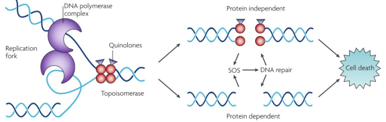
Quinolones are synthetic compounds inhibiting bacterial replication by blocking the replication pathway. The first quinolone introduced in the clinical field was Nalidixic Acid, in the 1960s, but due to its toxicity, it was substituted by second-generation quinolones with better antibacterial activity (i.e., norfloxacin, ciprofloxacin) (Allison & Lambert, 2015). The different quinolone generations are easily distinguished and have good tissue penetration, a broader spectrum of activity and high serum levels. In Gram-positive bacteria, quinolones target topoisomerase IV, trapping these enzymes at the DNA cleavage stage, preventing the strands to rejoin. By targeting the crucial process of generation of strand breaks induced by topo-IV and by binding non-covalently to the strand breaks, quinolones increase the concentration of enzyme-DNA cleavage complexes, becoming these antibiotics poisons for topoisomerases. This formation between topoisomerase, DNA complex and quinolone arrests the replication machinery, leading to inhibition of the DNA synthesis and eventually causing cell death (when the antibiotic is at bactericidal concentrations) (Figure 6) (Kohanski et al., 2010; Aldred et al., 2014; Bhattacharjee, 2016).
Figure 6. Quinolone target on the bacterial cell and its cell death effect (retrieved from Kohanski et al.,
(2010)).
Rifamycins were firstly isolated in 1957 from a soil bacterium, Amycolatopsis rifamycinica. The most commonly used antibiotic from this family is Rifampicin, a synthetic version of rifamycin and broad spectrum compound (Bhattacharjee, 2016). Rifamycins inhibit RNA synthesis by binding to RNA polymerase, the enzyme responsible for catalyzing the transcription pathway. These antibiotics bind in a small site of the β-subunit of RNA polymerase in the DNA/RNA channel thus blocking directly the elongation process of RNA. During
11 initiation, the transcription complex is unstable and the rifampicin bond promotes the dissociation of small RNA-DNA strands from the enzymatic complex (Chopra, 2010; van Bambeke et al., 2017).
1.4 Antibiotic resistance in enterococci
The increasing resistance to antibiotics is derived from multiple factors, including the widespread and inadequate use of these drugs, their extensive use in animal production, lack of sanitation and poor conditions, and increased routes that allow a greater transfer of resistances, allowing to cross the geographical barriers (Holmes et al., 2016).
Drug-resistant strains are more frequently found in hospitals and hospital-acquired infections, although the incidence of antibiotic-resistant pathogens in community-acquired infections has been rising in the last few years. The resistance to antimicrobials has been recognized for more than 50 years and has led to the rise of mortality, morbidity and health care costs. Despite all the efforts to control the infections caused by bacteria, these organisms developed mechanisms to avoid the antibiotic effects. Enterococci are one of the most dangerous microorganisms, responsible for many nosocomial infections that usually occur in patients that are in conditions that comprise risk factors (e.g. prolonged hospitalization, prior use of antimicrobials or compromised immune system). Antimicrobial resistance can occur in two ways: It can be intrinsic, which is chromosome-mediated, or it can be acquired, by a mutation of the existing DNA or acquisition of new DNA when bacteria are naturally susceptible to the antibiotic, obtaining the genes that encode to the resistance mechanism (Ang et al., 2004; Blair et al., 2015; Macgowan & Macnaughton, 2017). Resistance to antimicrobials occurs via the transmission of resistance genes, carried in mobile genetic elements (MGE), which can be transposons or plasmids (Levy & Bonnie, 2004) and the transfer can occur in three ways (Figure 7):
1. Conjugation: Direct contact between cells, occurring the plasmid transfer.
2. Transformation: Bacteria can acquire foreign genetic information through the incorporation of free DNA segments from the environment into their chromosome. The use of antibiotics exerts selective pressure, favouring bacteria possessing resistance
12 acquire survival advantages being capable of passing the resistance genes to other bacteria.
3. Transduction: Transfer of bacterial DNA by a bacteriophage, a virus affecting bacteria that replicates in the bacterial cell and incorporates fragments to the bacterial DNA in the assembled viral particles, which is then transferred to the next bacterial cell infected by the virus (Macgowan & Macnaughton, 2017).
The mutations that lead to antibiotic resistance (AbR) usually occur in three types of genes: genes encoding the antibiotic targets, genes encoding their transporters or genes encoding regulators that repress the expression of transporters or antibiotic-decontaminating elements (multidrug efflux pumps and antibiotic-modifying enzymes chromosomally encoded) (Martinez 2014; Blair et al., 2015).
Enterococci are characterized for having intrinsic resistance to a large number of antibiotics and for its capability of acquiring new resistances, as shown in Table 2. Their capacity to acquire resistance to many antibiotics and their resilient nature allows them to be well adapted and persistent in infections (Shepard & Gilmore, 2002).
Table 2. Intrinsic and acquired antibiotic resistance in enterococci (adapted from Shepard & Gilmore (2002); Martínez (2011)).
Intrinsic Resistance Acquired Resistance
β-lactams High concentrations of β-lactams
Fluoroquinolones High concentrations of Aminoglycosides
Clindamycin Glycopeptides
Trimethoprim-Sulfamethoxazole Tetracyclines
Low-level aminoglycosides Erythromycin
Sulfonamides Fluoroquinolones
Rifampicin Chloramphenicol
Oxazolidinones
The intrinsic resistance is a specific property of each bacterial species. In response to the expression of low-affinity PBP’s, enterococci show decreased susceptibility to β-lactams, in addition to intrinsic low-level resistance to aminoglycosides, glycopeptides, lincosamides and streptogramins (mainly E. faecalis). The excessive use of antibiotics against microorganisms other than enterococci to which they have intrinsic resistance or are susceptible can lead to the selection and increase of the incidence of infections caused by Enterococcus spp. (Martínez, 2011; Kristich et al., 2014).
13 1.4.1 Acquired resistances
1.4.1.1 Acquired resistance to β-lactams
High-level β-lactam resistance develops through the acquisition of β-lactamases or by an increment in the production of low-affinity PBP’s. β-lactamases are responsible for cleavage of the β-lactam ring, although these are rare and their production is constitutive. The bla genes, highly homologous to the Staphylococcus aureus blaZ genes, encode the β-lactamase production and, in enterococci, can be located chromosomally or in plasmids, being firstly described in 1983. Since then, the production of β-lactamases has only been reported in E. faecalis and E. faecium (Hollenbeck & Rice, 2012; Tang et al., 2014) The number of enterococci isolates resistant to β-lactams has been rising and the majority of E. faecium isolated clinically show high-level resistance to β-lactams due to the production of a modified PBP, the PBP5 (Shepard & Gilmore, 2002).
1.4.1.2 Resistance to aminoglycosides
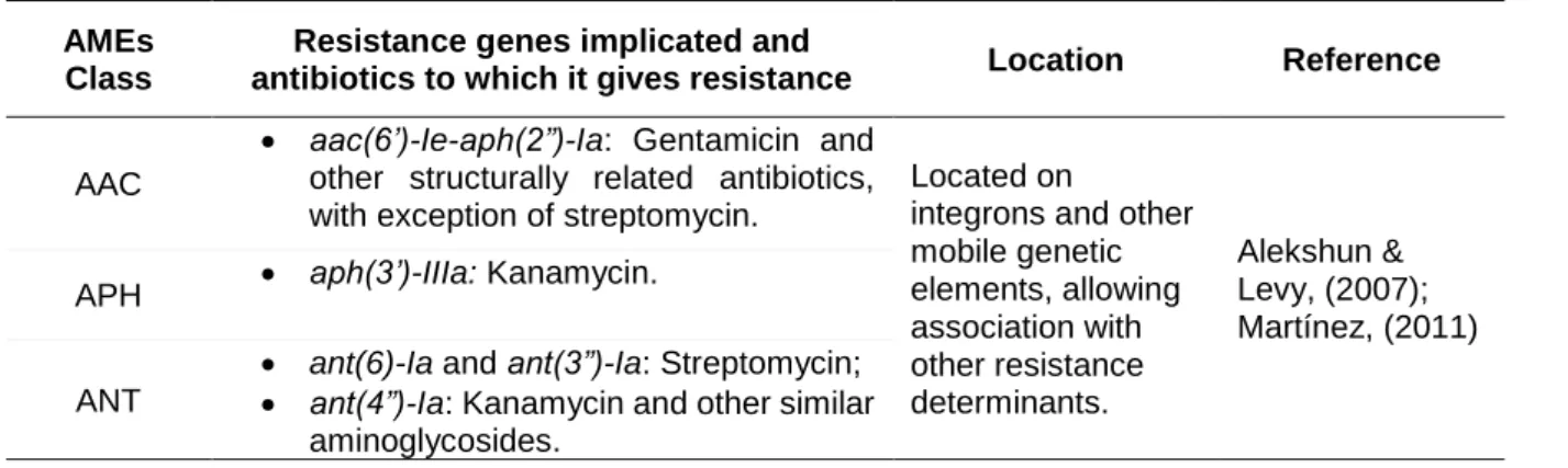
Besides the low-level resistance due to the low uptake of these agents, enterococci can also acquire mechanisms that allow inhibiting the effect of high concentrations of aminoglycosides. High-level resistance (HLR) to aminoglycosides can be triggered by i) modification of the ribosomal target; ii) alteration of the antibiotic transport (through efflux pumps); and iii) acquisition of aminoglycoside-modifying enzymes (AMEs) that are thought to be prevenient from aminoglycoside producing species, such as Streptomyces griseus, e.g. AMEs can be divided into three classes, according to their type of modification: acetyltransferases (AAC), phosphotransferases (APH) or nucleotidyltransferases (ANT) (Alekshun & Levy 2007; Rossolini et al., 2017). The most prevalent resistance mechanism leading to aminoglycoside resistance in enterococci are summarized in Table 3.
Table 3. Most prevalent aminoglycoside resistance genes in enterococci and the antibiotics to which they give resistance.
AMEs Class
Resistance genes implicated and
antibiotics to which it gives resistance Location Reference
AAC
• aac(6’)-Ie-aph(2”)-Ia: Gentamicin and other structurally related antibiotics, with exception of streptomycin.
Located on
integrons and other mobile genetic elements, allowing association with other resistance determinants. Alekshun & Levy, (2007); Martínez, (2011) APH • aph(3’)-IIIa: Kanamycin.
ANT
• ant(6)-Ia and ant(3”)-Ia: Streptomycin; • ant(4”)-Ia: Kanamycin and other similar
14 The most concerning genes encoding aminoglycoside resistance are those inhibiting the effects of both gentamicin and streptomycin, as these antibiotics are combined in order to treat severe enterococcal infections. Several other genes have been identified than those previously mentioned. The severity of aminoglycoside resistance derives from the fact that these resistances abolish the synergy between aminoglycosides and antibiotics targeting cell-wall, used in combination to enable the entrance of the aminoglycosides into the cell, solving the intrinsic resistance to these antibiotics (Shepard & Gilmore, 2002; Hollenbeck & Rice, 2012; Garrido et al., 2014).
1.4.1.3 Tetracyclines Resistance
Tetracycline resistance was described to occur by two major mechanisms: i) active efflux of the antibiotic through an integral membrane protein; and ii) ribosomal protection by a soluble protein (Wang et al., 2017). Enzyme inactivation can also occur although it is less frequent. Most of the resistance genes encoding tetracycline resistance are located on mobile genetic elements, allowing horizontal resistance transfer (Pogue et al., 2017). The efflux pumps involved in this resistance are encoded by the genes tetK, tetL and tetM and, on the other hand, the tetO and tetS genes are chromosomal resistance determinants encoding a protein that alters the ribosomal conformation, displacing the tetracycline bound (Miller et al., 2014). In enterococci, the most prevalent genes are tetM, tetL and tetK.
1.4.1.4 Resistance to the Macrolide- Lincosamide- StreptograminB (MLSB) group Acquired resistance to MLSB antibiotics, in enterococci, is mediated by three main mechanisms, that are:
i) Modification of the drug target, causing an affinity decrease with the binding site. The most frequent resistance mechanism consists in the modification of the 23S rRNA by methylases known as Erythromycin Resistance Methylase (ERM) which dimethylate a common binding site for MLSB antibiotics, causing cross-resistance to all drugs. The expression of erm genes is usually plasmid- and transposon-borne being ermA and ermB the most prevalent erm genes in enterococci (Leclercq 2002; Rossolini et al., 2017).
ii) Inactivation of the antibiotic by enzymatic modification, which includes resistance to quinupristin-dalfopristin (QD) in E. faecium, mediated by different mechanisms. The modification of dalfopristin occurs via the acetyltransferases vatD and vatE, annulling the synergy with quinupristin. The enzymatic cleavage of the streptogramin B ring is caused by vgbA and vgbB lactonases (Miller et al., 2014). Lincosamide resistance is mediated by a
15 nucleotidyltransferase, encoded by the linB gene, that catalyzes the adenylation of these antibiotics (Garrido et al., 2014).
iii) Active efflux of the drug allows the cell to keep a low antibiotic concentration, avoiding their effect. The pump mechanisms involve the genes mefA, mefE, msrA and mreA, leading to the efflux of macrolides. Mef and mreA genes are associated with macrolide resistance and msrA (ABC transporter family member) is associated with both macrolide and streptogramin B resistance (Portillo et al., 2000; Rossolini et al., 2017).
1.4.1.5 Chloramphenicol resistance
Chloramphenicol resistance is mostly caused by drug inactivation by a Chloramphenicol Acetyltransferase (CAT) enzyme that acetylates the antibiotic. The acetylated derivates of chloramphenicol are then unable to bind to the 50S subunit. The cat genes are usually carried on plasmids and in Gram-positive pathogens they may be inducible (Rossolini et al., 2017). Other mechanisms can also be found, such as efflux systems, inactivation by phosphotransferases or mutations of the target site (Schwarz et al., 2004). The most frequent gene found in enterococci is catA, although different variants of fex genes can also be implicated.
1.4.1.6 Oxazolidinones resistance
Oxazolidinone resistance occurs due to mutations in genes encoding the 23S rRNA, an important drug binding-site in the ribosome. These mutations were firstly described in S. aureus and, in enterococci, different acquired resistance genes have been described, such as cfr (codifies a methyltransferase of 23S rRNA), optrA (codifies an ABC transporter) or the recently described poxtA gene (implicated in ribosomal protection) (Hollenbeck & Rice, 2012; Miller et al., 2014; Antonelli et al., 2018). The cfr gene encodes a plasmid-borne methylase (Cfr) and as several antibiotics share this same specific binding location, cfr confers a multiresistant phenotype and impairs the ribosomal binding of phenicols, lincosamides, oxazolidinones and streptogramins A but not macrolides, differing from erm rRNA methylase genes (Roberts, 2008; Leclercq, 2009).
1.4.1.7 Quinolone resistance
Resistance to quinolones is based on two main mechanisms, chromosomally mediated, that are:
16 i) Alterations on the drug target, that is, mutations in the “Quinolone Resistance Determining Regions” (QRDR) of the genes encoding gyrase and topoisomerase IV, inhibiting the binding of the antibiotic to the enzymes, enabling the DNA replication. ii) Decreased accumulation of these antibiotics in the bacterial cell due to efflux pumps,
encoded by the emeA and efrAB genes.
Another mechanism of quinolone resistance was described and it implies the protection of both DNA gyrase and topoisomerase IV against quinolone inhibition. This mechanism is mediated by a protein of the Qnr family but its function remains unclear (Garrido et al., 2014; Kristich et al., 2014; Schindler et al., 2017).
1.4.1.8 Rifamycins resistance
Although rifampicin is not used routinely to treat enterococcal infections, these organisms are susceptible to this antibiotic. In most bacteria, resistance to rifampicin is due to mutations in the rpoB gene, which encodes the β-subunit of RNA polymerase, i.e., as previously said, the target of this drug (Enne et al., 2004). These mutations include deletions, insertions and point mutations, occurring in a small region that is less than 100bp (Lambert, 2005).
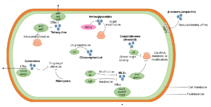
Figure 8 summarizes the acquired mechanisms to antimicrobial resistance, with exception of vancomycin, which will be covered in the next topic in more detail.
17 Figure 8. Acquired mechanisms of enterococcal antimicrobial resistance. Resistance to ampicillin
occurs through the production of low-affinity PBPs, such as the PBP5. HLR to aminoglycosides results from the acquisition of AMEs, alteration of the target binding or alterations on the drug transport. Enterococcal resistance to the MLSB class involves several pathways, including drug modification and
inactivation (by the vat and vgb genes, respectively), drug efflux (via the genes msrA, mefA, mefE and mreA) and ribosomal alteration caused by the erm genes. Resistance to linezolid involves a mutation in the 23S rRNA. Rifamycin and quinolone resistances involve the drug target alterations and, in the case of quinolones, it also includes resistance by drug efflux (caused by the emeA and efrAB genes). The cat genes inactivate chloramphenicol, leading to resistance to this antimicrobial and, tetracycline resistance involves ribosomal protection, conferred by tetO and tetS and drug efflux which occurs via the action of the tetK, tetL and tetM genes. Adapted from Arias & Murray (2012).
1.5 Vancomycin resistance mechanism and its epidemiology
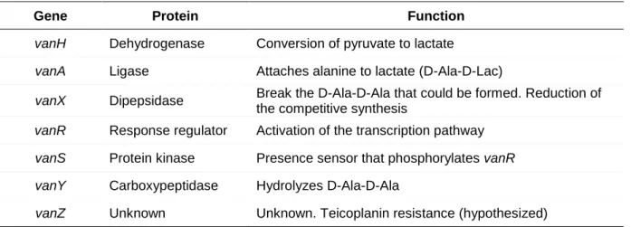
High-level enterococcal resistance to glycopeptides occurs due to the presence of operons inducing the synthesis of low-affinity precursors,
altering the antibiotic target, that is, substitution of the D-Ala:D-Ala terminal of the pentapeptide to other amino acid termination, D-Lactate (D-Lac) or D-Serine (D-Ser), resulting in low-affinity peptidoglycan precursors (Figure 9). The D-Lac replacement results in a 1000-fold decrease in glycopeptide affinity due to the loss of one of the hydrogen bonds needed for the interaction between drug and peptidoglycan precursor. In addition, the D-Ser replacement affects the action of the antibiotic in a similar but less effective way. Additionally, glycopeptide resistance also occurs by prevention or destruction of the
Figure 9. Mechanism or glycopeptide
resistance (retrieved from Hauser 2012).