Susana Raquel
Costa Barros
REDUÇÃO DO CITOCROMO C E RESISTÊNCIA AO
METOTREXATO EM CANCRO DA MAMA
CYTOCHROME C REDUCTION AND
METHOTREXATE RESISTANCE IN BREAST
CANCER
Ano 2012
Susana Raquel
Costa Barros
REDUÇÃO DO CITOCROMO C E RESISTÊNCIA AO
METOTREXATO EM CANCRO DA MAMA
CYTOCHROME C REDUCTION AND
METHOTREXATE RESISTANCE IN BREAST CANCER
Dissertação apresentada à Universidade de Aveiro para cumprimento dos requisitos necessários à obtenção do grau de Mestre em Biologia Aplicada, ramo de Biologia Molecular e Celular, realizada sob a orientação científica do Professor Doutor Carlos Julian Ciudad Gomez, Professor Catedrático do Departamento de Bioquímica e Biologia Molecular da Faculdade de Farmácia da Universidade de Barcelona e da Professora Doutora Maria da Conceição Lopes Vieira dos Santos, Professora Associada com Agregação do Departamento de Biologia da Universidade de Aveiro
SAF2011- 23582 from “Plan Nacional de Investigación Científica” (Spain)
I dedicate this work to my sister, Irina, to my brother in law, Danilo, to my uncle, Zé, and to my “scientific mother” Núria for all your efforts, in different ways, to make this project run.
o júri Professor Doutor João António de Almeida Serôdio (Presidente)
Professor Auxiliar, Departamento de Biologia, Universidade de Aveiro
Doutor José Miguel Pimenta Ferreira de Oliveira (Arguente Principal)
Investigador Pós-Doutoramento, CESAM - Centro de Estudos do Ambiente e do Mar, Universidade de Aveiro
Professor Doutor Carlos Julián Ciudad Gómez (Orientador)
Professor Catedrático, Departamento de Bioquímica e Biologia Molecular, Faculdade de Farmácia, Universidade de Barcelona
Professora Doutora Maria da Conceição Lopes Vieira dos Santos (Co-orientador)
agradecimentos I would like to express my deepest gratitude first and foremost to Prof. Dr. Maria da Conceição Santos for supporting my decision to go to Barcelona and joining this project. I’m also grateful for her friendship during these 2 years of Master.
I would also like to thank Prof. Dr. Carlos J. Ciudad and Dr. Veronique Noe for promptly accepting my application to join the CClab and this project and for everything they taught me: the scientific advices, knowledge and effort to make me a better scientist. I am especially very grateful to Carlos for providing me with enthusiastic scientific discussions and for his obvious care and friendship along the 6 months I spent in Barcelona. I consider myself very lucky to have had the opportunity of joining this project and meeting this great team at CCLab.
I am deeply indebted to Núria Mencía, my “scientific mother”, for her patience, for sharing her knowledge and experience, even in the simplest things, and for her effort to make this project work! It was a great privilege to be “supervised” by someone like Núria, who you would love suddenly and never forget and who will remain my role model as a scientist!
I would also like to thank Laura, Carlota, Xenia and Anna Solé for all the good times we shared in the lab even when I was bad mooded and obviously for all the help and support. They are genuinely nice and supportive and I’m very glad to have met them and worked with them. Also, I would like to thank Gaspar, Anna Camps and Iris.
I wish to thank André, Ricardo, Xavi, Raquel, Juanito, Georgina and Betta, for all the good memories in Barcelona, their friendship and for teaching my basic knowledge of Spanish!
Since this project was run between Barcelona and Aveiro, I also wish to thank my colleagues from the LabCyt and my master courses’ colleagues for their support and good moments.
I would also like to thank my friends Linda, Natacha, Nádia, Claudia, Joana Valdez, Maria and Joana Oliveira for their friendship and all the smiles they gave me in person or by phone/internet, especially during the last phase of this work the hardest one!
Last but not least, my deepest gratitude to my sister, Irina, Danilo and to my uncle Zé for making possible the dream of living and working in the great city of Barcelona for 6 months. Without you, this would have never been possible! Thanks to my family for the enthusiasm, patience and care during this period of my life.
palavras-chave citocromo c; estado redox; resistência ao metotrexato; apoptose; GSTs; GSH; cancro da mama
resumo O metotrexato é um agente quimioterápico usado para tratar uma variedade de
cancros. No entanto, a ocorrência de resistência limita a sua eficácia. A relação entre o estado redox do citocromo c, apoptose e o desenvolvimento de resistência ao metotrexato foi avaliada em células humanas de cancro da mama. O citocromo c no seu estado reduzido é menos capaz de induzir a cascata apoptótica. Aqui nós mostramos que células incubadas com agentes redutores de citocromo c, tais como tetrametilfenildiamina, ascorbato ou glutationa reduzida, demonstraram menos mortalidade e apoptose na presença do metotrexato. Por outro lado, a inibição de glutationa aumentou a acção apoptótica do metotrexato, demonstrando um envolvimento do estado redox do citocromo c na apoptose induzida por metotrexato. Usando ferramentas de genómica funcional detectámos que GSTM1 e GSTM4 foram sobreexpressos em células MCF7 resistentes ao metotrexato. A inibição destas isoformas causou um aumento na citotoxicidade em células sensíveis. Estes resultados sugerem que estas GSTs específicas juntamente com a glutationa reduzida endógena poderão ajudar a manter o citocromo c num estado mais reduzido o que por sua vez poderá causar uma diminuição na apoptose e contribuindo assim para a resistência ao metotrexato em células tumorais humanas.
keywords cytochrome c; redox state; methotrexate resistance; apoptosis; GSTs; GSH; breast cancer
abstract Methotrexate is a chemotherapeutic agent used to treat a variety of cancers.
However, the occurrence of resistance limits its effectiveness. The relationship among redox-state of cytochrome c, apoptosis and development of resistance to methotrexate was assessed in human breast cancer cells. Cytochrome c in its reduced state is less capable of triggering the apoptotic cascade. Here we show that cells incubated with cytochrome c-reducer agents, such as tetramethylphenylenediamine, ascorbate or reduced glutathione, showed less mortality and apoptosis in the presence of methotrexate. Conversely, depletion of glutathione increased the apoptotic action of methotrexate, demonstrating an involvement of cytochrome c redox state in methotrexate-induced apoptosis. Using functional genomics we detected that GSTM1 and GSTM4 were overexpressed in methotrexate-resistant MCF7 breast cancer cells. Inhibition of these isoforms caused an increase in methotrexate cytotoxicity in sensitive cells. These results suggest that these specific GSTs together with endogenous reduced glutathione would help to maintain a more reduced state of cytochrome c which in turn would cause a decrease in apoptosis thus contributing to methotrexate resistance in human cancer cells.
i
LIST OF TABLES ... V ACRONYM AND ABBREVIATIONS LIST ... VI
CHAPTER I - GENERAL INTRODUCTION ... 1
1. METHOTREXATE AND CANCER TREATMENT ... 1
1.1. Mechanism of action: cell cycle S phase arrest and apoptosis ... 1
1.1. Mechanisms of MTX resistance ... 3
1.2.1. Gene amplification of the DHFR locus ... 3
1.2.2. Deficiency in MTX transport ... 3
1.2.3. Reduction in the MTX polyglutamation ... 4
1.2.4. Increase of the multi-drug resistance phenotype ... 4
1.2.5. Mutations of the DHFR gene ... 5
1.2.6. Altered genes ... 5
1.2.6.1. Overexpression of AKR1C1 ... 5
1.2.6.2. Overexpression of S100A4 ... 6
1.2.6.3. Overexpression of caveolin-1, enolase-2 and PRKCA and decrease of E-Cadherin-1 expression… ... 7
1.2.6.4. Overexpression of DKK1 and EEF1A1 ... 8
1.2.6.5. Increase in UGT1A family expression ... 9
1.2.7. Altered miRNA expression: underexpression of miR-224 ... 10
2. APOPTOSIS IN CANCER AND CHEMOTHERAPY: P53 AS AN APOPTOTIC MEDIATOR ... 11
2.1. Apoptotic intrinsic pathway: cytochrome c release and MTX ... 12
3. REGULATION OF APOPTOSIS BY CYTOCHROME C REDOX-STATE ... 13
3.1. Cytosolic GSH and cytochrome c redox state ... 16
4. ROLE OF GSTS AND GSH IN ANTI-CANCER DRUG-RESISTANCE ... 17
5. REFERENCES ... 19
ii 1. Abstract ... 31 1.1. Background ... 31 1.2. Results ... 31 1.3. Conclusions ... 31 2. Introduction ... 32
3. Materials and Methods ... 33
3.1. Cell lines and cell culture ... 33
3.2. Cell viability assay ... 34
3.3. Microarrays ... 34
3.4. Microarrays data analysis ... 34
3.5. RT-Real-Time PCR ... 35
3.6. Inhibition of GSTM1 and GSTM4 expression levels ... 36
3.7. Apoptosis ... 37
3.8. Oxygen consumption assay ... 37
3.9. GSH endogenous levels ... 38
3.10. Exogenous cytochrome c reduction by cytosolic extracts ... 38
3.11. Statistical analysis ... 38
4. Results and Discussion... 39
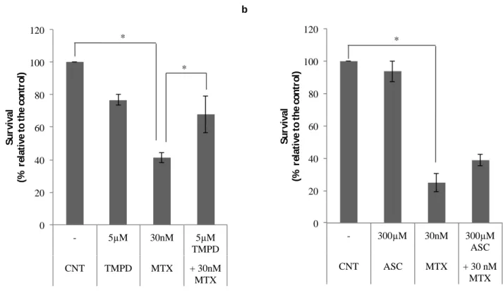
4.1. Effect of TMPD and ascorbate on the cytotoxicity produced by methotrexate tetramethylphenylenediamine (TMPD) and ascorbate ... 39
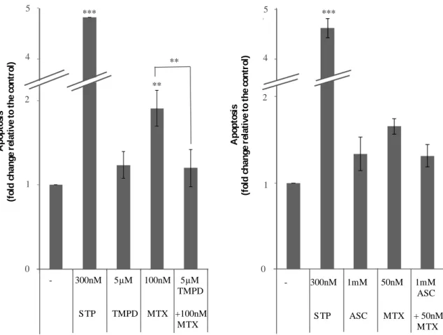
4.2. TMPD and ascorbate decrease the apoptotic effect of MTX ... 41
4.3. Effect of addition or depletion of GSH on MTX action ... 42
4.4. Endogenous levels of GST in sensitive and MTX resistant MCF7 cells ... 46
4.5. Inhibition of GSTM1 and GSTM4 increases the cytotoxicity produced by MTX ... 48
5. Conclusions ... 51
Acknowledgments ... 51
References ... 52
CHAPTER III - GENERAL CONCLUSION ... 31
ANNEX – EXPERIMENTAL PROTOCOLS ... 49
Annex I ... 67
Annex II ... 69
Annex III ... 71
Annex IV ... 73
iii Annex VIII ... 81
iv
Figure 1 – Schematic representation of folates (F) metabolism 2
Figure 2 – Chemical structure of natural folates (a) and methotrexate (antifolate) (b)
2
Figure 3– Regulation of apoptosis by the redox state of cytosolic cytochrome 15
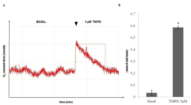
Figure 4 – O2 consumption analysis upon treatment with TMPD 32
Figure 5 – Effect of TMPD or ascorbate (ASC) in combination with MTX on cell viability
33
Figure 6 – Effect of TMPD and ascorbate (ASC) on apoptosis induced by MTX 34
Figure 7 – Effect of GSH treatment on the cytotoxicity caused by MTX 35
Figure 8 - Effect of veratridine (VERA) and GSH on MTX induced apoptosis 36
Figure 9 - Endogenous GSH levels and cytochrome c redox capacity in cytosolic extracts
38
Figure 10 – Validation of GSTM1 and GSTM4 overexpression in MCF7 MTX-resistant cells
40
Figure 11 - Effect of GSTM1 and GSTM4 inhibition on GSTM1 and GSTM4 mRNA levels, and on sensitivity towards MTX
v
Table 1 – Cellular changes during the apoptotic process 13
Table 2 - Primer sequences 28
Table 3 – Hairpin sequences 29
vi ALDH AKR ASK CAV1 DKK1 DHF DHFR DMSO dTMP dUMP EEF1A1 ENO2 ETC FPGS GPx GR GSH GSSG GST HPRT IAP IMM JNK MAPK MTT miRNA MDR MRP mtGPx MTX Aldehyde dehydrogenase Aldo-keto reductase
Apoptosis signal-regulating kinase Caveolin-1 Dikkofp homolog 1 Dihydrofolate Dihydrofolate reductase Dimethyl sulfoxide Thymidine monophosphate Deoxyuridine monophosphate
Eukaryotic translation elongation factor 1A1 Enolase-2
Electron transport chain Folil polyglutamate synthase Glutathione peroxidase Glutathione Reductase Glutathione Glutathione disulfide Glutathione-S-transferase Hypoxanthine-guanine phosphoribosyltransferase Inhibitor of apoptosis
Inner mitochondrial membrane c-Jun N-terminal kinase
Mitogen-activated protein kinase
3-(4,5-Dimethylthiazol-2-yl)-2,5-diphenyltetrazolium bromide microRNA
Multi-drug resistance
Multi-drug resistance protein
Mitochondrial glutathione peroxidase Methotrexate
vii PKCα PPRH PTP RFC ROS siRNA SDS SN STP THF TMPD TS UDPGA UGT VERA Protein kinase c α
Polypurine reverse-hoogsteen hairpin Permeability transition pore
Reduced folate carrier Reactive oxygen species Small interfering RNA Sodium Dodecyl Sulfate Supernatant
Staurosporine Tetrahydrofolate
N,N,N’,N’-tetramethyl-p-phenylenediamine Thymidilate synthase
Uridine diphosphoglucoronic acid UDP-glucoronosyl transferase Veratridine
1
1. Methotrexate and cancer treatment
1.1. Mechanism of action: cell cycle S phase arrest and apoptosis Methotrexate (MTX) is an antifolate chemotherapeutic agent widely used, alone or in combination with other agents for the treatment of a range of cancers, such as breast cancer, osteosarcoma, head and neck cancer, lymphoma and acute lymphoblastic leukemia (Jolivet et al. 1983).
This type of drug and its polyglutamates inhibit competitively and reversely the dihydrofolate reductase (DHFR) affecting the synthesis de novo of purines and pirimidines, thus the treatment with MTX results in the DNA synthesis inhibition, causing p53 mediated cell cycle arrest in S phase and death cell generally by triggering apoptosis in several cell lines (Kaufmann 1989; Barry et al. 1990; da Silva et al. 1996; el Alaoui et al. 1997; Hattangadi et al. 2004). The absence of folates and nucleotides precursors causes changes in the DNA synthesis, once the carbon atoms exchange reactions are blocked. In this case, following the DNA damage caused by this type of anticancer drug, apoptosis may occur as a major event, where apoptotic regulating genes will influence the lethal effect of this drug or may be a secondary response to the induced drug cytotoxic effect (Brown and Wouters 1999; Tannock and Lee 2001). Tumor cells which proliferate abnormally
are particularly susceptible to the lethal effects of the MTX.
DHFR catalyzes the reduction of dihydrofolate (DHF) to tetrahydrofolate (THF) which is necessary, as well as its derivatives, as cofactor in different biochemical mechanisms involved in carbonyl groups transference, such as aminoacids (e.g. methionine) and the purines and pyrimidines synthesis as well. Thus, DHFR is a very important enzyme in the DNA replication and cell division, by keeping the intracellular reduced folate levels. Also thymidylate synthase (TS) has an important role in DNA synthesis, since it catalyzes thymidine monophosphate formation (dTMP) from deoxyuridine monophosphate (dUMP), precursor of pyrimidines.
2
Figure 1– Schematic representation of folates (F) metabolism. Reduction of dihydrofolate (DHF) to tetrahydrofolate (THF) catalyzed by dihydrofolate reductase (DHFR). THF.C1 represents THF active derivatives that participate in specific folate-dependent reactions. (Adapted and modified from unknown source) The natural folates are formed by three structural components: a ptheridin ring, an acid p-aminobenzoic and a glutamate (Figure 2a). The antifolates also share this structure however with slight variations (Figure 2b).
a
b
Figura 2– Chemical structure of natural folates (a) and methotrexate (antifolate) (b).
3
1.1. Mechanisms of MTX resistance
The occurrence of resistance to MTX upon treatment compromises its effectiveness, limiting its use in chemotherapy (Hryniuk and Bertino 1969) and its occurrence can be either by a single mechanism or by combination of several of them (Sharma et al. 1991), thus drug resistance is a complex process in which several pathways can be involved. Several resistance mechanisms in vitro have been described using mammalian cells as model. Following, a brief description of each mechanism:
1.2.1. Gene amplification of the DHFR locus
One explanation for the amplification of the DHFR gene copy number could be due to unequal sister chromatid exchanges generating stable chromossomic regions. It has also been described that the generation of extrachromossomic copies of the DHFR gene, normally lost in daughter cells because of the absence of the centromere, is associated to the amplification process (Schimke et al., 1981). Other studies suggested a direct relationship between the amplication of the DHFR gene and mutated p53 gene (Livingstone et al. 1992).
1.2.2. Deficiency in MTX transport
MTX transport into the cell occurs mainly through a specific folate/antifolate transporter named reduced folate carrier (RFC). RFC is a membrane glycoprotein encoded by SLC19A1 gene. Underexpression of human SLC19A1 gene seems to be the central mechanism associated to the deficiency in MTX transport (Gorlick et al. 1997; Belkov et al. 1999; Guo et al. 1999; Ferreri et al. 2004) probably caused by the loss or deletion of locus SLC19A1 (Ding et al. 2001; Zhao et al. 2004). Another common mechanism associated to resistance development is the occurrence of
SLC19A1 gene mutations, changing the transporter function, thus causing a
decrease in MTX transport and originating the drug resistance (Roy et al. 1998; Rothem et al. 2002).
4
1.2.3. Reduction in the MTX polyglutamation
Polyglutamation is a biochemical process with an important role in the MTX pharmacology (Rosowsky et al. 1982). Like for the natural folates, folil polyglutamate synthase (FPGS) has high affinity to MTX adding sequentially five groups of glutamate to its molecule. Once it has more than three glutamate groups, MTX is no longer substrate for the RFC transport system, causing the accumulation of polyglutamated MTX inside the cell (Cowan and Jolivet 1984). This polyglutamated form of MTX is further a stronger inhibitor of the synthesis of purines and pyrimidines (Kimura et al. 2004) by DHFR and other THF-dependent enzymes inhibition (Cheok and Evans 2006). This inhibition can still remain even after the chemotherapeutic treatment, increasing the cytotoxic activity of the MTX (Cowan and Jolivet 1984).
The occurrence of less polyglutamation had been described in resistant cells (McCloskey et al. 1991; Mauritz et al. 2002; Liani et al. 2003). It is already described that several tumor cell lines resistant to antifolates show a suppression in the FPGS activity, probably due to post-translational alterations or gene mutations (Roy et al. 1997), thus the decrease in the accumulation of polyglutamated MTX can be associated to a low activity of FPGS.
1.2.4. Increase of the multi-drug resistance phenotype
The development of drug resistance in chemotherapy has been related to several mechanisms and it may occur as a multifactorial event where several mechanisms are simultaneously activated. The appearance of the multi-drug resistance (MDR) phenotype is a great concern regarding the chemotherapeutic treatment in several types of cancer (Sharom 1997). This phenotype can be inherited, an innate property of tumor cells, or acquired upon drug treatment (Sharom 1997). Its development can be associated to classic mechanisms, directly involved in DNA replication or detoxification processes: 1) the classic MDR phenotype can result from the overexpression of the p-glycoprotein, encoded by
mdr1 gene, and from other multidrug resistance proteins (MRP) involved in the
5 p-glycoprotein is an ATP-dependent translocase known for its ability to carry chemotherapeutic drugs, such as MTX (Sharom 1997); 2) the development of non-classic MDR phenotype is normally associated to alterations in the levels or activity of topoisomerase II and overexpression of detoxification enzymes phase II such as glutathione S-transferases (GSTs), UDP-glucuronosyl transferases (UGTs), aldehyde dehydrogenases (ALDHs) and aldo-keto reductases (AKRs) (Ax, 2000; Inoue, 1993). However, this type of MDR phenotype could be masked by the activation of the drug efflux via MDR or MDP.
Hereupon, it has been supposed that the increase of the MDR phenotype could be associated to alterations in the functional p-glycoprotein caused by the exposure to high doses of MTX (Assaraf et al. 1989). It is also described that the overexpression of different members of MRP family provides resistance to antifolates, although it is not described MRP overexpression in antifolate selected cell lines (Assaraf 2007).
1.2.5. Mutations of the DHFR gene
Mutations in DHFR gene in resistant cell lines decrease the affinity of DHFR protein to MTX compromising its inhibition by this drug, thus favoring the occurrence of the resistance (Srimatkandada et al. 1989).
1.2.6. Altered genes
Besides DHFR gene, other genes have been identified also important in the development of the MTX resistant phenotype, such as AKR1C1, UGT1A6, DKK1, E-cadherin, caveolin 1, enolase-2, PRKCA, EEF1A1 and S100A4.
1.2.6.1. Overexpression of AKR1C1
Aldo-keto reductase (AKR) superfamily proteins are monomeric cytoplasmic proteins and have been suggested to be involved in detoxification processes (Maser 1995; Burczynski et al. 1999; O'Connor et al. 1999; Penning
6 2005) by catalyzing the NAD(P)H-dependent oxido-reduction of a variety of substrates (Jez et al. 1997).
Overexpression of AKR1C1 has been related to drug resistance in a range of cancers. Some anticancer drugs share similar chemical structure with some compounds metabolized by AKR1C1, suggesting that these drugs may be subject to this enzyme activity (Hsu et al. 2001). Several studies using different anti-cancer drugs are in concordance with this, altogether suggesting an association between AKR1C1 and drug detoxification (Ciaccio et al. 1993; Shen et al. 1997; Ax et al. 2000; Deng et al. 2004; Chen et al. 2005; Hung et al. 2006). Also MTX-resistant colon cancer cells showed an overexpression of AKR1C1 (Selga et al. 2008). In this study, AKR1C1 overexpression seems to represent a mechanism, parallel to DHFR amplification, to the development of MTX resistance, once AKR1C1 up-regulation counteracted MTX-induced S phase arrest of cell cycle and apoptosis caused by the drug.
1.2.6.2. Overexpression of S100A4
S100A4 belongs to the S100 calcium binding protein family and as many members of this family, S100A4 is a symmetric homodimer characterized by the presence of two calcium binding sites of EF-hand type (helix-loop-helix) (Dukhanina et al. 1997) that allow S100 proteins to respond to calcium stimulus induced by cell signaling. It has been involved in the regulation of a wide variety of cellular processes, such as protein phosphorylation, dynamics of cytoskeleton
constituents or Ca2+ homeostasis (Donato 2001; Donato 2003). S100A4 has been
described to have a function in cell cycle progression, in cell motility and as modulator of intracellular adhesion and of the invasive properties of cells (Sherbet and Lakshmi 1998; Donato 2001). Its overexpression has been associated to tumor malignancy (Parker et al. 1994), metastasis (Lloyd et al. 1998), angiogenesis (Ambartsumian et al. 2001) and drug resistance as well (Mahon et al. 2007).
Mencia et al. (2010) demonstrated that S100A1 is overexpressed in several MTX-resistant colon cancer, breast cancer, pancreatic cancer, leukemia and osteosarcoma cell lines. This study also showed that cellular knockdown of S100A4 in parental cells leads to chemosensitization toward MTX and overexpression
7 desensitizes the cells toward this drug, suggesting a role for S100A1 in MTX-resistance in colon cancer cells.
1.2.6.3. Overexpression of caveolin-1, enolase-2 and PRKCA
and decrease of E-Cadherin-1 expression
Caveolin-1 (CAV1) is the main component of the caveolae membrane system and is known to control cell proliferation and viability by suppressing survivin, a member of IAP (inhibitor of apoptosis) family via a transcriptional mechanism involving β-catenin-Tcf/Lef-1 pathway (Torres et al. 2007). This protein function has been found to be involved in tumor progression and invasiveness (Thompson 1998; Yang et al. 1998; Ho et al. 2002) and also
associated to multidrug resistance (Lavie, 1998). Studies have demonstrated that
CAV1 is overexpressed in MTX-resistant colon cancer cell lines, showing a role for
CAV1 in MTX resistance (Bender et al. 2000; Selga et al. 2008).
Enolase-2 (ENO2) is glycolysis-related and is induced by hypoxia, an
intrinsic tumorigenic property.It has been described that ENO2 plays an important
role in tumorogenesis of colorectal cancers (Yeh et al. 2008) and it is upregulated in a variety of cancers (Fujiwara et al. 2002; Karnak et al. 2005; Kitakata et al. 2007). Selga et al. (2008) showed a role for ENO2 in the MTX resistance when
noticed an increase in sensitivity to MTX after transfected with a siENO2.
Protein kinase c α (PKCα) has also been associated with multidrug resistance (Yu et al. 1991). The α-isoenzyme PKC phosphorylates different proteins promoting a wide variety of cellular responses including proliferation, differentiation, membrane transport, gene expression and tumor promotion (Martelli et al. 1999; Wang et al. 1999). Using chemical inhibitors of PKC activity, Noé et al. (1995) have been proposed PKC as resistance modulators in MTX chemotherapy. Moreover, reducing PKCα mRNA levels diminishes the MDR phenotype in tumor cells (Ahmad and Glazer 1993) and sensitizes cells to anticancer drugs, both in vitro (Wang and Liu 1998; Isonishi et al. 2000; Lahn et al.
8 increase of sensitization toward MTX with a decrease in PKCα mRNA levels, revealing a role for PKCα in the MTX resistance.
A direct interaction has been described between PKCα and CAV1 (Oka, Yamamoto et al. 1997). PKCα overexpression may represent an important cellular event in tumor progression, once in MCF-7 breast cancer cells transfected with
PKCα, the expression of E-cadherin and β-catenin decrease, resulting in a loss of
cell-cell adhesion and thus in a more aggressive tumor phenotype (Lahn et al. 2004).
The loss of E-cadherin is frequently associated with tumor progression (Behrens et al. 1989; Perl et al. 1998) and is considered a central event that stimulates metastasis and invasiveness of the tumors (Frixen et al. 1991; Cavallaro
and Christofori 2004). MTX-resistant colon cancer cells showed an
underexpression of E-cadherin (Selga et al. 2008).
1.2.6.4. Overexpression of DKK1 and EEF1A1
Dikkofp homolog 1 (DKK1) is a protein involved in embryonic development (Forget et al. 2007) and has also been described as an inhibitor of Wnt signaling (Rothbacher and Lemaire 2002). A role of its overexpression has been suggested in cancer (Monaghan et al. 1999; Gregory et al. 2003; Koch et al. 2005; Forget et al. 2007; Voorzanger-Rousselot et al. 2007) however the precise mechanism is poorly understood. In MTX-resistant colon cancer cells the role of DKK1 is still unclear, but it seems to be related to the resistant phenotype (Selga et al. 2009). Also Katula et al. (2007) demonstrated that MTX inhibited DKK1 transcription, corroborating with the hypothesis that DKK1 overexpression could constitute a mechanism to overcome the MTX inhibitory effect over the transcription.
Eukaryotic translation elongation factor 1A1 (EEF1A1) is an elongation protein factor that recruits aminoacetylated tRNAs to the A site of the ribosome (Thornton et al. 2003) and also its overexpression has been found in a variety of cancers (Alon et al. 1999; Thornton et al. 2003). It is already described that
9
EEF1A1 expression is involved in increased cell proliferation (Hassell and
Engelhardt 1976; Grassi et al. 2007), oncogenic transformation (Tatsuka et al. 1992), delayed senescence (Shepherd et al. 1989) and metastasis (Edmonds et al. 1996). Furthermore, its overexpression has also been associated with MTX resistance (Beyer-Sehlmeyer et al. 1999) and to drug resistance toward other chemotherapeutic agents as well (Bertram et al. 1998; Johnsson et al. 2000), probably due to its inhibitory activity over apoptosis (Talapatra et al. 2002). Selga et al. (2009) verified that the knockdown of EEF1A1 by small interfering RNA (siRNA) technology in pancreatic cancer cell line sensitized the cell to MTX, revealing a role for EEF1A1 in MTX resistance.
1.2.6.5. Increase in UGT1A family expression
UDP-glucoronyltransferases are a family of enzymes involved in phase II metabolism, responsible for the glucoronidation of many lipophilic endogenous substrates such as bilirubin, estrogens, and xenobiotics. The addition of a glycosyl group from uridine diphosphoglucoronic acid (UDPGA) returns hydrophobic compounds more soluble for their elimination via bile and urine.
Selga et al. (2009) showed that there is an overexpression in UGT1A family in human breast cancer cells resistant to MTX. UGT1A6, among different members of UGT1A family, could be the main responsible for the rise of UGT1A expression (Selga et al. 2009). UGT1A6 is the main UGT that mediates glucoronidation in human (Krishnaswamy et al. 2005). As MTX shares a phenolic structure common to other UGT1A substrates, UGT1A family could somehow contribute to MTX metabolism, thus contributing to resistance. It is also described that UGT1A overexpression in MTX resistant cell lines is associated with an increase in UGT1A transcription caused by MTX action as an inducer of increased mRNA levels and not due to gene amplification. This induction could occur through MTX action on some transcription factors involved in UGT1A induction, such as ARNT and AhR/ARNT (Selga et al. 2009).
10
1.2.7. Altered miRNA expression: underexpression of miR-224 MicroRNAs (miRNAS) are a new class of small non-coding RNAs involved in RNA silencing that play a role in many biological processes (Carrington and Ambros 2003; Bartel 2004). They are involved in the development of many diseases, including cancer (Calin et al. 2002; Metzler et al. 2004; Cimmino et al. 2005; Hayashita et al. 2005; Iorio et al. 2005; Murakami et al. 2006; Voorhoeve et al. 2006). Extensive studies show that miRNAs play a role in cancer pathogenesis and in the development of drug resistance as well (Ma et al. 2010).
Mencia et al. (2011) showed that miR-224 is greatly underexpressed in MTX-resistant colon cancer cells. In this work, the underexpression of miR-224 seems to be responsible for the increase of mRNA levels of SLC4A4, CDS2 and HSPC159 genes in colon cancer MTX-resistant cells, thus contributing to less MTX cytotoxicity. This was confirmed when using Polypurine reverse-Hoogsteen hairpins (PPRHS) and siRNAs to knock down the miR-224 targets the cells became more sensitive to the MTX. SLC4A4 is a membrane transport protein responsible for transport of sodium and bicarbonate across the membrane in epithelial cells (Romero et al. 1997). As described above, alterations in membrane transport have shown to be a determining factor for the development of MTX resistance (Zhao and Goldman 2003). Differential expression of SLC4A4 can induce changes in pH affecting the optimal activity of RFC and therefore decreasing the amount of MTX inside the cell, favoring the resistance phenotype (Mencia et al. 2011). CDS2 encodes an enzyme that is responsible for the conversion of phospatidic acid to CDP-diaglycerol, thus regulating the amount of phosphatidylinositol available for signaling (Weeks et al. 1997). Breakdown products of phosphoinositides are ubiquitous second messengers involved in important cellular processes such as cell growth and protein kinase C activity. The role of PKCα has already been associated with resistance to MTX (Selga et al. 2008). Its inhibition caused chemosensitization to MTX, thus playing an important role in MTX resistance in colon cancer cells (Mencia et al. 2011). HSPC159 (galectin-related protein) is part of the galectin family but it lacks the capacity to bind β-galactosides (Cooper and Barondes 1999; Cooper 2002) and its biological function still remains unknown.
11 These results demonstrate that besides all the mechanisms described before, the underexpression of miR-224 in its targets expression SLC4A4, CDS2 and
HSPC159 leads to insensitivity towards MTX, favoring the resistant phenotype.
2. Apoptosis in cancer and chemotherapy: p53 as an apoptotic mediator Deregulated proliferation and inhibition of apoptosis are the main alterations during cancer development inherent to an accumulation of oncogenic mutations and obviously these two altered processes represent important targets for therapy (Evan and Vousden 2001). Deregulation of both phenomena is a complex process involving numerous coupled key mechanisms of oncogenic proliferative signals and suppression of apoptosis.
Originally apoptosis was described by Kerr et al. (1972) as a defined highly regulated form of programmed cell death characterized by progressive activation of specific pathways toward precise morphological and biochemical changes in the cell without involving an inflammatory response (Table 1).
These morphological changes lead to the packaging of the dead cells into apoptotic bodies. Apoptotic bodies are subsequently recognized and swallowed by phagocytic cells. As a gene-directed program, apoptosis is a crucial process to maintain the cell´s homeostasis under physiological conditions, since it seems to be induced when injury exceeds the repair ability, as well as during the normal tissue development and like any other metabolic or developmental program, it can be disrupted by mutation. In fact, its deregulation is associated either as a cause or consequence of several pathologies ranging from neurodegenerative disorders to malignancy.
Alterations in p53 pathway appear to be central in cancer development. p53 ability to eliminate damaged cells by apoptosis is vital to regulate the cell proliferation in multi-cellular organisms (Huang and Strasser 2000) and can be activated by external or internal stimuli. Due to damaged DNA, expression of oncogenes, hypoxia and nucleotide depletion (Giaccia and Kastan 1998), p53 is activated and its transcriptional activity can induce the expression of pro-apoptotic proteins and activate proteins involved in cell growth arrest.
12 Apoptotic cell death can occur by at least two distinct molecular signaling pathways based on the origin of apoptotic signal: extrinsic pathway activated by extracellular signals and intrinsic pathway that involves mitochondrial mediation and subsequent cytochrome c release, both activating the caspases, proteolytic enzymes responsible for dismantling the cells.
Most of chemotherapeutic DNA-damaging agents act primarily as apoptotic-inducers in the target cells. DNA-damaging agents apoptotic action is associated to activation of p53, which in turn stimulates the expression of Bcl-2 family members involved in the mitochondrial pathway (Fisher 1994; Kaufmann and Earnshaw 2000).
2.1. Apoptotic intrinsic pathway: cytochrome c release and MTX MTX, as DNA-damaging agent, activates the p53 pathway triggering apoptosis by induction of some proteins involved in the mitochondrial or intrinsic pathway, such as Bax and Bid.
Currently, it is widely accepted that mitochondria play a key role in the regulation of apoptosis and has a main role in the apoptotic intrinsic pathway. Upon anticancer drugs treatment, mitochondria are induced to release cytochrome c. This process is dominated by the Bcl-2 protein family (Cory and Adams 2002;
Kuwana et al. 2002). This family includes anti-apoptotic proteins, such as Bcl-XL,
and pro-apoptotic proteins, such as Bax and Bid. The later proteins are p53 targets. In response to induced-stress, Bax forms a homo-oligodimer and interacts
with of permeability transition pore (PTP) components causing the opening of non-specific pores in outer mitochondrial membrane (OMM) and subsequent cytochrome c release (Narita et al. 1998). Mitochondrial membrane permeabilization therefore appears to be a key initiative step in the apoptotic process. Once released from the mitochondria into cytosol cytochrome c interacts with Apaf-1 recruiting and activating pro-caspase-9, forming the apoptosome (Zou et al. 1999).
13 Table 1 – Cellular changes during the apoptotic process
(Kerr et al. 1972)
Activated caspase-9, in turn, cleaves and activates caspase-3 and -7 (Kim et al. 2005). These effector caspases are responsible for the activation of several key proteins leading to apoptotic characteristic biochemical and morphological changes (Robertson et al. 2000).
3. Regulation of apoptosis by cytochrome c redox-state
Cytochrome c release from the mitochondria into the cytosol is the key event in the apoptotic intrinsic pathway and even though it is irreversible, some evidences suggest that the execution phase of apoptosis is highly regulated after cytochrome c release (Twiddy et al. 2004; Martin et al. 2005).
Apoptosis Morphological Asymmetric membrane without loss of integrity
Aggregation of chromatin in the nuclear envelope Condensation of cytoplasm and nuclei
Vesicle formation Apoptotic bodies
Mitochondria become permeable
Biochemical Active process
Signals transduction cascades activation: activation of caspase cascade Non aleatory fragmentation of DNA
Physiological Individual cells affected
Induced by physiological stimuli Phagocytosis mostly by macrophages With no inflammatory response
14 Cytochrome c is a heme-protein traditionally localized in the mitochondrial inter-membrane and it is an essential component of the electron transport chain (ETC), being responsible for shuttling electrons between complexes III and IV.
Cytochrome c exists in interconvertible reduced (heme Fe2+) or oxidized (haem
Fe3+) forms. These two forms have different physical and biochemical properties
(Martin et al. 2005). Reduced cytochrome c is less capable of binding to anions and binds less tightly to negatively charged membrane (Hancock et al. 2001) and this may influence the kinetics of activation of the apoptosome (Brown and Borutaite 2008).
Several studies where cytochrome c in its two different redox states was added to cell extracts demonstrated that oxidized cytochrome c induced apoptotic activity and reduced cytochrome c had no effect, measured by nuclear fragmentation or caspase-9 and -3 activities (Pan et al. 1999; Suto et al. 2005; Borutaite and Brown 2007).
Borutaite and Brown (2007) demonstrated that cytochrome c when added to cytosolic extracts, was partially reduced and when further reduced using agents such as tetramethylphenylenediamine (TMPD) or ascorbate also the caspase activation was inhibited, whereas when cytochrome c oxidase was added to oxidize cytochrome c, caspase activation was enhanced.
All these studies are in agreement with each other, showing that reduced cytochrome c is little or not able to induce caspase activation in cytosol, whereas oxidized cytochrome c induces caspase activation – Figure 3.
15
Figure 3– Regulation of apoptosis by the redox state of cytosolic cytochrome c. Cytochrome c in its oxidized form (Cyt. Cox) is released, binding to Apaf-1 forming the apoptosome which activates pro-caspase-9 leading to apoptosis. Cytochrome c can be reduced by several reductants, which cannot activate the apoptosome and therefore cannot promote apoptosis. (Adapted from Brown&Borutaite, 2008)
Also the different redox forms of cytochrome c might have different binding affinities for Apaf-1 and different abilities to activate Apaf-1, after binding, whereas the reduced form of cytochrome c would have lower affinity to Apaf-1. Activation of Apaf-1 could be blocked by reduced cytochrome c when it binds to ATP needed for Apaf-1 activation (Chandra et al. 2006), thus the apoptosome formation, inhibiting apoptosis.
However, Hampton (1998) and Kluck (1997) have demonstrated that cytochrome c redox state did not affect the caspase activation, thus the apoptosis by caspase; still they found that its activation was dependent on some structural features of cytochrome c.
Furthermore, it appears that the redox state of cytochrome c may also regulate apoptosis upstream of caspase activation. Redox state of cytochrome c is
16 strongly regulated by the cellular redox system. In mitochondria, cytochrome c is in its proper location, under physiological conditions, due to an interaction with cardiolipin, preventing its release to the cytosol (Ott et al. 2007). Since cardiolipin is sensitive to lipid peroxidation, when the ROS-scavenging activity of mitochondrial GSH and mitochondrial glutathione peroxidase (mtGPx) is compromised, excessive ROS can activate the peroxidase activity of cytochrome c, leading to cardiolipin peroxidation (Kagan et al. 2005). Oxidized cardiolipin loses its affinity to cytochrome c, favoring its release from the mitochondria. This could be one way in which the cytochrome redox state regulates apoptosis prior cytochrome c release, since the reduced form of cytochrome c is unlikely to be capable of oxidizing cardiolipin.
3.1. Cytosolic GSH and cytochrome c redox state
Glutathione (L-g-glutamy-L-cysteinylglycine - GSH) is the most abundant intracellular tripeptide thiol and it is well established that it is one of the most important components in the antioxidant defense system and a major detoxification agent in cells via catalysis by GST and glutathione peroxidases (GPx) (Clark et al. 1984), maintaining the optimal redox environment for normal activity of the cellular proteins. Under oxidative conditions, reduced GSH is oxidized to glutathione disulfide (GSSG) and then reverted to GSH by glutathione reductase (GR) activity (Ames 1989; Ames et al. 1993).
Beyond its antioxidant and detoxification activity, it has been also described the role of GSH in apoptosis in a variety of cell types (Pias et al. 2003; Okouchi et al. 2006; Circu and Aw 2008). Apoptosis can be induced by many factors, including
the presence of reactive oxygen species (ROS), such as hydrogen peroxide (H2O2)
or superoxide (O2-) that can arise from a variety of cellular sources, such as
electron leakage in mitochondria. Generally, ROS-mediated apoptosis is associated with a decrease in GSH levels in the cell, including mitochondrial GSH and the loss of intracellular redox balance. ROS production associated to the decrease of mitochondrial GSH levels provokes the loss of mitochondrial membrane potential inducing cytochrome c release. It has also been described that the intracellular GSH decrease may induce the cytochrome c release, through redox regulation of PTP
17 opening and Bax translocation to mitochondria (Ghibelli et al. 1999; D'Alessio et al. 2005).
The released cytochrome c, is a key component for the triggering of the intrinsic pathway of apoptosis and, since it can exist in two redox forms, reduced or oxidized, it can also be influenced by the redox environment in the cytosol (Hancock et al. 2001). As stated before, once in the cytosol, cytochrome c needs to be in its oxidized state to induce apoptosis. Depletion of cytosolic GSH would induce the pro-apoptotic action of cytochrome c, since the cytosolic environment will be more oxidized and therefore the cytochrome c may be oxidized, triggering the apoptosis (Brown and Borutaite 2008). Under physiological conditions, the high GSH in cytosol will keep cytochrome c in a reduced state, whereas if the cell has been exposed to oxidizing conditions the cytochrome c becomes oxidized. Vaughn and Deshmukh (2008) have demonstrated that in healthy neurons and cancer cells, cytochrome c is kept in its reduced state, thus held inactive by increased GSH levels, generated by glucose metabolism by pentose phosphate pathway.
GSH also protects proteins from irreversible oxidation by forming mixed disulfide linkages with cysteines in proteins. This is called s-glutathionylation and it is important to regulate protein function mediated by GSTs (Fratelli et al. 2002; Dalle-Donne et al. 2005; Shelton et al. 2005). Although there is no evidence of s- glutathionylation of cytochrome c in vivo, it has been demonstrated a direct interaction between cytochrome c and GSH in vitro (Deng 2006).
4. Role of GSTs and GSH in anti-cancer drug-resistance
GST family is part of cellular Phase II detoxification system composed by enzymes that catalyse the conjugation of GSH to a variety of endogenous and exogenous electrophilic compounds, protecting cellular macromolecules from their attack (Hayes et al. 2004).
Human GSTs can be divided into three main super-families: cytosolic, mitochondrial and membrane-bound microsomal. Human cytosolic GSTs are extremely polymorphic and are distributed into seven classes initially based on Greek alphabet (newer nomenclature uses Latin script) alpha (A), Mu (M), Omega
18 (O), Pi (P), Sigma (S), Theta (T) and Zeta (Z) (Hayes and Pulford 1995; Armstrong 1997; Hayes and McLellan 1999; Hayes, Flanagan et al. 2004). This classification is essentially based on > 60% share sequence identity within a class, focused on the more highly conserved N-terminal domains containing catalytically active tyrosine, cysteine or serine residues.
The catalytic functions of GSTs in conjugating GSH with a variety of electrophilic substrates is well described (Boyland and Chasseaud 1969). Apart from its functions in detoxification, GSTs also play a role in death signaling regulation via mitogen-activated protein kinase (MAPK) pathway, inducing either extrinsic or intrinsic apoptotic pathway when the cell is under oxidative stress (Adler et al. 1999). The GSTs regulation lies in the interaction between c-Jun N-terminal kinases (JNK) and apoptosis signal-regulating kinase (ASK). This interaction is activated by GST-mediated negative regulation in response to cellular stress. ASK1 is a mitogen-activated protein kinase kinase kinase responsible for activation of JNK and p38 pathways leading to stress-induced apoptosis. Both JNK and ASK1 are maintained in a low level in non-stressed cells due to the interaction with GST. GST overexpression blocked ASK1 oligomerization, repressing ASK1-dependent apoptotic cell death via JNK-pathway, since JNK is responsible for phosphorylation of transcription factors involved in apoptosis (Cho et al. 2001). This can possibly explain the resistant phenotype toward chemotherapy agents in some drug resistant cells, even when the drug is not a substrate for GST-mediated conjugation to GSH. A variety of anticancer agents induce apoptosis via JNK and p38 pathways.
Therefore, GSTs can have two different roles in the development of drug resistance: via direct detoxification as well as inhibiting MAPK-induced apoptosis, thus a large number of tumor types exhibit high levels of GSTs and GSH as well.
19
5. References
Adler, V., Z. Yin, et al. (1999). "Regulation of JNK signaling by GSTp." EMBO J 18(5): 1321-1334. Ahmad, S. and R. I. Glazer (1993). "Expression of the antisense cDNA for protein kinase C alpha
attenuates resistance in doxorubicin-resistant MCF-7 breast carcinoma cells." Mol Pharmacol 43(6): 858-862.
Alon, U., N. Barkai, et al. (1999). "Broad patterns of gene expression revealed by clustering analysis of tumor and normal colon tissues probed by oligonucleotide arrays." Proc Natl Acad Sci U S A 96(12): 6745-6750.
Ambartsumian, N., J. Klingelhofer, et al. (2001). "The metastasis-associated Mts1(S100A4) protein could act as an angiogenic factor." Oncogene 20(34): 4685-4695.
Ames, B. N. (1989). "What are the major carcinogens in the etiology of human cancer? Environmental pollution, natural carcinogens, and the causes of human cancer: six errors." Important Adv Oncol: 237-247.
Ames, B. N., M. K. Shigenaga, et al. (1993). "Oxidants, antioxidants, and the degenerative diseases of aging." Proc Natl Acad Sci U S A 90(17): 7915-7922.
Armstrong, R. N. (1997). "Structure, Catalytic Mechanism, and Evolution of the Glutathione Transferases." Chemical Research in Toxicology 10(1): 2-18.
Assaraf, Y. G. (2007). "Molecular basis of antifolate resistance." Cancer Metastasis Rev 26(1): 153-181.
Assaraf, Y. G., A. Molina, et al. (1989). "Cross-resistance to the lipid-soluble antifolate trimetrexate in human carcinoma cells with the multidrug-resistant phenotype." J Natl Cancer Inst 81(4): 290-294.
Ax, W., M. Soldan, et al. (2000). "Development of daunorubicin resistance in tumour cells by induction of carbonyl reduction." Biochem Pharmacol 59(3): 293-300.
Barry, M. A., C. A. Behnke, et al. (1990). "Activation of programmed cell death (apoptosis) by cisplatin, other anticancer drugs, toxins and hyperthermia." Biochem Pharmacol 40(10): 2353-2362.
Bartel, D. P. (2004). "MicroRNAs: genomics, biogenesis, mechanism, and function." Cell 116(2): 281-297.
Behrens, J., M. M. Mareel, et al. (1989). "Dissecting tumor cell invasion: epithelial cells acquire invasive properties after the loss of uvomorulin-mediated cell-cell adhesion." J Cell Biol 108(6): 2435-2447.
Belkov, V. M., E. Y. Krynetski, et al. (1999). "Reduced folate carrier expression in acute lymphoblastic leukemia: a mechanism for ploidy but not lineage differences in methotrexate accumulation." Blood 93(5): 1643-1650.
Bender, F. C., M. A. Reymond, et al. (2000). "Caveolin-1 levels are down-regulated in human colon tumors, and ectopic expression of caveolin-1 in colon carcinoma cell lines reduces cell tumorigenicity." Cancer Res 60(20): 5870-5878.
Bertram, J., K. Palfner, et al. (1998). "Overexpression of ribosomal proteins L4 and L5 and the putative alternative elongation factor PTI-1 in the doxorubicin resistant human colon cancer cell line LoVoDxR." Eur J Cancer 34(5): 731-736.
Beyer-Sehlmeyer, G., W. Hiddemann, et al. (1999). "Suppressive subtractive hybridisation reveals differential expression of serglycin, sorcin, bone marrow proteoglycan and prostate-tumour-inducing gene I (PTI-1) in drug-resistant and sensitive tumour cell lines of haematopoetic origin." Eur J Cancer 35(12): 1735-1742.
Borutaite, V. and G. C. Brown (2007). "Mitochondrial Regulation of Caspase Activation by Cytochrome Oxidase and Tetramethylphenylenediamine via Cytosolic Cytochrome c Redox State." Journal of Biological Chemistry 282(43): 31124-31130.
Boyland, E. and L. F. Chasseaud (1969). "The role of glutathione and glutathione S-transferases in mercapturic acid biosynthesis." Adv Enzymol Relat Areas Mol Biol 32: 173-219.
Brown, G. C. and V. Borutaite (2008). "Regulation of apoptosis by the redox state of cytochrome c." Biochim Biophys Acta 1777(7-8): 877-881.
Brown, J. M. and B. G. Wouters (1999). "Apoptosis, p53, and Tumor Cell Sensitivity to Anticancer Agents." Cancer Res 59(7): 1391-1399.
Burczynski, M. E., H. K. Lin, et al. (1999). "Isoform-specific induction of a human aldo-keto reductase by polycyclic aromatic hydrocarbons (PAHs), electrophiles, and oxidative stress: implications for the alternative pathway of PAH activation catalyzed by human dihydrodiol dehydrogenase." Cancer Res 59(3): 607-614.
20 Calin, G. A., C. D. Dumitru, et al. (2002). "Frequent deletions and down-regulation of micro- RNA genes miR15 and miR16 at 13q14 in chronic lymphocytic leukemia." Proc Natl Acad Sci U S A 99(24): 15524-15529.
Carrington, J. C. and V. Ambros (2003). "Role of microRNAs in plant and animal development." Science 301(5631): 336-338.
Cavallaro, U. and G. Christofori (2004). "Cell adhesion and signalling by cadherins and Ig-CAMs in cancer." Nat Rev Cancer 4(2): 118-132.
Chandra, D., S. B. Bratton, et al. (2006). "Intracellular nucleotides act as critical prosurvival factors by binding to cytochrome C and inhibiting apoptosome." Cell 125(7): 1333-1346.
Chen, Y. J., C. C. Yuan, et al. (2005). "Overexpression of dihydrodiol dehydrogenase is associated with cisplatin-based chemotherapy resistance in ovarian cancer patients." Gynecol Oncol 97(1): 110-117.
Cheok, M. H. and W. E. Evans (2006). "Acute lymphoblastic leukaemia: a model for the pharmacogenomics of cancer therapy." Nat Rev Cancer 6(2): 117-129.
Cho, S. G., Y. H. Lee, et al. (2001). "Glutathione S-transferase mu modulates the stress-activated signals by suppressing apoptosis signal-regulating kinase 1." J Biol Chem 276(16): 12749-12755.
Ciaccio, P. J., J. E. Stuart, et al. (1993). "Overproduction of a 37.5-kDa cytosolic protein structurally related to prostaglandin F synthase in ethacrynic acid-resistant human colon cells." Mol Pharmacol 43(6): 845-853.
Cimmino, A., G. A. Calin, et al. (2005). "miR-15 and miR-16 induce apoptosis by targeting BCL2." Proc Natl Acad Sci U S A 102(39): 13944-13949.
Circu, M. L. and T. Y. Aw (2008). "Glutathione and apoptosis." Free Radic Res 42(8): 689-706. Clark, A. G., G. L. Dick, et al. (1984). "Kinetic studies on a glutathione S-transferase from the larvae of
Costelytra zealandica." Biochem J 217(1): 51-58.
Cooper, D. N. (2002). "Galectinomics: finding themes in complexity." Biochim Biophys Acta 1572(2-3): 209-231.
Cooper, D. N. and S. H. Barondes (1999). "God must love galectins; he made so many of them." Glycobiology 9(10): 979-984.
Cory, S. and J. M. Adams (2002). "The Bcl2 family: regulators of the cellular life-or-death switch." Nat Rev Cancer 2(9): 647-656.
Cowan, K. H. and J. Jolivet (1984). "A methotrexate-resistant human breast cancer cell line with multiple defects, including diminished formation of methotrexate polyglutamates." J Biol Chem 259(17): 10793-10800.
D'Alessio, M., M. De Nicola, et al. (2005). "Oxidative Bax dimerization promotes its translocation to mitochondria independently of apoptosis." FASEB J 19(11): 1504-1506.
da Silva, C. P., C. R. de Oliveira, et al. (1996). "Apoptosis as a mechanism of cell death induced by different chemotherapeutic drugs in human leukemic T-lymphocytes." Biochem Pharmacol 51(10): 1331-1340.
Dalle-Donne, I., D. Giustarini, et al. (2005). "S-glutathionylation in human platelets by a thiol-disulfide exchange-independent mechanism." Free Radic Biol Med 38(11): 1501-1510. Deng, H. (2006). "Characterization of the reaction products of cytochrome c with glutathione by
mass spectrometry." Biochem Biophys Res Commun 342(1): 73-80.
Deng, H. B., M. Adikari, et al. (2004). "Ubiquitous induction of resistance to platinum drugs in human ovarian, cervical, germ-cell and lung carcinoma tumor cells overexpressing isoforms 1 and 2 of dihydrodiol dehydrogenase." Cancer Chemother Pharmacol 54(4): 301-307.
Ding, B. C., T. L. Witt, et al. (2001). "Association of deletions and translocation of the reduced folate carrier gene with profound loss of gene expression in methotrexate-resistant K562 human erythroleukemia cells." Biochem Pharmacol 61(6): 665-675.
Donato, R. (2001). "S100: a multigenic family of calcium-modulated proteins of the EF-hand type with intracellular and extracellular functional roles." Int J Biochem Cell Biol 33(7): 637-668.
Donato, R. (2003). "Intracellular and extracellular roles of S100 proteins." Microsc Res Tech 60(6): 540-551.
Dukhanina, E. A., A. S. Dukhanin, et al. (1997). "Spectral studies on the calcium-binding properties of Mts1 protein and its interaction with target protein." FEBS Lett 410(2-3): 403-406.
21 Edmonds, B. T., J. Wyckoff, et al. (1996). "Elongation factor-1 alpha is an overexpressed actin binding protein in metastatic rat mammary adenocarcinoma." J Cell Sci 109 ( Pt 11): 2705-2714.
el Alaoui, S., J. Lawry, et al. (1997). "The cell cycle and induction of apoptosis in a hamster fibrosarcoma cell line treated with anti-cancer drugs: its importance to solid tumour chemotherapy." J Neurooncol 31(1-2): 195-207.
Evan, G. I. and K. H. Vousden (2001). "Proliferation, cell cycle and apoptosis in cancer." Nature 411(6835): 342-348.
Ferreri, A. J., S. Dell'Oro, et al. (2004). "Aberrant methylation in the promoter region of the reduced folate carrier gene is a potential mechanism of resistance to methotrexate in primary central nervous system lymphomas." Br J Haematol 126(5): 657-664.
Fisher, D. E. (1994). "Apoptosis in cancer therapy: crossing the threshold." Cell 78(4): 539-542. Forget, M. A., S. Turcotte, et al. (2007). "The Wnt pathway regulator DKK1 is preferentially
expressed in hormone-resistant breast tumours and in some common cancer types." Br J Cancer 96(4): 646-653.
Fratelli, M., H. Demol, et al. (2002). "Identification by redox proteomics of glutathionylated proteins in oxidatively stressed human T lymphocytes." Proc Natl Acad Sci U S A 99(6): 3505-3510. Frixen, U. H., J. Behrens, et al. (1991). "E-cadherin-mediated cell-cell adhesion prevents
invasiveness of human carcinoma cells." J Cell Biol 113(1): 173-185.
Fujiwara, H., N. Arima, et al. (2002). "Clinical significance of serum neuron-specific enolase in patients with adult T-cell leukemia." Am J Hematol 71(2): 80-84.
Geiger, T., M. Muller, et al. (1998). "Antitumor activity of a PKC-alpha antisense oligonucleotide in combination with standard chemotherapeutic agents against various human tumors transplanted into nude mice." Anticancer Drug Des 13(1): 35-45.
Ghibelli, L., S. Coppola, et al. (1999). "Glutathione depletion causes cytochrome c release even in the absence of cell commitment to apoptosis." FASEB J 13(14): 2031-2036.
Giaccia, A. J. and M. B. Kastan (1998). "The complexity of p53 modulation: emerging patterns from divergent signals." Genes Dev 12(19): 2973-2983.
Gorlick, R., E. Goker, et al. (1997). "Defective transport is a common mechanism of acquired methotrexate resistance in acute lymphocytic leukemia and is associated with decreased reduced folate carrier expression." Blood 89(3): 1013-1018.
Grassi, G., B. Scaggiante, et al. (2007). "The expression levels of the translational factors eEF1A 1/2 correlate with cell growth but not apoptosis in hepatocellular carcinoma cell lines with different differentiation grade." Biochimie 89(12): 1544-1552.
Gregory, C. A., H. Singh, et al. (2003). "The Wnt signaling inhibitor dickkopf-1 is required for reentry into the cell cycle of human adult stem cells from bone marrow." J Biol Chem 278(30): 28067-28078.
Guo, W., J. H. Healey, et al. (1999). "Mechanisms of methotrexate resistance in osteosarcoma." Clin Cancer Res 5(3): 621-627.
Hancock, J. T., R. Desikan, et al. (2001). "Does the redox status of cytochrome C act as a fail-safe mechanism in the regulation of programmed cell death?" Free Radic Biol Med 31(5): 697-703.
Hassell, J. A. and D. L. Engelhardt (1976). "The regulation of protein synthesis in animal cells by serum factors." Biochemistry 15(7): 1375-1381.
Hattangadi, D. K., G. A. DeMasters, et al. (2004). "Influence of p53 and caspase 3 activity on cell death and senescence in response to methotrexate in the breast tumor cell." Biochem Pharmacol 68(9): 1699-1708.
Hayashita, Y., H. Osada, et al. (2005). "A polycistronic microRNA cluster, miR-17-92, is overexpressed in human lung cancers and enhances cell proliferation." Cancer Res 65(21): 9628-9632.
Hayes, J. D., J. U. Flanagan, et al. (2004). "GLUTATHIONE TRANSFERASES." Annual Review of Pharmacology and Toxicology 45(1): 51-88.
Hayes, J. D. and L. I. McLellan (1999). "Glutathione and glutathione-dependent enzymes represent a co-ordinately regulated defence against oxidative stress." Free Radic Res 31(4): 273-300. Hayes, J. D. and D. J. Pulford (1995). "The Glut athione S-Transferase Supergene Family: Regulation
of GST and the Contribution of the lsoenzymes to Cancer Chemoprotection and Drug Resistance Part I." Critical Reviews in Biochemistry and Molecular Biology 30(6): 445-520. Ho, C. C., P. H. Huang, et al. (2002). "Up-regulated caveolin-1 accentuates the metastasis capability
22 Hryniuk, W. M. and J. R. Bertino (1969). "Rationale for the selection of chemotherapeutic agents."
Adv Intern Med 15: 267-297.
Hsu, N. Y., H. C. Ho, et al. (2001). "Overexpression of dihydrodiol dehydrogenase as a prognostic marker of non-small cell lung cancer." Cancer Res 61(6): 2727-2731.
Huang, D. C. and A. Strasser (2000). "BH3-Only proteins-essential initiators of apoptotic cell death." Cell 103(6): 839-842.
Hung, J. J., K. C. Chow, et al. (2006). "Expression of dihydrodiol dehydrogenase and resistance to chemotherapy and radiotherapy in adenocarcinoma cells of lung." Anticancer Res 26(4B): 2949-2955.
Iorio, M. V., M. Ferracin, et al. (2005). "MicroRNA gene expression deregulation in human breast cancer." Cancer Res 65(16): 7065-7070.
Isonishi, S., K. Ohkawa, et al. (2000). "Depletion of protein kinase C (PKC) by 12-O-tetradecanoylphorbol-13-acetate (TPA) enhances platinum drug sensitivity in human ovarian carcinoma cells." Br J Cancer 82(1): 34-38.
Jez, J. M., T. G. Flynn, et al. (1997). "A new nomenclature for the aldo-keto reductase superfamily." Biochem Pharmacol 54(6): 639-647.
Johnsson, A., I. Zeelenberg, et al. (2000). "Identification of genes differentially expressed in association with acquired cisplatin resistance." Br J Cancer 83(8): 1047-1054.
Jolivet, J., K. H. Cowan, et al. (1983). "The Pharmacology and Clinical Use of Methotrexate." New England Journal of Medicine 309(18): 1094-1104.
Kagan, V. E., V. A. Tyurin, et al. (2005). "Cytochrome c acts as a cardiolipin oxygenase required for release of proapoptotic factors." Nat Chem Biol 1(4): 223-232.
Karnak, D., S. Beder, et al. (2005). "Neuron-specific enolase and lung cancer." Am J Clin Oncol 28(6): 586-590.
Katula, K. S., A. N. Heinloth, et al. (2007). "Folate deficiency in normal human fibroblasts leads to altered expression of genes primarily linked to cell signaling, the cytoskeleton and extracellular matrix." J Nutr Biochem 18(8): 541-552.
Kaufmann, S. H. (1989). "Induction of endonucleolytic DNA cleavage in human acute myelogenous leukemia cells by etoposide, camptothecin, and other cytotoxic anticancer drugs: a cautionary note." Cancer Res 49(21): 5870-5878.
Kaufmann, S. H. and W. C. Earnshaw (2000). "Induction of apoptosis by cancer chemotherapy." Exp Cell Res 256(1): 42-49.
Kerr, J. F., A. H. Wyllie, et al. (1972). "Apoptosis: a basic biological phenomenon with wide-ranging implications in tissue kinetics." Br J Cancer 26(4): 239-257.
Kim, R., M. Emi, et al. (2005). "Caspase-dependent and -independent cell death pathways after DNA damage (Review)." Oncol Rep 14(3): 595-599.
Kimura, E., K. Nishimura, et al. (2004). "Methotrexate differentially affects growth of suspension and adherent cells." Int J Biochem Cell Biol 36(5): 814-825.
Kitakata, H., K. Yasumoto, et al. (2007). "A case of primary small cell carcinoma of the breast." Breast Cancer 14(4): 414-419.
Koch, A., A. Waha, et al. (2005). "Elevated expression of Wnt antagonists is a common event in hepatoblastomas." Clin Cancer Res 11(12): 4295-4304.
Krishnaswamy, S., Q. Hao, et al. (2005). "UDP glucuronosyltransferase (UGT) 1A6 pharmacogenetics: I. Identification of polymorphisms in the 5'-regulatory and exon 1 regions, and association with human liver UGT1A6 gene expression and glucuronidation." J Pharmacol Exp Ther 313(3): 1331-1339.
Kuwana, T., M. R. Mackey, et al. (2002). "Bid, Bax, and lipids cooperate to form supramolecular openings in the outer mitochondrial membrane." Cell 111(3): 331-342.
Lahn, M., G. Kohler, et al. (2004). "Protein kinase C alpha expression in breast and ovarian cancer." Oncology 67(1): 1-10.
Liani, E., L. Rothem, et al. (2003). "Loss of folylpoly-gamma-glutamate synthetase activity is a dominant mechanism of resistance to polyglutamylation-dependent novel antifolates in multiple human leukemia sublines." Int J Cancer 103(5): 587-599.
Livingstone, L. R., A. White, et al. (1992). "Altered cell cycle arrest and gene amplification potential accompany loss of wild-type p53." Cell 70(6): 923-935.
Lloyd, B. H., A. Platt-Higgins, et al. (1998). "Human S100A4 (p9Ka) induces the metastatic phenotype upon benign tumour cells." Oncogene 17(4): 465-473.