Elsevier Editorial System(tm) for Forensic Science International
Manuscript Draft Manuscript Number: FSI-D-15-01043R1
Title: Sequencing CYP2D6 for the detection of poor-metabolizers in post-mortem blood samples with tramadol
Article Type: SI: TIAFT 2015 Section/Category:
Keywords: CYP2D6; poor metabolizers; tramadol; post-mortem; method validation; forensic toxicology.
Corresponding Author: Dr. Suzana Fonseca,
Corresponding Author's Institution: National Institute of Legal Medicine and Forensic Science
First Author: Suzana Fonseca
Order of Authors: Suzana Fonseca; António Amorim; Heloísa Afonso Costa; João Franco; Maria João Porto; Jorge Costa Santos; Mário Dias
Abstract: Tramadol concentrations and analgesic effect are dependent on the CYP2D6 enzymatic activity. It is well known that some genetic
polymorphisms are responsible for the variability in the expression of this enzyme and in the individual drug response. The detection of allelic variants described as non-functional can be useful to explain some
circumstances of death in the study of post-mortem cases with tramadol. A Sanger sequencing methodology was developed for the detection of genetic variants that cause absent or reduced CYP2D6 activity, such as *3, *4, *6, *8, *10 and *12 alleles. This methodology, as well as the GC/MS method for the detection and quantification of tramadol and its main metabolites in blood samples was fully validated in accordance with
international guidelines. Both methodologies were successfully applied to 100 post-mortem blood samples and the relation between toxicological and genetic results evaluated. Tramadol metabolism, expressed as its
metabolites concentration ratio
(N-desmethyltramadol/O-desmethyltramadol), has been shown to be correlated with the poor-metabolizer phenotype based on genetic characterization. It was also demonstrated the importance of enzyme inhibitors identification in toxicological analysis. According to our knowledge, this is the first study where a CYP2D6 sequencing methodology is validated and applied to post-mortem samples, in Portugal. The developed methodology allows the data collection of post-mortem cases, which is of primordial importance to enhance the application of these genetic tools to forensic toxicology and pathology.
Lisbon, 30th September of 2015
Dear Prof. Drummer,
We are submitting our paper “Sequencing CYP2D6 for the detection of poor-metabolizers in post-mortem blood samples with tramadol” for peer review and
subsequent publication in the Forensic Science International, as a contribution for the special issue “TIAFT 2015” in the form of an Original Research Article.
We further state that this paper reports original work and is not under consideration for publication elsewhere.
Yours sincerely, Suzana Fonseca
e-mail: sfonseca@dlinml.mj.pt
Especialista Superior de Medicina Legal
Serviço de Química e Toxicologia Forenses
Instituto Nacional de Medicina Legal e Ciências Forenses, I.P. – Delegação do Sul
Rua Manuel Bento de Sousa, n.º 3 | 1169-201 Lisboa, Portugal
telef: +351 21 881 18 00| fax: +351 21 886 44 93 | web: www.inml.mj.pt
Sede - Largo da Sé Nova | 3000-213 Coimbratelef: +351 239854220 | fax: +351 239836470
Este e-mail pretende ser amigo do ambiente. Pondere antes de o imprimir!
Original Research Article
Sequencing CYP2D6 for the detection of poor-metabolizers in
post-mortem blood samples with tramadol
Authors:
Suzana Fonseca; António Amorim; Heloísa Afonso Costa; João Franco; Maria João Porto; Jorge Costa Santos; Mário Dias
Affiliation:
National Institute of Legal Medicine and Forensic Sciences
Corresponding author:
Suzana Fonseca
Contacts:
Email: sfonseca@dlinml.mj.pt Mobile number: +351969945406
Adress: Rua Manuel Bento de Sousa, nº 3, 1169-201 LISBOA, Portugal
Sequencing CYP2D6 for the detection of poor-metabolizers in
post-mortem blood samples with tramadol
Highlights:
Validation of a Sanger sequencing method to detect null alleles of CYP2D6 in post-mortem samples
Application to 100 samples of forensic cases with tramadol
Poor-metabolizers correlated with the post-mortem concentrations of tramadol and metabolites
CYP2D6 inhibitors must be included in the toxicological screening in cases with tramadol
1 Original Research Article
1
Sequencing CYP2D6 for the detection of poor-metabolizers in
2post-mortem blood samples with tramadol
3 4
Abstract
5
Tramadol concentrations and analgesic effect are dependent on the CYP2D6 enzymatic 6
activity. It is well known that some genetic polymorphisms are responsible for the 7
variability in the expression of this enzyme and in the individual drug response. The 8
detection of allelic variants described as non-functional can be useful to explain some 9
circumstances of death in the study of post-mortem cases with tramadol. A Sanger 10
sequencing methodology was developed for the detection of genetic variants that cause 11
absent or reduced CYP2D6 activity, such as *3, *4, *6, *8, *10 and *12 alleles. This 12
methodology, as well as the GC/MS method for the detection and quantification of 13
tramadol and its main metabolites in blood samples was fully validated in accordance 14
with international guidelines. Both methodologies were successfully applied to 100 15
post-mortem blood samples and the relation between toxicological and genetic results 16
evaluated. Tramadol metabolism, expressed as its metabolites concentration ratio (N-17
desmethyltramadol/O-desmethyltramadol), has been shown to be correlated with the 18
poor-metabolizer phenotype based on genetic characterization. It was also demonstrated 19
the importance of enzyme inhibitors identification in toxicological analysis. According 20
to our knowledge, this is the first study where a CYP2D6 sequencing methodology is 21
validated and applied to post-mortem samples, in Portugal. The developed methodology 22
allows the data collection of post-mortem cases, which is of primordial importance to 23
enhance the application of these genetic tools to forensic toxicology and pathology. 24
25 26
Keywords: pharmacogenetics; CYP2D6; poor metabolizers; tramadol; post-mortem; 27
forensic toxicology 28
29
*Manuscript (without author details) Click here to view linked References
2 30
1. Introduction
31
Tramadol is a centrally acting opioid analgesic commonly prescribed for treatment of 32
postoperative, dental, cancer, neuropathic and acute musculoskeletal pain control , with 33
high clinical efficacy, low incidence of adverse effects and low abuse potential. 34
Tramadol is administrated in a racemic mixture and undergoes extensive phase I and II 35
metabolization to 23 metabolites, mostly excreted in the urine. The main metabolites 36
resulting from the phase I metabolization are O-desmethyltramadol (ODT), catalyzed by 37
CYP2D6 enzyme, and N-desmethyltramadol (NDT), catalyzed by CYP3A4 and 38
CYP2B6 enzymes. Tramadol acts as a norepinephrine and serotonergic re-uptake 39
inhibitor, possesses low affinity for µ opioid receptors and no affinity for δ or κ opioid 40
receptors. The main opioid analgesic effect is attributed to ODT because it has 41
approximately 300 times more affinity to -opioid receptors than the parent compound 42
[1–3]. 43
Post-mortem concentrations of tramadol are difficult to compare with reference values 44
of therapeutic and toxic levels. There are variables as post-mortem redistribution, 45
variations on the sample collection site and the time between the administration and the 46
death that can influence the post-mortem concentrations. Tolerance in chronic users, 47
drug interactions and individual genetic factors are also specific aspects of each case 48
that must be considered in the interpretation of toxicological results. Genotyping can be 49
a useful tool to post-mortem toxicology to explain some unexpected concentrations of 50
tramadol and parent/metabolite ratios. 51
1.1. CYP2D6 52
CYP2D6 enzyme is coded by a gene with the same name that is located on the human 53
chromosome 22 (22q13.1) and is part of a cluster with 2 pseudogenes, CYP2D7 and 54
CYP2D8. These three genes have a high genetic homology but can be distinguished by 55
some well characterized sequence variants that are responsible for the incapacity of the 56
pseudogenes to produce a functional enzyme [4]. CYP2D6 gene is highly polymorphic 57
and more than 20 allelic variants have been already correlated with the enzyme 58
inactivation, usually called null alleles (http://www.cypalleles.ki.se/cyp2d6.htm), some 59
of them with high prevalence in Caucasian population. When these genetic variants are 60
present the metabolic activity is compromised. The characteristics of CYP2D6 gene 61
locus, possible genotypes and phenotypes, as well as the difficulties of the genetic 62
3 analysis and interpretation were extensively reviewed by Andrea Gaedigk in 2013 [4]. 63
1.2. Genotype and metabolism 64
The metabolic capability can be distinguished in four different groups: poor (PM), 65
intermediate (IM), extensive (EM) and ultra-rapid metabolizers (UM). Poor 66
metabolizers have very low metabolic capacity and higher metabolic ratios. Genetically 67
they are characterized by the presence of 2 null alleles and 5 to 10% of the European 68
population are considered to be PM [5]. The alleles CYP2D6 *3, *4, *5 and *6 are 69
responsible for 93-98% of the PM [6]. The relationship between CYP2D6 genetic 70
variation and enzymatic activity has already been studied by many authors and recently 71
reviewed by Zanger and Schwab [5]. 72
In tramadol positive cases, the relation between low metabolism phenotype and PM 73
genotype of CYP2D6 has already been demonstrated by several studies: PM have a 74
lower concentration of ODT, the main active metabolite, reducing the opioid analgesic 75
effect as well as the opioid related adverse effects [2,7–13]. The half-life and 76
concentration of tramadol can be higher in PMs [11] and the alternative metabolic ways 77
can be stimulated. CYP2B6 and CYP3A4 enzymes catalyze the biotransformation of 78
tramadol to N-desmethyltramadol (NDT), an inactive metabolite. The inhibition of the 79
CYP2D6 metabolism can conduce to an increment of the concentration of NDT 80
[1,7,10]. 81
High blood concentrations of tramadol, due to accumulation or to increasing dosage, 82
can lead to adverse reactions, not directly related with the opioid depression of the 83
central nervous system, but specially with the inhibition of serotonin and 84
norepinephrine reuptake [1]. 85
1.3. Forensic application 86
Forensic pharmacogenetics is a relatively new and growing area of research [9,14–16]. 87
The application of genotyping methodology to post-mortem forensic cases is dependent 88
on the level of the DNA degradation, the existence of reliable methodologies that can be 89
applied to routine analysis and on the gene characteristics. The interpretation of the 90
results depends on the knowledge based on scientific research with statistical coverage, 91
reason why further studies and the compilation of post-mortem data are needed to fully 92
understand the relation between toxicological and genetic results. This information is 93
important to enable the use of genotyping in the evaluation of some cases, specially 94
when the concentration of drugs and metabolites can be considered suspect of an acute 95
4 intoxication.
96 97
To aid the interpretation of unexpected high concentrations of tramadol in post-mortem 98
cases, it is important to have a method capable to detect genetic variants of CYP2D6 99
responsible for the enzyme inactivation. The purpose of this study was to develop and 100
validate a Sanger sequencing method to detect the more prevalent null variants in post-101
mortem blood samples, using the technology usually existent in the forensic genetics 102
laboratories without further costs. 103
104
2. Materials and methods
105
2.1.Samples 106
100 post-mortem peripherical blood samples, positive for tramadol, were selected from 107
forensic toxicological cases that were analyzed between 2012 and 2015 in the south 108
branch of the National Institute of Legal Medicine and Forensic Science (INMLCF), in 109
Portugal. In the selected cases, 56 were male and the mean age of the subjects was 65 110
years old (range: 30 to 93). The probable cause of death mentioned in the toxicological 111
request was: violent traumatism (34 cases); intoxication (10 cases); natural death (16 112
cases) and unknown (40 cases). 113
2.2.CYP2D6 genetic analysis 114
Blood spots were collected in Whatman® FTA cards according to manufacturer’s 115
recommendations. DNA was extracted using Chelex100® method [17] and quantified by 116
Real-Time PCR using the Quantifiler® Trio DNA Quantification kit from Applied 117
Biosystems (AB) [18], according to the standard protocol . 118
The PCR amplification method was modified and optimized from Levo et al [7] and 119
Hersberger et al [6]. For each sample, three fragments of 200bp, 437bp and 736bp were 120
directly amplified, to detect the main null alleles CYP2D6 *3 (2549delA); CYP2D6 *4 121
(100C>T and 1846G>A) and CYP2D6 *6 (1707delT), but also other variants located in 122
these fragments, such as CYP2D6 *8 (1758G>T), *10 (100C>T), *12 (124 G>A), *14 123
(1758G>A), *15 (137_138insT), *40 (1863_1864 ins), *43 (77G>A), *44 (82C>T), *47 124
(73C>T), *49 (1611T>A) and *50 (1720A>C). PCR was prepared to a final volume of 125
25 L, using Multiplex PCR Master Mix 2x (QIAGEN); dimethylsulfoxide 5% 126
(DMSO); 200nM of primers and approximately 5 ng of DNA. Thermocycling 127
conditions were adjusted to obtain the better results for each fragment (Table 1). After 128
5 purification with ExoSAP-IT® (Affymetrix) the amplification was confirmed by SDS-129
PAGE Electrophoresis with Silver Staining in a Phastsystem (GE Healthcare). 130
The modified Sanger sequencing reaction was performed with BigDye Terminator v.3.1 131
Cycle Sequencing Kit of AB, using 4 μL of Better Buffer (Microzone); DMSO; 500nM 132
of each of the PCR primers and 1μL of amplified template, to a final volume of 10 L. 133
Thermocycling conditions used are in Table 1. After purification with the BigDye 134
XTerminator Purification Kit (AB), the sequencing products were analyzed by capillary 135
electrophoresis in a Genetic Analyzer 3130 of AB. 136
137
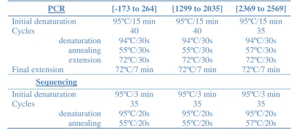
Table 1. Thermocycling conditions for the PCR and sequencing methods. 138
PCR [-173 to 264] [1299 to 2035] [2369 to 2569]
Initial denaturation 95ºC/15 min 95ºC/15 min 95ºC/15 min
Cycles 40 40 35
denaturation 94ºC/30s 94ºC/30s 94ºC/30s
annealing 55ºC/30s 55ºC/30s 57ºC/30s
extension 72ºC/30s 72ºC/30s 72ºC/30s
Final extension 72ºC/7 min 72ºC/7 min 72ºC/7 min
Sequencing
Initial denaturation 95ºC/3 min 95ºC/3 min 95ºC/3 min
Cycles 35 35 35
denaturation 95ºC/20s 95ºC/20s 95ºC/20s
annealing 55ºC/20s 55ºC/20s 57ºC/20s
139
The results were verified using the Sequencing Analysis v.5.2 software. The sequence 140
alignment was done according to the Human Cytochrome P450 Allele Nomenclature 141
Committee (http://www.cypalleles.ki.se) and the allelic variants were detected 142
comparing the sequences obtained with the reference sequence (entry M33388.1 at 143
Genbank) using SeqScape v.3 software. 144
2.3.Validation of the genetic method 145
The method was validated according to the general SWGDAM guidelines, with a 146
special approach for Sanger sequencing methodology [21], and fulfilling the ENFSI 147
recommended minimum criteria for the following parameters: specificity, accuracy, 148
repeatability, reproducibility and sensitivity [19–23]. 149
Reference materials were selected from the Coriell Cell Repositories (National Institute 150
of General Medical Sciences) based on genotypic characterization with the more 151
prevalent variants (ref: NA17226; NA17280 and NA17300) that included: 100C>T; 152
1707 del T; 1846G>A, 2549delA [24]. The genotypes were correctly assessed and the 153
6 peak balance ratios of heterozygote alleles were above 60%. These samples were used 154
as positive controls in the analysis. 155
All the sequences obtained were well aligned with the reference sequence. NCBI’s 156
BLAST (basic local alignment search tool) analysis for sequence similarity was also 157
used in the primers and in the sequences of 3 different samples to evaluate the 158
specificity and to check for homology to other genes or pseudogenes that may interfere 159
with the analysis. The search was made using “Standard Nucleotide BLAST”, in the 160
“nucleotide collection nr/nt; human (taxid:9606)”, with Megablast [29]. The 18 161
sequences verified had matches of 99-100% with the “Homo sapiens CYP2D6 162
(CYP2D6) gene, complete cds, Sequence ID: gb|JF307778.1|Length: 6587”, depending 163
on the variants of each sample. The search using NCBI Genomes (chromosome) 164
database only match with the Homo sapiens Chromossome 22 Primary Assembly with 165
100% identity. 166
The Limit of Detection was determined by performing dilution experiments of a high-167
quality genomic DNA with a known concentration for the following final 168
concentrations: 10, 1, 0.5, 0.1, 0.05, 0.01ng/uL. Signal/Noise and variant identification 169
were the parameters evaluated. The minimum concentration at which was possible to 170
correctly identify the genotype in all the fragments was 0.1ng/L. The limits of 171
detection of the smaller fragments were lower (0.05ng/L for the fragment with 437bp 172
and 0.01ng/L for the fragment with 200bp), as expected. Nevertheless one postmortem 173
sample with a DNA concentration of 0.06ng/L was successfully analyzed. 174
The precision of the method was verified by repeatability and reproducibility 175
experiments. To test the repeatability, three replicates of five samples with different 176
genotypes were simultaneously analyzed, only varying the location in the thermocycling 177
equipment and in the sequencing plate. With 2 sequences for each of the 3 fragments, a 178
total of 30 replicates for each variant were evaluated, with 100% of success. 179
To test the reproducibility, 15 samples were analyzed in three different days using the 180
same technique, including the normal variables of the routine work in the lab, such as 181
room temperature, reagents and equipments. With 2 sequences for each of the 3 182
fragments, a total of 90 replicates in each variant were evaluated for quality and the 183
correct variant identification. Results are given in the Table 2. 184
185
Table 2. Reproducibility results of the genetic method validation 186
7
Allele *4, *10 *12 *6 *8 *4 *3
Variant 100C>T 124G>A 1707delT 1758G>T 1846G>A 2549delA
Day 1 15/15 15/15 15/15 15/15 14/142 15/15
Day 2 15/15 15/15 14/141 14/141 14/141 15/15
Day 3 15/15 15/15 15/15 15/15 15/15 15/15
1
– Failure in the amplification of the fragment. 2 – variant not sequenced. 187
188
Most fragments were consistently amplified and sequenced, with only two failures: one 189
in the sequence (day1) and one in the amplification (day2). The validation was made 190
with routine post-mortem samples. The reported failures are in the larger fragment (with 191
736bp) and the 2 samples with problems had a degradation index of 1.3 and 1.6, which 192
are above 1 (the cut-off of the Quantifiler trio kit). 193
The quality of the sequences was evaluated with the following criteria, as referenced in 194
the Userguide for DNA Sequencing by Capillary Electrophoresis of the Applied 195
Biosystems: Signal > 50, Signal/noise > 25 and Sample score between 20 and 50. 196
Selecting 5 samples of one day of the reproducibility study, the values were calculated 197
for 30 sequences and fulfilled the criteria: minimum Signal was 278, minimum 198
signal/noise was 120 and minimum Sample score was 23. 199
2.4. Toxicological analysis 200
All the samples were analyzed by a general toxicological screening for pharmaceutical 201
drugs as antidepressants, antipsychotics, opioids and others. The screening comprised 202
the more prescribed compounds that are known to inhibit CYP2D6 enzymatic action, 203
such as fluoxetine, paroxetine, sertraline, citalopram, haloperidol, methadone or 204
ticlopidine [5,25]. The confirmation analysis of tramadol and its metabolites, O-205
desmethyltramadol (ODT) and N-desmethyltramadol (NDT) was done in 500 L of 206
peripherical blood stored at -10ºC in test tubes containing 1% of sodium fluoride. 207
Blood samples were prepared by solid phase extraction using Oasis® HLB 3cc 60 mg 208
cartridges (Waters) and GC-MS analysis was performed using an Agilent 6890 Gas 209
Chromatograph equipped with a HP-5MS (30mx0.25mmx0.25mm) capillary column 210
and a 5973 Mass Detector. The injector was set a 280ºC and the injection (1 µL) was 211
made in split mode with 10:1 split ratio. The oven temperature was held at 150ºC for 1 212
min, increased to 290ºC at a rate of 5ºC/min with a final hold time of 8 min. Data was 213
acquired using selected ion monitoring mode (Error! Reference source not found.). 214
Using a positive control prepared and analyzed simultaneously to the samples, the 215
8 identification criteria for positivity was: retention time within 2% or ±0.1 min; the 216
presence of 3 ionic fragments per compound with S/N>3; the maximum allowed 217
tolerances for the relative ion intensities were as required by the World Anti-Doping 218
Agency [26]. 219
2.5. Validation of the analytical method 220
The method was fully validated according to international parameters. Experiments 221
were conducted as described in the SWGTOX guidelines in terms of selectivity, 222
interference studies, recovery, limit of detection, limit of quantification, linearity and 223
calibration model, repeatability, reproducibility, accuracy and carryover [27,28]. All 224
validation experiments were conducted using fortified samples of blank post-mortem 225
blood using LGC and Lipomed standards. 226
Selectivity was evaluated by analyzing 40 blank samples pooled. Two aliquots of each 227
of the 10 pools were prepared: one was analyzed as blank and the other was spiked with 228
all the analytes (100ng/mL). The chromatograms were compared, the identification 229
criteria applied and the existence of interferences by matrix constituents was checked in 230
the blank chromatograms. The method proved to be selective, fulfilled the criteria for all 231
the samples and without interferences. For the recovery studies, six replicates were 232
prepared at three concentrations (150, 500 e 850 ng/mL), three of them were spiked 233
before extraction and the others 3 after. The internal standard was only added after the 234
extraction procedure. The obtained peak area ratios were compared and the results are in 235
the Table 3 . Five calibration curves were measured over a period of 15 days, using 236
seven levels of spiked blood samples in the working range (between 50 and 237
1000ng/mL) and three independent controls were prepared each day with the 238
concentrations of 150, 500 and 850ng/mL. The calibration model was chosen as 239
explained by Almeida et al [28] using as criteria the correlation coefficient higher than 240
0.99 and the best calibrators’ accuracy (obtained by back calculating their 241
concentrations). The method was linear over the working range using a weighting factor 242
of 1/x2. Repeatability (within-day precision) was determined by analyzing six spiked 243
samples at the low, medium and high concentration levels simultaneously. The accuracy 244
and the precision were determined by the calculation of BIAS and the coefficient of 245
variation (% CV), using the concentration obtained for the triplicates of controls (see 246
Table 3). The limit of detection (LOD) was determined by the analysis of blood samples 247
spiked with decreasing amounts of the analytes, being the lowest concentration that 248
9 fulfilled the identification criteria, with the signal/noise of all the peaks above 3, in the 249
replicates (Table 3). The limit of quantitation (LOQ) was validated by analyzing six 250
replicates of spiked samples with a concentration of 50ng/mL (the first point of the 251
calibration curve) and verifying the coefficient of variation (<10%). Dilution of the 252
sample was tested for 1:2 and 1:5 using 10 real samples, covering a concentration range 253
between 50ng/mL to 5000ng/mL. The main results of the method validation are 254
summarized in Table 3Error! Reference source not found.. 255
256
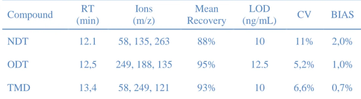
Table 3. Summary of the main results obtained in the validation of the confirmation method for
257
tramadol (TMD), N-demethyltramadol (NDT) and O-demethyltramadol (ODT).
258 Compound RT (min) Ions (m/z) Mean Recovery LOD (ng/mL) CV BIAS NDT 12.1 58, 135, 263 88% 10 11% 2,0% ODT 12,5 249, 188, 135 95% 12.5 5,2% 1,0% TMD 13,4 58, 249, 121 93% 10 6,6% 0,7% 259 2.6. Statistical analysis 260
The genetic and toxicological results were graphically and statistically compared, using 261
SPSS 17.0 software. The distribution of the results was tested for normality with 262
Kolmogorov-Smirnova test and the hypothesis was rejected. Non-parametric tests were 263
then used and the statistical differences between the medians of the genotype groups 264
were calculated using the Mann–Whitney test with 95% of confidence interval. 265
266
3. Results and discussion
267
Post-mortem blood samples were studied searching for CYP2D6 genetic variants 268
responsible for the enzyme inactivation, and the results obtained were then compared 269
with the concentration of tramadol and its main metabolites: NDT and ODT. 270
3.1. Genetic Results 271
Among the 100 post-mortem samples analyzed in this study, amplification failed only in 272
3 samples, which is comparable to other studies [9]. The DNA degradation was 273
probably the main limitant factor as was demonstrated by the degradation index (DI) 274
obtained with the Quantifiler trio kit. The degradation index of these samples was 4.2, 275
6.5 and 12.1 whereas it was under 1.6 for all the others samples. 276
10 The sequencing methodology allowed the detection of 4 different alleles: CYP2D6*3 277
(2549delA); CYP2D6*4 (100C>T and 1846G>A); CYP2D6*6 (1707delT) and 278
CYP2D6*10 (100C>T). Sanger sequencing methodology can’t detect the copy number 279
variation (CNV) of the gene, so it was not possible to identify the gene complete 280
deletion (allele *5). Nevertheless, in cases with allele *5 the PM phenotype assignment 281
is not necessarily compromised. In heterozigotes, this methodology will assign the 282
individual as if he was homozygote for the other allele: if it is null, the individual will 283
be designated as PM; if it’s functional, he will not. On the other hand, in *5/*5 284
homozygotes, there will be no amplification product because there is no gene. So, when 285
the amplification fails, there may be two main explanations: the low quantity or the 286
degradation of the DNA in the sample, which can be evaluated using the degradation 287
index given by Quantifiler trio kit; or maybe the individual is homozygote for the 288
CYP2D6*5 allele, and this genotype should then be confirmed by a suitable method. 289
290
The allele with the higher prevalence was CYP2D6 *4, with a frequency of 19.6%. The 291
allele *10 was detected with a prevalence of 16,5%, which is higher than the expected 292
based on large European population studies and according to CYP2D6 Allelic Variation 293
Summary Table in http://www.cypalleles.ki.se/cyp2d6.htm [30,31] but is in accordance 294
with a recent study for Portuguese population [32]. CYP2D6*10 isn’t a null allele but it 295
can decrease the enzymatic activity and change the substrate specificity of the enzyme 296
[13,33–35]. Only one allele *3 and one allele *6 have been detected. All the alleles that 297
hadn’t any variant in the studied fragments were considered as wild type (WT). 298
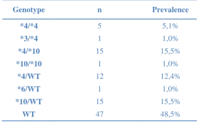
The genotype distribution is presented in the Table 4. 299
300
Table 4. Genotype distribution for the 97 post-mortem samples. 301 Genotype n Prevalence *4/*4 5 5,1% *3/*4 1 1,0% *4/*10 15 15,5% *10/*10 1 1,0% *4/WT 12 12,4% *6/WT 1 1,0% *10/WT 15 15,5% WT 47 48,5% 302
11 Six persons were predicted as poor metabolizers (PM) according to their CYP2D6 303
genotype: 5 individuals were found as homozygotes for the allele *4 and one was 304
heterozygote with one allele *3 and one allele *4. 305
3.2. Toxicology results 306
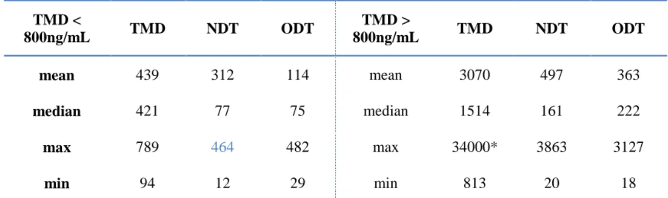
Among the post-mortem samples selected for this study, half presented tramadol 307
concentration below 800 ng/mL, within the therapeutic range according to the published 308
reference tables [36,37]. 27 of these cases had a negative result for at least one of the 309
metabolites: 4 of them in the second group (tramadol concentrations above 800 ng/mL). 310
Descriptive statistics of the concentration of tramadol and metabolites are presented in 311
the Table 5. 312
313
Table 5. Descriptive statistics for the concentration of tramadol (TMD), N-desmethyltramadol
314
(NDT) and O-desmethyltramadol (ODT) in 100 post-mortem blood samples.
315 TMD < 800ng/mL TMD NDT ODT TMD > 800ng/mL TMD NDT ODT mean 439 312 114 mean 3070 497 363 median 421 77 75 median 1514 161 222 max 789 464 482 max 34000* 3863 3127 min 94 12 29 min 813 20 18
* values < 50 ng/mL and > 5000 ng/mL were obtained by extrapolation of de calibration curves. 316
317
3.3. Correlation results 318
The genetic and toxicological results were correlated. Cases where was not possible to 319
confirm the presence of at least one of the metabolites were excluded, remaining a total 320
of 73 cases. The concentration ratio of TMD and its metabolites was plotted in a 321
boxplot graphic with a decimal logarithmic scale and the samples were grouped in two 322
categories according to the genotypes: PM and others. A third group of cases with 323
positive result to inhibitors compounds, such as fluoxetine, paroxetine, sertraline, 324
citalopram, ticlopidine and methadone [25,38,39] was also plotted (INIB). Graphics are 325
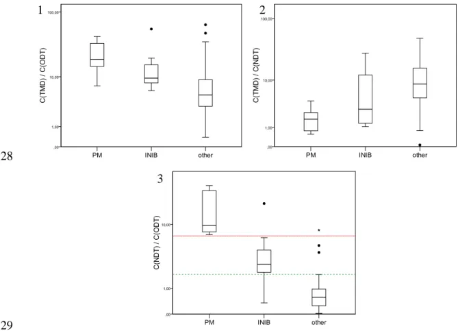
presented in Fig. 1. 326
12 328
329
Fig. 1. Distribution of the concentration ratios tramadol/O-demethyltramadol (graphic 1), 330
tramadol/N-demethyltramadol (graphic 2) and N-demethyltramadol/O-demethyltramadol 331
(graphic 3) according to the poor-metabolizer predicted phenotype (PM; n=6), the cases that 332
were positive for substances that are considered enzymatic inhibitors (INIB; n=10)) and the 333
other cases (n=57).
334 335
The metabolic ratios TMD/ODT or TMD/NDT used by other authors [7,9], as well as 336
the concentration ratio NDT/ODT were tested. The best correlation was obtained using 337
NDT/ODT ratio, as is shown in the graphics presented in Fig. 1. This observation can 338
be explained by the complementarity of the two tramadol metabolic pathways [1,7,10]. 339
In the presence of high substrate concentrations, low CYP2D6 concentrations or when 340
this enzyme is inhibited, a metabolic switch in favor of enhanced N-demethylation can 341
be observed. On the other hand, the possible involvement of CYP2D6 in the elimination 342
process of NDT may explain the increase in its concentration. So, in these cases the 343
ratio between the two metabolites will be higher, allowing to differentiate the PM 344
phenotype and the possible presence of a CYP2D6 inhibitor. 345
346
Using the concentration ratio NDT/ODT, the poor metabolizers (PM) are completely 347
separated from the others (INIB and Others), with a NDT/ODT concentration ratio 348
1 2
13 above 7. The INIB group has a wide concentration ratio interval, but more than 3 349
quarters are between 2,5 and 7. In these cases, regardless of genotype, the interaction of 350
the inhibitors leads to a different metabolic behavior. These results are in accordance 351
with other previous studies [15,38,40,41]. The medians of the groups were statistically 352
compared using a Mann-Whitney test with a level of significance of 0.05 and proved to 353
be significantly different using the NDT/ODT ratio. 354
355
In post-mortem cases, the information about the administration is oftentimes unknown, 356
like the time, dosage, route and the time until death [9]. The concentration range is very 357
wide for both the parent compound and the metabolites and is not correct to compare it 358
with the results obtained in clinical studies. A high TMD/ODT does not necessarily 359
means that there is a deficient metabolization. Many factors can explain it, like co-360
medication, existence of pathologies, or if the death occurred right after the 361
administration. NDT/ODT ratio can be useful to reduce the impact of those unknown 362
variables, as the degree of metabolization at the time of death. Further evaluation of 363
these data might be important and should be considered in future studies. 364
3.4.Case Results 365
In this study, six individuals were found to be poor-metabolizers (PM). All the available 366
information concerning these six cases is presented in the Table 6. 367
368
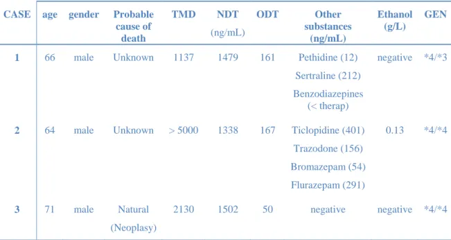
Table 6. Case information: Age, gender, probable cause of death, toxicological findings and 369
genotype of the 6 PM cases. 370
CASE age gender Probable
cause of death TMD NDT (ng/mL) ODT Other substances (ng/mL) Ethanol (g/L) GEN
1 66 male Unknown 1137 1479 161 Pethidine (12)
Sertraline (212) Benzodiazepines
(< therap)
negative *4/*3
2 64 male Unknown > 5000 1338 167 Ticlopidine (401)
Trazodone (156) Bromazepam (54) Flurazepam (291) 0.13 *4/*4 3 71 male Natural (Neoplasy) 2130 1502 50 negative negative *4/*4
14
4 71 male Accident
(traumatic)
528 221 30 negative negative *4/*4
5 55 male Unknown 205 103 <25 negative negative *4/*4
6 83 male Suicide
(hanging)
147 262 <25 Paroxetine (160)
Alprazolam (6)
negative *4/*4
(in the table: TMD, NDT and ODT are tramadol, N-desmethyltramadol 371
and O-desmethyltramadol concentrations; GEN is the genotype) 372
373
The first three cases have tramadol concentrations higher than the therapeutic range 374
according to reference tables [36,37] but the ODT concentrations are comparatively 375
low, considering the data published by Grond et Sablotzki and Stamer et al [1, 11, 12]. 376
Namely in the case number 3 the ODT concentration is comparable to the obtained as a 377
therapeutic concentration in the pharmacokinetics studies. In this particular case, the 378
individual was a cancer patient. The absence of a functional genetic variant for CYP2D6 379
can explain the concentrations found in post-mortem peripherical blood. The higher 380
concentration of tramadol may be due to accumulation or to an increment of the dosage, 381
which can be related with a decrease in the opioid analgesic effect associated with the 382
lower ODT concentration. However, we cannot exclude the possibility of the lack of 383
analgesic effect be due to the development of tolerance in a chronic user. Only one pill 384
was found at the stomach content and the cause of death was determined as natural. 385
386
Predicting the analgesic effect of tramadol based on the pharmacogenetics results is 387
tempting but there are multiple factors, some of them still unknown, that can influence 388
the interpretation [42,43]. Additional fundamental research and collection of routine 389
data is still needed before using pharmacogenetics results as evidence in court. 390
However, in particular post-mortem cases, these approaches, together with all the 391
autopsy findings and clinical information, can be very useful in the investigation of 392 cause of death. 393 394 4. Conclusions 395
This study proved that Sanger sequencing methodology can be successfully applied to 396
the detection of genetic polymorphisms at CYP2D6 in post-mortem blood samples. The 397
method proved to be specific, accurate, with a good precision and limit of detection for 398
the null variants analyzed. 399
15 The results showed a good correlation between the PM genotype and the toxicology 400
results of the tramadol metabolic ratio NDT/ODT, appearing to be an alternative 401
parameter in the evaluation of the degree of metabolization of tramadol in post-mortem 402
cases. 403
The presence of enzymatic inhibitors affects significantly the degree of metabolization, 404
which can be seen in the results obtained. By this reason, is very important to include 405
that compounds in the toxicology screening. 406
The detection of allelic variants described as non-functional can be useful to explain 407
some circumstances of death in the study of tramadol positive cases and the results 408
obtained demonstrate the importance of this genetic tool to forensic toxicology and 409
pathology. 410
Sanger sequencing methodology applied in this study can also be applied to cases with 411
other substances with the same metabolic pathway (CYP2D6), such as codeine, 412
antidepressants and neuroleptics. 413
414
5. References
415
[1] S. Grond, A. Sablotzki, Clinical Pharmacology of Tramadol, Clin. 416
Pharmacokinet. 43 (2004) 879–923. 417
[2] J.K. Coller, L.L. Christrup, A. a Somogyi, Role of active metabolites in the use 418
of opioids., Eur. J. Clin. Pharmacol. 65 (2009) 121–39. doi:10.1007/s00228-008-419
0570-y. 420
[3] G. Li, U.M. Stamer, M. V. Tzetkov, R.B. Altman, T.E. Klein, PharmGKB 421
summary: tramadol pathway, Pharmacogenet. Genomics. 24 (2014) 374–380. 422
doi:10.1016/j.micinf.2011.07.011.Innate. 423
[4] A. Gaedigk, Complexities of CYP2D6 gene analysis and interpretation., Int. Rev. 424
Psychiatry. 25 (2013) 534–53. doi:10.3109/09540261.2013.825581. 425
[5] U.M. Zanger, M. Schwab, Cytochrome P450 enzymes in drug metabolism: 426
regulation of gene expression, enzyme activities, and impact of genetic variation., 427
Pharmacol. Ther. 138 (2013) 103–41. doi:10.1016/j.pharmthera.2012.12.007. 428
[6] M. Hersberger, J. Marti-jaun, K. Rentsch, E. Hanseler, CRapid Detection of the 429
CYP2D6*3, CYP2D6*4, and CYP2D6*6 Alleles by Tetra-Primer PCR and of 430
the CYP2D6*5 Allele by Multiplex Long PCR, Clin. Chem. 46 (2000) 1072– 431
1077. 432
[7] A. Levo, A. Koski, I. Ojanperä, E. Vuori, A. Sajantila, Post-mortem SNP 433
analysis of CYP2D6 gene reveals correlation between genotype and opioid drug 434
(tramadol) metabolite ratios in blood, Forensic Sci. Int. 135 (2003) 9–15. 435
http://www.sciencedirect.com/science/article/pii/S0379073803001592 (accessed 436
September 26, 2013). 437
[8] R.S. Pedersen, P. Damkier, K. Brosen, Tramadol as a new probe for cytochrome 438
16 P450 2D6 phenotyping: a population study., Clin. Pharmacol. Ther. 77 (2005) 439
458–67. doi:10.1016/j.clpt.2005.01.014. 440
[9] A. Koski, INTERPRETATION OF POSTMORTEM TOXICOLOGY RESULTS
441
Pharmacogenetics and Drug-Alcohol Interaction, University of Helsinki, Finland, 442
2005. https://helda.helsinki.fi/handle/10138/20385. 443
[10] E. García-Quetglas, J.R. Azanza, E. Cardenas, B. Sábada, M.A. Campanero, 444
Stereoselective Pharmacokinetic Analysis of Tramadol and its Main Phase I 445
Metabolites in Healthy Subjects after Intravenous and Oral Administration of 446
Racemic Tramadol, Biopharm. Drug Dispos. 28 (2007) 19–33. doi:10.1002/bdd. 447
[11] E. García-Quetglas, J.R. Azanza, B. Sádaba, M.J. Muñoz, I. Gil, M.A. 448
Campanero, Pharmacokinetics of tramadol enantiomers and their respective 449
phase I metabolites in relation to CYP2D6 phenotype., Pharmacol. Res. 55 450
(2007) 122–30. doi:10.1016/j.phrs.2006.11.003. 451
[12] U.M. Stamer, F. Musshoff, M. Kobilay, B. Madea, a Hoeft, F. Stuber, 452
Concentrations of tramadol and O-desmethyltramadol enantiomers in different 453
CYP2D6 genotypes., Clin. Pharmacol. Ther. 82 (2007) 41–47.
454
doi:10.1038/sj.clpt.6100152. 455
[13] S.H. Gan, R. Ismail, W.A. Wan Adnan, W. Zulmi, Impact of CYP2D6 genetic 456
polymorphism on tramadol pharmacokinetics and pharmacodynamics., Mol. 457
Diagn. Ther. 11 (2007) 171–181. doi:10.1007/BF03256239. 458
[14] T.C. Kupiec, V. Raj, N. Vu, Pharmacogenomics for the Forensic Toxicologist, J. 459
Anal. Toxicol. 30 (2006) 65–72. doi:10.1093/jat/30.2.65. 460
[15] J. Sistonen, PHARMACOGENETIC VARIATION AT CYP2D6 , CYP2C9 , 461
AND CYP2C19 : Population Genetic and Forensic Aspects, University of 462
Helsinki, Finland, 2008. https://helda.helsinki.fi/handle/10138/20340. 463
[16] A.-L. Zackrisson, Pharmacogenetics from a Forensic Perspective – CYP2D6 and 464
CYP2C19 genotype distributions in autopsy cases, Linköping University, 465
Sweden, 2009. http://swepub.kb.se/bib/swepub:oai:DiVA.org:liu-17936. 466
[17] P.S. Walsh, D.A. Metzger, R. Higuchi, Chelex 100 as a medium for simple 467
extraction of DNA for PCR-based typing from forensic material, Biotechniques. 468
10 (1991) 506—513. http://europepmc.org/abstract/MED/1867860. 469
[18] S. Vernarecci, E. Ottaviani, A. Agostino, E. Mei, L. Calandro, P. Montagna, 470
Genetics Quantifiler Trio Kit and forensic samples management : A matter of 471
degradation, Forensic Sci. Int. Genet. 16 (2015) 77–85.
472
doi:10.1016/j.fsigen.2014.12.005. 473
[19] W. Branicki, T.C. Kupiec, R. Pawlowski, Validation of Cytochrome b Sequence 474
Analysis as a method of Species Identification, J. Forensic Sci. 48 (2003). 475
www.astm.org. 476
[20] B. Budowle, N.D. Connell, A. Bielecka-oder, R.R. Colwell, C.R. Corbett, J. 477
Fletcher, et al., Validation of high throughput sequencing and microbial forensics 478
applications, Investig. Genet. 5 (2014) 1–18. doi:10.1186/2041-2223-5-9. 479
[21] G. Pont-kingdon, F. Gedge, W. Wooderchak-Donahue, I. Schrijver, K. Weck, J. 480
Kant, et al., Design and Analytical Validation of Clinical DNA Sequencing 481
Assays, Arch Pathol Lab Med. 136 (2012) 41–46. doi:10.5858/arpa.2010-0623-482
17 OA.
483
[22] Scientific Working Group on, D.A. Methods, SWGDAM Validation Guidelines 484
for DNA Analysis Methods, (2012) 1–13. http://www.swgdam.org. 485
[23] ENFSI DNA WORKING GROUP, Recommended Minimum Criteria for the 486
Validation of Various Aspects of the DNA Profiling Process, (2010) 11. 487
http://www.enfsi.eu/sites/default/files/documents/minimum_validation_guideline 488
s_in_dna_profiling_-_v2010_0.pdf. 489
[24] V.M. Pratt, B. Zehnbauer, J.A. Wilson, R. Baak, N. Babic, M. Bettinotti, et al., 490
Characterization of 107 genomic DNA reference materials for CYP2D6, 491
CYP2C19, CYP2C9, VKORC1, and UGT1A1: a GeT-RM and Association for 492
Molecular Pathology collaborative project., J. Mol. Diagn. 12 (2010) 835–846. 493
doi:10.2353/jmoldx.2010.100090. 494
[25] D. Flockhart, Cytochrome P450 Drug Interaction Table, Indiana Univ. Sch. Med. 495
(2007) 1–2. http://medicine.iupui.edu/clinpharm/ddis/main-table (accessed 496
January 21, 2016). 497
[26] World Anti-Doping Agency, WADA Technical Document – TD2015IDCR 498
SPECTROMETRIC CONFIRMATION OF THE IDENTITY OF ANALYTES 499
WADA Technical Document – TD2015IDCR, 2015. https://www.wada-500
ama.org/en/resources/science-medicine/td2015-idcr. 501
[27] Scientific working group for forensic toxicology, Standard practices for method 502
validation in forensic toxicology (SWGTOX), (2013) 52. doi:10.1093/jat/bkt054. 503
[28] A.M. Almeida, M.M. Castel-Branco, A.C. Falcao, Linear regression for 504
calibration lines revisited: weighting schemes for bioanalytical methods, J. 505
Chromatogr. B. 774 (2002) 215–222. 506
[29] A. Morgulis, G. Coulouris, Y. Raytselis, T.L. Madden, R. Agarwala, A. a. 507
Schäffer, Database indexing for production MegaBLAST searches, 508
Bioinformatics. 24 (2008) 1757–1764. doi:10.1093/bioinformatics/btn322. 509
[30] S. Bernard, K. a Neville, A.T. Nguyen, D. a Flockhart, Interethnic differences in 510
genetic polymorphisms of CYP2D6 in the U.S. population: clinical implications., 511
Oncologist. 11 (2006) 126–35. doi:10.1634/theoncologist.11-2-126. 512
[31] J. Sistonen, A. Sajantila, O. Lao, J. Corander, G. Barbujani, S. Fuselli, CYP2D6 513
worldwide genetic variation shows high frequency of altered activity variants and 514
no continental structure., Pharmacogenet. Genomics. 17 (2007) 93–101. 515
doi:10.1097/01.fpc.0000239974.69464.f2. 516
[32] V. Gaio, I. Picanço, B. Nunes, A. Fernandes, F. Mendonça, F. Horta Correia, et 517
al., Pharmacogenetic Profile of a South Portuguese Population: Results from the 518
Pilot Study of the European Health Examination Survey in Portugal, Public 519
Health Genomics. 18 (2015) 139–150. doi:10.1159/000373920. 520
[33] A. Bagheri, B. Kamalidehghan, M. Haghshenas, P. Azadfar, L. Akbari, M. 521
Hossein, et al., Prevalence of the CYP2D6*10 (C100T), *4 (G1846A), and *14 522
(G1758A) alleles among Iranians of different ethnicities, Drug Des. Devel. Ther. 523
9 (2015) 2627–2634. 524
[34] W. Leppert, CYP2D6 in the metabolism of opioids for mild to moderate pain., 525
Pharmacology. 87 (2011) 274–85. doi:10.1159/000326085. 526
18 [35] Q. Li, R. Wang, Y. Guo, S. Wen, L. Xu, S. Wang, Relationship of CYP2D6 527
genetic polymorphisms and the pharmacokinetics of tramadol in Chinese 528
volunteers., J. Clin. Pharm. Ther. 35 (2010) 239–47. doi:10.1111/j.1365-529
2710.2009.01102.x. 530
[36] M.R. Repetto, M. Repetto, Tabla de Concentraciones de xenobioticos en fluidos 531
biologicos humanos como referencia para el diagnostico toxicologico, Sevilla, 532
2015. http://busca-tox.com. 533
[37] M. Schulz, S. Iwersen-bergmann, H. Andresen, A. Schmoldt, Therapeutic and 534
toxic blood concentrations of nearly 1 , 000 drugs and other xenobiotics, Crit. 535
Care. 16 (2012) 2–5. http://ccforum.com/content/16/4/R136. 536
[38] P.H. Saladores, J.C. Precht, W. Schroth, H. Brauch, M. Schwab, Impact of 537
Metabolizing Enzymes on Drug Response of Endocrine Therapy in Breast 538
Cancer, Expert Rev Mol Diagnosis. 13 (2013) 349–65.
539
http://www.medscape.com/viewarticle/804598. 540
[39] J.K. Coller, J.R. Michalakas, H.M. James, A.L. Farquharson, J. Colvill, J.M. 541
White, et al., Inhibition of CYP2D6-mediated tramadol O-demethylation in 542
methadone but not buprenorphine maintenance patients., Br. J. Clin. Pharmacol. 543
74 (2012) 835–41. doi:10.1111/j.1365-2125.2012.04256.x. 544
[40] M. Jin, S.B. Gock, P.J. Jannetto, J.M. Jentzen, S.H. Wong, Pharmacogenomics as 545
Molecular Autopsy for Forensic Toxicology: Genotyping Cytochrome P450 546
3A4*1B and 3A5*3 for 25 Fentanyl Cases, J. Anal. Toxicol. 29 (2005) 590–598. 547
doi:10.1093/jat/29.7.590. 548
[41] V. Stearns, M.D. Johnson, J.M. Rae, A. Morocho, A. Novielli, P. Bhargava, et 549
al., Active tamoxifen metabolite plasma concentrations after coadministration of 550
tamoxifen and the selective serotonin reuptake inhibitor paroxetine., J. Natl. 551
Cancer Inst. 95 (2003) 1758–1764. doi:10.1093/jnci/djg108. 552
[42] F. Musshoff, U.M. Stamer, B. Madea, Pharmacogenetics and forensic 553
toxicology., Forensic Sci. Int. 203 (2010) 53–62.
554
doi:10.1016/j.forsciint.2010.07.011. 555
[43] A. Sajantila, J.U. Palo, I. Ojanperä, C. Davis, B. Budowle, Pharmacogenetics in 556
medico-legal context., Forensic Sci. Int. 203 (2010) 44–52. 557
doi:10.1016/j.forsciint.2010.09.011. 558
We would like to thank both reviewers for the deep appreciation of our manuscript and for all the suggestions. We tried to fulfill all the requirements and we will answer point-by point to the comments
Reviewer #1:
1. The introduction of this manuscript oversimplifies the metabolism and pharmacological properties of tramadol. The metabolism of tramadol is complex and mediated by several polymorphic cytochrome P450 enzymes [Gong Li et al. Pharmacogenetics and genomics (2014)].
* Secondly, tramadol analgesia is mediated not only by the polymorphic mu opioid receptor but also through modulation of norepeniphrine ad serotonergic re-uptake.
A: I have reformulated the introduction as suggested.
* Thirdly, tramadol is not always given in controlled doses (as stated), but commonly prescribed in outpatient settings. Therefore, the assumption that higher than expected tramadol doses would arise from malpractice or neglect is short-sighted and should probably be removed from the manuscript althogether as it is outside the scope of this work.
A: Removed as suggested.
2. Please provide a reference to support the statement (page 3) that PM of tramadol have reduced analgesic effects with tramadol and reduced adverse effects with tramadol.
A: Done.
3. The postmortem concentrations should not, in general, be compared to those in clinical studies/therapeutic studies. Please discuss at length the various issues with postmortem findings (post-mortem redistribution, variances in drug collection sites (femoral blood, etc), death interval) as they pertain to the population in this study, and as they pertain to tramadol specifically.
A: We have added the information required.
4. The methods section should clearly state which concomitant medications were considered in this study as CYP2D6 substrates. Furthermore, not all CYP2D6 substrates are "inhibitors". Please refer to the page by David Flockhart in Indiana University for correct classification of medication as CYP2D6 substrate or inhibitor.
A: The analytical method has the most common medication, including anti-depressants, antipsychotic and other opioids. For the INIB group we have focused in the substances that are reported as inhibitors (also in the Flockhart chart). The outliers of the “others” group on the NDT/ODT graphic are, curiously,
positive for compounds that are also substrates of CYP2D6, as venlafaxine and nortriptiline. Done as suggested.
5. There are over 100 allelic variants of CYP2D6. In this study, the author's method only categorizes 4 of these variants. The authors then subscribe individuals to either poor, intermediate, or extensive metabolizers based on only 4 variants and make several assumptions on their tested population to rationalize this approach. As their testing approach is not the gold standard in pharmacogenetic testing, given its limited scope of testing, they need to compare their methodology with a platform that tests for the majority of CYp2D6 alleles in order to justify these assumptions. A false positive and false negative rate of attributing an individual to the poor metabolizer phenotype based on only 4 variants needs to be provided.
A: We have clarified in the manuscript the aims of our work.
The focus was to detect the PM. We have tried to explain better the exceptions. We changed also the assignment of the genotypes based in the variants searched to restrict it to the PM.
On the other hand, we described in more detail the validation of the method. It is not possible for us to compare our results with other methodology as suggested, but we had used reference materials to evaluate the accuracy of the method.
We would like to thank both reviewers for the deep appreciation of our manuscript and for all the suggestions. We tried to fulfill all the requirements and we will answer point-by point to the comments
Reviewer #2:
1. The present manuscript describes a Sanger sequencing method for detection of some CYP2D6 polymorphisms causing a reduced or absent enzyme function, as well as a GC/MS method for the quantitation of tramadol, O-desmethyltramadol and N-desmethyltramadol. The methods were applied to 100 tramadol positive post-mortem samples. The purpose of doing that is however not clearly stated. Several papers on CYP2D6 sequencing methods and quantitative tramadol methods have been published previously. Furthermore, many of those are better described in terms of validation and performance and are using techniques able to detect also the CYP2D6 ultra-rapid metabolizers and quantifying the enantiomers of tramadol and its metabolites. If the authors clearly state the aim of identifying poor metabolizers and further describe and discuss the six cases being poor metabolizers the present manuscript could however add on to the current knowledge within this field.
A: We have clarified in the manuscript the aims of our work.
2. Abstract:
* "A Sanger sequencing methodology was developed for the detection of genetic variants that cause absence of CYP2D6 activity, such as *3, *4, *6, *8, *10 and *12 alleles". As mentioned later in the manuscript *10 causes a decreased function of the enzyme, not a total absence. Consider to reformulate, for example "…genetic variants that cause absent or reduced CYP2D6 activity".
A: Done.
* Why is not all alleles possible to identify with the present method mentioned?
A: The amplification of the entire gene is very difficult (> 5000pb), specially in post-mortem samples with high degradation index. On the other hand, Sanger sequencing only allows the analysis of amplicons with a maximum of 1000 pb. To detect copy number variation is necessary to use other methods, with other equipments such as real-time PCR or platforms as AmpliChip CYP450 test from Roche. The method that we have used is simple, rapid, low cost and available in forensic genetics labs and can detect the main variants. We had clarified it on the manuscript as required.
3. Introduction:
* The aim of the study is not clearly stated. Why was the study conducted? What was the research question? Any hypothesis?
A: We have clarified in the manuscript the aims and scope of our work.
* In the first paragraph it is written that "The information about therapeutic concentrations of ODT is scarce". There are however several publications covering both tramadol and ODT concentrations.
A: The sentence was removed. There are several publications with clinical studies but the toxic or lethal concentrations aren’t well defined, to our knowledge.
* UMs are usually referred to as rapid metabolizers but in this paper the term ultra-metabolizers are used instead. Consider changing that.
A: Changed
* I also suggest that explanations of abbreviations, for example "poor metabolizer (PM)", are given the first time the word is written. Subsequently only the abbreviation is used.
A: Changed
4. Materials and methods:
* If the toxicological analysis has been published previously a reference referring to that publication could be added to section 2.2. If there is no previous publication the sample preparation and validation could be further described. The validation parameters investigated are given but no information on how the experiments were conducted. What chemicals, reagents and reference compounds were used? What quality control levels were used? How many replicates were utilized? What kind of blank blood was used? What were the predetermined acceptance criteria for each validation parameter? Please clarify in the manuscript.
* It is also important that results are given for all the validation parameters investigated, and they could with advantage be given in the results and discussion section.
* It is stated that the coefficient of variation is under 11% for the three compounds. Clarify what parameters that are referred to.
* Was dilution integrity part of the validation? Since calibration ranges between 0.05-1 mg/L and some samples have significantly higher concentrations I assume some dilution was made? Please clarify in the manuscript.
A: we described in more detail the validation of the method.
* What transitions were used in SIM mode? Add those to the manuscript.
A: added as requested.
* The two first comments for this section are applicable also to the validation of the sequencing method. There is no information on how the validation experiments were conducted and results are scarce. "The quality of the sequences was considered good", in terms of what? Accuracy is an especially important parameter when it comes to Sanger sequencing; how was the accuracy experiment conducted and what was the result of it? Please describe in the manuscript.
* Should pb be bp instead, bp for base pairs?
A: Changed
* Which polymorphisms were used for the identification of allele *8, *12, *14, *15, *40 etc., mentioned on page 5? Add those to the manuscript.
A: Done
* The thermocycling conditions used for each fragment could be described in the manuscript.
A: Done
5. Results and discussion:
* The first sentence on page 6 is in my opinion not necessary.
A: Removed
* Check the maximum concentration for NDT in table 1, should it be 5889?
A: Corrected
* It does not emerge from table 1 that 27 cases had metabolite concentrations below the detection limit (is the limit of detection and the limit of quantitation the same?), as it does in the text. Consider if it should since minimum values for the metabolites are presented in the table, and the table text says descriptive statistics for all the 100 cases.
A: Corrected.
Values < 50 ng/mL and > 5000 ng/mL were obtained by extrapolation of de calibration curves.
* Concerning the first and second sentence in section 3.2: What was the degradation index of the three samples that failed amplification in comparison to the degradation index of the other samples? That information would be of interest to add to the manuscript.
A: added as requested.
as suggested * If it is desirable to shorten the manuscript figure 1 and 2 could be deleted since the information is given in the text as well.
A: Removed
* In figure 2 and table 2 the text implies results for all the 100 post-mortem samples. However, since 3 samples failed amplification the results are only for 97 samples, right? Prevalences in table 2 as well as allele frequencies in figure 2 need to be slightly adjusted in case of calculating with 100 samples, which seems to be the case since the sum of prevalences in table 2 should be 100% but is only 97%.
A: Corrected.
* The allele frequency of the null allele *5 (which was not searched for) is higher than that of *3 and *6 (which was searched for) in a Caucasian population. Therefore consider to reformulate the following sentences on page 7 "All the alleles that hadn´t any variant in the studied fragments were considered as wild type. This assumption was considered acceptable because the prevalence of the other null alleles that weren´t searched for is very low in Caucasian population".
A: The sentence was removed and we have tried to explain better the exceptions, focusing in the identification of the PM, which is the main purpose of the study.
* In section 3.2 it is written "Since this method is not able to detect CNV events, the ultra-metabolizers were included in the group of EM. This assumption was considered acceptable once the final objective of the present work was to detect the PM". What was the purpose of constructing figure 3 if only PMs were important to find? Consider to reformulate.
A: We changed the assignment of the genotypes based in the variants searched to restrict it to the PM. Thank you for your suggestions.
* A further description of the INIB group would be valuable. Were those cases PM, IM or EM? What was the concentrations of the inhibitors? To be able to draw conclusions about the significance of the inhibitors a comparison between for example EMs with and without inhibitors seems more valuable than putting all cases with inhibitors in one group, regardless of genotype.
A: Since we had focused in the PM cases, we removed the EM and IM assignment. On the other hand, and unfortunately, the available information of the cases is scarce. We were not able to do this.
* Table 3 is in my opinion not necessary. It would be more interesting to compare all the measured values for each group, than just comparing medians between groups. Statistically significant differences based on all measured values for each group could be indicated in figure 3, a separate table is not necessary. The number of individuals that are given in table 3 could be given in the text below figure 3.
A: Done as suggested.
* Abbreviations are not explained in the text below figure 3 which would be good.
A: corrected as suggested.
* Why does the authors conclude that the NDT/ODT ratio is a better measure of TMD to ODT metabolism than the TMD/ODT ratio? The correlation seemed better for the NDT/ODT ratio, yes, although other factors than the CYP2D6 genotype might affect this ratio. A discussion concerning the impact of CYP2B6 and CYP3A4 genotype for the formation of NDT would be meaningful. If the authors think that the correlation between the TMD/ODT ratio and CYP2D6 genotype was less than expected a discussion concerning reasons for this would be highly valuable. Could for example the inability of the sequencing method to detect allele *5 and multiple copies of the gene have had an impact on the results? Or could the classification into IMs and EMs have had an impact? The present classification is not wrong although according to other definitions EMs have two functional alleles. The present EM group includes individuals with both one, two and multiple copies of CYP2D6 alleles.
A: We have tried to explain it better, especially for post-mortem cases.
* The information in table 4 is interesting, as well as the measured concentrations in table 5, although the concentration unit must be given in table 5. This information could however be compiled in one table, while DNA-concentration, degradation index and polymorphisms are left
out. The important thing is that all individuals are PMs and that information is given in the table text.
A: Done as suggested.
* The results of table 4 and 5 are only discussed with a few sentences on page 11, saying that in three cases the tramadol concentration was higher than the therapeutic range but ODT concentrations were acceptable. What does that mean, what are the conclusions drawn? Case 2 and 3 have "unknown" and "natural" stated as the cause of death, respectively, in spite of high tramadol concentrations. Was the tramadol concentrations not considered toxic (because of low ODT concentrations)? What were the circumstances of death? Any comments regarding the other cases? Please discuss the results in more detail.
* What substances were included in the toxicological screening? It could be interesting to describe case 3,4 and 5 (without other substances present) in more detail.
* It could perhaps be of interest to show the concentrations and CYP2D6 genotypes for the 10 intoxication cases mentioned in section 2.1.
A: Done as suggested, although we cannot discuss better the results because of the lack of information. The study of the autopsy results of these cases together with the pathologists would be indeed very interesting but it was not possible yet.
6. Conclusions:
* "The Sanger sequencing method proved to be specific, accurate, with a good precision and limit of detection". As mentioned previously results for all validation parameters are not presented in the manuscript, for what reason it is difficult for the reader to be able to draw the same conclusion.
A: We have reformulated the manuscript and included the validation parameters.
* "The results of genotypes showed a good correlation with toxicology results of the tramadol metabolic ratio NDT/ODT, appearing to be a better parameter to know the degree of metabolization of TMD to ODT, than the usual metabolic ratio as TMD/ODT". See also the comments above in the "results and discussion" section. In my opinion this is not a correct conclusion.
A: We have reformulated the results and discussion and also the conclusions in the manuscript.
* "The presence of enzymatic inhibitors affects significantly the degree of metabolization" See also the comments above in the "results and discussion" section. It is known that some drugs acts as inhibitors of CYP2D6 and therefore affect the metabolism of tramadol. In my opinion it is however not satisfactory shown in this manuscript.
A: We have reformulated the conclusions in the manuscript.
* "The detection of allelic variants described as non-functional were useful to explain some circumstances of death in the study of tramadol positive cases and demonstrate the importance of this genetic tool to forensic toxicology and pathology". What circumstances were clarified in
this study? A more profound discussion regarding the results is desirable.
A: As said above, it was not possible yet. Nevertheless, the cases number 1 and 3 can be good examples of the future applications of this approach.
7. References:
* Is reference 8, 12 and 13 complete? Where can one find them?
A: Corrected
* Where is reference 19 published?
A: Corrected
* I am not sure if user guides (reference 20 in this case) are appropriate to refer to in the reference list?