U
NIVERSIDADE DE LISBOAF
ACULDADE DEM
EDICINA DENTÁRIAT
ISSUE HEALING OF IMMEDIATEIMPLANT PLACEMENT IN EXTRACTION SOCKETS WITH TITANIUM VERSUS ZIRCONIUM OXIDE IMPLANTS
:
AN EXPERIMENTAL STUDY IN THE BEAGLE DOG
H
ELENAC
RISTINAD
EO
LIVEIRAF
RANCISCOOrientadores:
Prof. Doutor João Manuel Mendez Caramês Prof. Doutor António Duarte Sola Pereira da Mata
Tese especialmente elaborada para a obtenção do grau de Doutor em Medicina Dentária, especialidade de Periodontologia
UNIVERSIDADE DE LISBOA
FACULDADE DE MEDICINA DENTÁRIA
TISSUE HEALING OF IMMEDIATE IMPLANT PLACEMENT IN EXTRACTION SOCKETS WITH TITANIUM
VERSUS ZIRCONIUM OXIDE IMPLANTS: AN EXPERIMENTAL STUDY IN THE BEAGLE DOG
HELENA CRISTINA DE OLIVEIRA FRANCISCO
Orientadores:
Prof. Doutor João Manuel Mendez Caramês Prof. Doutor António Duarte Sola Pereira da Mata
Tese especialmente elaborada para a obtenção do grau de Doutor em Medicina Dentária, especialidade de Periodontologia
Júri Presidente:
Doutor Mário Filipe Cardoso de Matos Bernardo, Professor Catedrático e Presidente do
Conselho Científico da Faculdade de Medicina Dentária da Universidade de Lisboa;
Vogais:
Dennis Perry Tarnow, Clinical Professor of Dental Medicine do College of Dental Medicine
da Columbia University, USA;
Doutor António Cabral de Campos Felino, Professor Catedrático da Faculdade de Medicina
Dentária da Universidade do Porto;
Doutor Ricardo Manuel Casaleiro Lobo de Faria Almeida, Professor Catedrático da
Faculdade de Medicina Dentária da Universidade do Porto;
Doutor Femando Alberto Deométrio Rodrigues Alves Guerra, Professor Associado com
Agregação da Faculdade de Medicina da Universidade de Coimbra;
Doutor João Manuel Mendes Caramês, Professor Catedrático da Faculdade de Medicina
Dentária da Universidade de Lisboa;
Doutor António Duarte Sola Pereira da Mata, Professor Catedrático da Faculdade de
Medicina Dentária da Universidade de Lisboa;
Doutor Paulo Alexandre Mascarenhas Lopes, Professor Auxiliar da Faculdade de Medicina
Dentária da Universidade de Lisboa;
A
CKNOWLEDGEMENTSThe research presented in this thesis would not have been possible without the help of many people to whom I am gratefully indebted. It will be simple to name all the people that helped to get this done, but it will be tough to thank them enough. First and foremost, my sincere thanks go to my two Ph.D. supervisors Professor João Manuel Mendez Caramês and Professor António Duarte Sola Pereira da Mata.
I would like to express my sincerest gratitude to my supervisor Professor João Manuel Mendez Caramês, Full Professor at the University of Lisbon College of Dentistry, who has supported me throughout my thesis with his patience and knowledge. Professor Caramês welcomed me to his office when I arrived from New York and has been a strong guiding influence on my professional career ever since. I have been extremely lucky to have a supervisor whose care and attention has made this such a thoughtful and rewarding journey. His expertise in animal research in particular was a great asset to the research. His continuing effort, commitment and mentorship has helped me to bring this study to its conclusion. His sincere enthusiasm, absolute commitment and generous support continue to inspire me.
Professor António Duarte Sola Pereira da Mata, Full Professor at the University of Lisbon College of Dentistry, introduced me to academic and research life in my second year of dental school when I became a member of GIBO (Oral Biology and Research Group) at ISCS-S (Instituto Superior de Ciências da Saúde Sul). He influenced my graduate and postgraduate career in an ineffably positive way. His thoroughness and understanding of biology, his passion for academia and research remain a tremendous source of inspiration, support and an example to follow. Moreover, his patient guidance and valued friendship were crucial in helping me to reach my goals.
During my training at New York University College of Dentistry I had the chance to meet and work with a number of wonderful people. There are however, two people that had a profound impact on my academic and professional life. I will always be
expertise and thoughtful criticism. More than a mentor, he is an extraordinary friend. My sincere thanks for inspiring me to pursue my ideas, go further and keep it simple. I will always cherish his lectures. To Dr Gary Greenstein whose everlasting support and encouragement, excellent sense of humor and dedication to the profession throughout the years was truly priceless. My work at New York University College of Dentistry would not have been as enjoyable without him.
I am very grateful and wish to acknowledge the administration of FMDUL for the institutional support, in particular to the Director Professor Luís Pires Lopes, Vice-Director Professor Jaime Portugal and Past-Vice-Director Professor João Aquino and who always expressed interest in this research project and also to o the Scientific Board at FDMUL, and its President, Professor Mário Bernardo.
My deepest gratitude to the team of Professor Ramiro Mascarenhas, Dr João Maria Nobre and Paulo Dias at the Experimental Surgery Center at National Zoothecnical Station in Santarém. Without their help this work could not have taken place.
I would like to extend my sincere thanks to Professor Fernando Guerra and his team at the University of Coimbra. My personal thanks to Professor Paulo Palma and Dr João Martins for their skillful support. I cannot thank Cláudia Brites enough for spending long hours struggling to have the zirconia sections ready on time.
My sincere gratitude to Professor José Rino, responsible for microscopy at Champalimaud Foundation who willingly shared his expertise and gave me access to the laboratory and research facilities.
I am grateful to my dear long time friend Professor Joana Rita da Silva Fialho for her help in the phase of the conclusion of this thesis. Her statistical assistance during the final stretch was greatly appreciated.
This thesis would not have been possible without the help of Franz Berghaenel from Metoxit who I would like to thank specially for his help and commitment and also to Ulrich Voltz whose help in the initial phases of this project and knowledge on zirconia implants was crucial in bringing this thesis to fruition.
My thanks also to Professor Duarte Marques for being a loyal friend and colleague and for his continuous encouragement and friendship throughout our years working together. His sound advice and support have been invaluable on an academic level for which I am extremely grateful.
I would like to express my gratitude to my dear friends and colleagues at the University of Lisbon College of Dentistry, Dr André Chen and Dr João Canta, for the great support and friendship that they have given me over the years.
To Professor Joana Marques and Professor João Silveira my dearest friends I would like to thank them for all the support and mostly for their friendship since we started working in the Oral Biology Research Group (GIBO) in 2001.
To all the staff and friends at the Implantology Institute. I would like to thank Manuela André especially who was very important during the experimental phase. Her human qualities, help and enthusiasm were always present during experimental surgery. To my dental assistant Filipa Teixeira for being patient, for organizing everything at the office and for taking everything she could off my shoulders so that I could focus on this thesis.
The dedication needed to complete this work would not have been possible without the understanding and support of my family and friends. First and foremost, I cannot thank my parents Celeste and Manuel enough. They have encouraged me in my studies and gave me both the freedom and support that I needed to become the person I am today. They were excellent teachers and role models, but still trusted me enough to let me choose my own goals. They have served as my guiding light throughout my education and I hope that I have lived up to their expectations. Last
A
BSTRACTObjectives: To compare and evaluate the biomechanical, radiographic and
histological behavior of zirconia and titanium implants placed into extraction sockets.
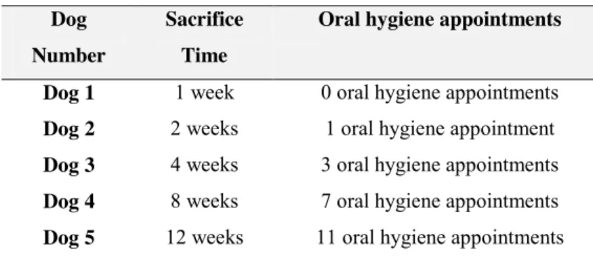
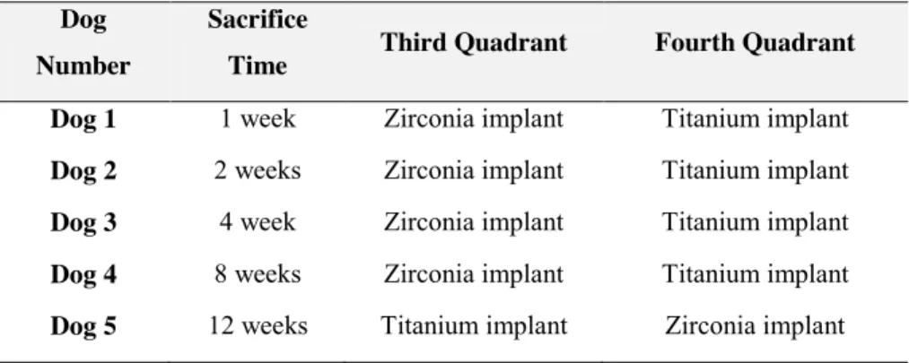
Materials and methods: Five Beagle dogs received 15 titanium implants (Ti) and
15 zirconia implants (Zr) immediately placed into the distal socket of the second, third and fourth premolars. Implant stability and radiographic evaluation was performed at the time of implant placement and sacrifice. Animals were sacrificed at 1, 2, 4, 8 and 12 weeks. Peri-implant mucosa dimensions, marginal bone loss and bone-to-implant contact were evaluated. Kruskall-Wallis test, Mann-Whitney test and Spearman correlation analysis were used when appropriate. Values of p < 0.05 were taken as significant.
Results: The primary stability values were 82.53 ± 1.10 ISQ (Ti) and 57.6 ± 3.29
ISQ (Zr) (p = .05). After 12 weeks, implant stability was 79.33 ± 0.58 ISQ (Ti) and 84.67 ± 6.11 (Zr) (p > .05). The buccal peri-implant mucosa ranged between 3.54 ± 0.23 mm (Zr) and 3.93 ± 0.49 mm (Ti) (p > .05). The buccal bone crest was located 1.53 ± 0.15 mm (Ti) and 1.55 ± 0.12 mm (Zr) (p > .05) below the implant shoulder (p = .05). The BIC, NBF and TBA were 59.4 ± 0.75 % (Ti) and 57.8 ± 2.26 % (Zr), 65.37 ± 3.05 % (Ti) and 63.63 ± 3.79 % (Zr), 77.97 ± 2.08 % (Ti) and 75.1 ± 2.31 % (Zr) respectively (p > 0.05).
Conclusions: Even though zirconia implants exhibited less primary stability when
compared to titanium implants they reach a similar degree of stability over time. Zirconia implants did not prevent the remodeling of the extraction socket. Zirconia implants rendered similar peri-implant soft tissue dimensions, ridge alterations and osseointegration when compared to titanium implants.
R
ESUMOIntrodução: A colocação de implantes imediatamente após extração dentária
tornou-se, nos últimos anos, um protocolo clínico frequente. A literatura descreve taxas de sobrevivência elevadas e semelhantes às encontradas em implantes convencionais. Nos últimos 50 anos, o titânio tem constituído o material de eleição para o fabrico de implantes dentários, devido à sua biocompatibilidade, elevada resistência à corrosão e propriedades mecânicas. No entanto, a procura de materiais não metálicos para utilização em reabilitação oral, tem permitido o desenvolvimento recente de novos materiais cerâmicos. A zircónia, caracterizada por uma elevada dureza, resistência e estabilidade, a par de uma excelente biocompatibilidade, tem sido considerada na literatura atual, uma alternativa válida ao titânio. A sua utilização tem sido indicada em várias situações clínicas, inclusive na colocação imediata de implantes no alvéolo pós-extracional. Do nosso conhecimento não existe na literatura nenhuma publicação, que descreva a evolução dos eventos biológicos iniciais na cicatrização de implantes em zircónia colocados imediatamente após extração dentária e a sua comparação com a de implantes em titânio colocados nas mesmas condições.
Objectivo: O principal objetivo desta investigação experimental foi estudar, no
modelo animal, a evolução do processo de cicatrização, nas doze primeiras semanas, de implantes de zircónia, colocados em alvéolos pós-extracionais, analisando o seu comportamento biomecânico, radiográfico e histológico e compará-la com a evolução do processo de cicatrização de implantes em titânio. Os objetivos secundários foram: avaliar a estabilidade e as alterações radiográficas de implantes em titânio e em zircónia, colocados em alvéolos pós-extracionais; descrever as fases iniciais de cicatrização dos tecidos moles e duros em torno de implantes em titânio e em zircónia; determinar e comparar as dimensões dos tecidos moles em torno de implantes em titânio e em zircónia colocados em alvéolos pós-extracionais; avaliar as alterações da crista óssea e formação óssea de implantes em titânio e em zircónia colocados em alvéolos pós-extracionais; correlacionar a estabilidade implantar com a percentagem de contacto ósseo com o implante dentário; correlacionar os achados radiológicos com os histológicos.
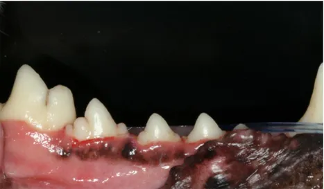
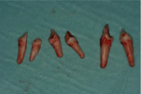
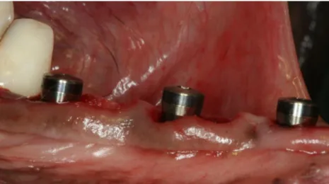
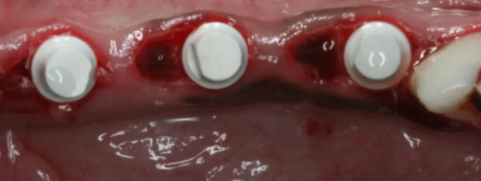
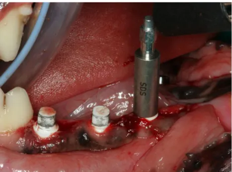
género masculino com 2 anos de idade. Foi feita extração dos quatro pré-molares de cada quadrante mandibular, de cada um dos cinco animais experimentais. Nos alvéolos distais do segundo, terceiro e quarto pré-molares foram colocados 15 implantes em titânio (Ti) (Astra OsseoSpeed TX 4.0 S de 4 x 11 mm; Astra Tech AB, Molndal, Sweden) e 15 implantes em zircónia (Zr) (4 x 11 mm; ref. SDScd401411; SDS Swiss Dental Solutions AG Switzerland). Cada cão recebeu seis implantes, três em titânio (grupo de controlo) de um lado da mandíbula e três em zircónia (grupo teste) do lado contralateral. Durante o período de osteointegração foi seguido um protocolo de higiene oral semanal e a uma dieta mole. Os animais foram sacrificados em diferentes períodos de tempo (1, 2, 4, 8 e 12 semanas) para análise histológica e histomorfométrica. A resposta tecidular foi avaliada utilizando técnicas biomecânicas (análise da frequência de ressonância, Ostell® ISQ), radiográficas, e histológicas. Os testes Kruskall-Wallis e
Mann-Whitney foram utilizados quando apropriado. As médias das várias variáveis foram calculadas e valores de p < 0.05 foram considerados significativos.
Resultados: A estabilidade primária avaliada no dia da colocação, através da
análise da frequência de ressonância (Ostell® ISQ) dos implantes em titânio (82.53 ±
1.10 ISQ), foi superior à dos implantes em zircónia (57.6 ± 3.29 ISQ) (p = .05). Essa tendência inverteu-se significativamente na segunda semana do período de cicatrização, com diminuição da estabilidade dos implantes em titânio (68.33 ± 5.13 ISQ) e aumento da estabilidade nos implantes de zircónia (86.67 ± 5.51 ISQ) (p = .05). Da segunda à quarta semana a estabilidade dos implantes em titânio aumentou progressivamente (82.67 ± 0.58 ISQ), enquanto a estabilidade dos implantes em zircónia apresentou ligeira diminuição (84.0 ± 1.73 ISQ). A partir da oitava semana ambos os tipos de implantes apresentaram estabilidades semelhantes sem diferenças significativas (Titanio, 80.33 ± 3.06 ISQ às oito semanas e 79.33 ± 0.58 ISQ às 12 semanas; Zirconia, 77.33 ± 1.53 ISQ às 8 semanas e 84.67 ± 6.11 ISQ às 12 semanas.
O nível ósseo marginal radiográfico no dia da colocação dos implantes, foi em mesial -1.50 ± 0.04 mm (Ti) e -1.17 ± 0.05 (Zr); em distal foi - 1.45 ± 0.03 mm (Ti)
e -1.13 ± 0.06 mm (Zr). Após 12 semanas de cicatrização o nível ósseo radiográfico diminuiu significativamente, sendo em mesial - 0.48 ± 0.04 mm (Ti) e - 0.28 ± 0.04 mm (Zr) (p = 0.05) e em distal - 0.44 ± 0.05 mm (Ti) (p = 0.05) e - 0.23 ± 0.04 mm (Zr).
Em vestibular o nível ósseo marginal radiográfico registado às 12 semanas foi 1.57 ± 0.14 mm (Ti) e 1.54 ± 0.02 mm (Zr) (p > 0.05). Em lingual foi - 0.42 ± 0.05 mm (Ti) e - 0.17 ± 0.05 mm (Zr) (p = 0.05).
O espaço biológico em vestibular após período de 12 semanas foi 3.54 ± 0.23 mm (Zr) e 3.93 ± 0.49 mm (Ti) e em lingual 2.35 ± 0.31 mm (Zr) e 2.6 ± 0.36 mm (Ti) (p > .05). As dimensões do epitélio em vestibular foram 1.32 ± 0.19 mm (Zr) e 1.34 ± 0.18 mm (Ti) (p > .05), enquanto que em lingual foram 0.94 ± 0.11 mm (Zr) e 1.73 ± 0.23 mm (Ti) (p = .05). As dimensões do tecido conjuntivo em vestibular foram 2.22 ± 0.27 mm (Zr) (p > .05) e 2.15 ± 0.16 mm (Ti) e em lingual 1.45 ± 0.23 mm (Zr) e 0.87 ± 0.25 mm (Ti) (p = .05). Em vestibular a crista óssea estava localizada a 1.53 ± 0.15 mm (Ti) e 1.55 ± 0.12 mm (Zr) (p > .05) apical ao ombro do implante, enquanto que em lingual estava localizada a - 0.39 ± 0.06 mm (Ti) e - 0.13 ± 0.06 mm (Zr) coronal ao ombro do implante (p = .05). A percentagem de contato osso/ implante, formação óssea e área óssea total foram de 59.4 ± 0.75 % (Ti) e 57.8 ± 2.26 % (Zr), 65.37 ± 3.05 % (Ti) e 63.63 ± 3.79 % (Zr), 77.97 ± 2.08 % (Ti) e 75.1 ± 2.31 % (Zr), respetivamente, não existindo diferenças estatisticamente significativas entre os dois grupos de implantes (p > 0.05). O coeficiente de correlação de Spearman entre a estabilidade e percentagem de contacto osso/ implante foi de - 0.011 (Ti) e - 0.441 (Zr). O coeficiente de correlação de Spearman entre os resultados radiológicos e histomorfométricos foi de 1 para ambos os grupos de implantes.
Conclusões: Neste estudo demonstrámos que os implantes em zircónio, colocados
após exodontia, não evitam a remodelação fisiológica do alvéolo, afetando maioritariamente a dimensão vertical da parede vestibular, tendo um comportamento semelhante aos implantes em titânio colocados nas mesmas condições. As comparações entre os implantes em zircónia e titânio deverão ser interpretadas como tendências e não como conclusões definitivas, devido ao número reduzido de amostras. No entanto, os resultados biomecânicos, radiológicos e
investigações futuras. Dentro das limitações deste estudo podemos concluir que: a sobrevivência de implantes em zircónia é semelhante á encontrada em implantes em titânio não tendo sido perdido nenhum implante em ambos os grupos; a estabilidade biomecânica dos implantes em zircónia parece ser comparável à dos implantes em titânio; todos os implantes obtiveram valores de estabilidade semelhantes ao fim de um período de 12 semanas, independentemente dos valores da estabilidade primária; os resultados radiológicos demonstraram que após exodontia e colocação imediata de implantes, a remodelação óssea marginal continuou durante a fase de cicatrização em mesial, distal, vestibular e lingual, sendo mais acentuada em vestibular; a colocação de implantes imediatos em zircónia não evitou as alterações dimensionais do alvéolo pós-extracional; a perda óssea na tábua vestibular após um período de 12 semanas nos implantes em zircónia foi semelhante aos implantes em titânio; quanto às dimensões dos tecidos moles, os implantes em zircónia apresentaram valores semelhantes aos implantes em titânio; os resultados histológicos demonstraram que os implantes em zircónio não alteraram o padrão de cicatrização do alvéolo pós-extracional e que a maior perda óssea se deu ao nível da tábua vestibular; os dois tipos de implantes utilizados neste estudo apresentaram níveis semelhantes de osteointegração relativamente à área de contacto osso-implante. Este estudo experimental demonstrou que não existe uma correlação entre a estabilidade implantar medida através de análise da frequência de ressonância e a percentagem de contacto osso/ implante. Investigação futura poderá complementar este estudo na compreensão do processo de cicatrização alveolar e validar a utilização de implantes de zircónia, em alvéolos pós-extracionais,
Palavras Chave: implantes imediatos, titânio, zirconia, histologia, estabilidade do
T
ABLE OF CONTENTSList of abbreviations ... xix
CHAPTER 1. INTRODUCTION ... 1
1.1 Osseointegration ... 3
1.1.1 Peri-implant hard tissue healing ... 4
1.1.1.1 Bone response to implant placement ... 4
1.1.1.2 Bone modeling ... 5
1.1.1.3 Bone remodeling... 6
1.1.2 Dental implant materials ... 8
1.1.2.1 Titanium ... 8
1.1.2.2 Ceramics ... 13
1.1.3 Implant stability ... 18
1.1.3.1 Implant stability testing ... 19
1.1.3.1.1 Destructive methods ... 19
1.1.3.1.2 Non-destructive methods ... 21
1.2 Soft tissues around dental implants ... 26
1.2.1 Peri-implant biologic width ... 28
1.2.2 Peri-implant epithelium ... 36
1.2.3 Connective tissue around dental implants ... 41
1.3 Healing of extraction sockets ... 46
1.3.1 Histologic events ... 48
1.3.2 Dimensional changes at extraction sockets ... 56
1.3.3 Dimensional changes of the mucosa ... 62
1.4 Implant placement into fresh extraction sockets ... 64
1.4.1 Classification of the timing of implant placement after tooth extraction ... 67
1.4.2 Tissues healing after immediate implant placement ... 69
1.4.2.1 Morphogenesis and integration of the peri-implant mucosa ... 69
1.4.2.2 Bone healing and osseointegration ... 74
1.4.2.3 Ridge alterations after immediate implant placement ... 78
1.4.3 Survival and success rates of implants placed into fresh extraction sockets ... 84
1.5 Scope of this project and aims ... 90
2.2 Animal experimental model ... 95
2.2.1 Transportation of experimental animals ... 96
2.2.2 Facilities for the experimental animals ... 96
2.2.3 Diet of the experimental animals ... 99
2.2.4 Oral hygiene protocol ... 99
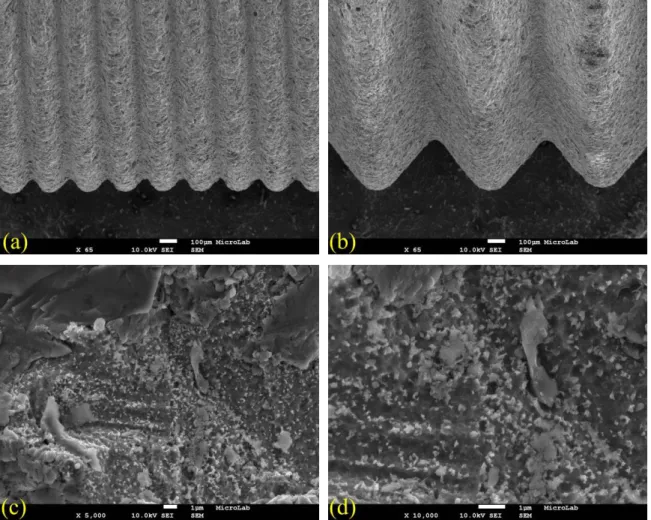
2.4 Implants used in the experimental study ... 102
2.4.1 Titanium dental implants ... 102
2.4.2 Zirconia dental implants ... 103
2.5 Experimental surgery on beagle dogs ... 105
2.5.1 Anesthetic protocol ... 105
2.5.2 Dental extractions ... 106
2.5.3 Implant placement ... 109
2.5.3.1 Drilling protocol for control group ... 110
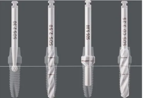
2.5.3.2 Drilling protocol for the test group ... 111
2.5.4 Resonance frequency measurements with Osstell® ISQ device ... 113
2.5.5 Radiographic analysis ... 116
2.5.6 Postoperative care and follow-up visits ... 118
2.6 Sacrifice of experimental animals ... 119
2.7 Preparation of histologic sections for microstructural analysis ... 122
2.7.1 Fixation ... 122
2.7.2 Dehydration and Infiltration ... 122
2.7.3 Inclusion/ photopolymerization ... 123
2.7.4 Preparation of acrylic blocks ... 124
2.7.5 Initial cuts of the blocks ... 124
2.7.6 Sandwich preparation ... 125
2.7.7 Sample facing ... 126
2.7.8 Preparation and calibration of the glass slide ... 127
2.7.9 Toluidine blue staining method ... 130
2.8 Histometric evaluation ... 131
2.8.1 Descriptive histological observations ... 131
2.8.2.1 Peri-implant soft tissues ... 132
2.8.2.2 Ridge alterations ... 133
2.8.2.3 Bone-to-implant contact (BIC) ... 134
2.8.2.4 Total bone area and new mineralized bone tissue ... 135
2.9 Statistical analysis ... 135
CHAPTER 3. RADIOGRAPHIC AND RESONANCE FREQUENCY ANALYSES OF PERI-IMPLANT TISSUES: TITANIUM VS. ZIRCONIA IMPLANTS ... 137
3.1 Introduction... 139
3.2 Materials and methods ... 143
3.3 Results ... 143
3.3.1 Resonance frequency analysis measurements ... 143
3.3.2 Radiographic analysis ... 148
3.3.2.1 Titanium implants ... 148
3.3.2.1.1 Mesial and distal ... 148
3.3.2.1.2 Buccal and lingual ... 151
3.3.2.2 Zirconia implants ... 153
3.3.2.2.1 Mesial and Distal ... 154
3.3.2.2.1 Buccal and Lingual ... 157
3.3.2.3 Titanium vs. Zirconia implants ... 159
3.4 Discussion ... 161
3.4.1 Implant Stability ... 162
3.4.2 Radiographic analysis ... 171
3.5 Conclusions ... 177
CHAPTER 4. MORPHOGENESIS OF THE PERI-IMPLANT MUCOSA OF IMPLANTS PLACED IN EXTRACTION SOCKETS: TITANIUM VS. ZIRCONIA ... 179
4.1 Introduction... 181
4.2 Materials and methods ... 186
4.3 Results ... 186
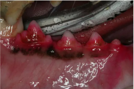
4.3.1 Clinical observations ... 186
4.3.2 Histological observations ... 186
4.3.2.1 One week of healing ... 186
4.3.2.2 Two weeks of healing ... 191
4.3.2.3 Four weeks of healing ... 195
4.3.3.1 Biological width ... 207
4.3.3.1.1 Titanium ... 207
4.3.3.1.2 Zirconia ... 209
4.3.3.1.3 Titanium vs. zirconia ... 211
4.3.3.2 Length of the barrier epithelium ... 212
4.3.3.2.1 Titanium ... 212
4.3.3.2.2 Zirconia ... 214
4.3.3.2.3 Titanium vs. Zirconia ... 216
4.3.3.3 Length of the connective tissue ... 217
4.3.3.3.1 Titanium ... 218
4.3.3.3.2 Zirconia ... 219
4.3.3.3.3 Titanium vs. zirconia ... 221
4.4 Discussion ... 223
4.5 Conclusions ... 239
CHAPTER 5. BONE HEALING AND RIDGE ALTERATIONS OF IMPLANTS PLACED IN EXTRACTION SOCKETS: TITANIUM VS. ZIRCONIA ... 240
5.1 Introduction... 242
5.2 Materials and methods ... 245
5.3 Results ... 245
5.3.1Histological results ... 245
5.3.1.1 One week of healing ... 246
5.3.1.2 Two weeks of healing ... 252
5.3.1.3 Four weeks of healing ... 257
5.3.1.4 Eight weeks of healing ... 261
5.3.1.5 Twelve weeks of healing ... 265
5.3.2 Histometric analysis ... 269
5.3.2.1 Ridge alterations ... 269
5.3.2.1.1 Distance from the implant shoulder to the bone crest (IS-BC) ... 269
5.3.2.1.1.1 Titanium ... 269
5.3.2.1.1.2 Zirconia ... 272
5.3.2.1.1.3 Titanium vs. zirconia ... 274
5.3.2.1.2 Distance from the implant shoulder to the first bone-to-implant contact (IS-B) ... 276
5.3.2.1.2.1 Titanium ... 276
5.3.2.1.2.2 Zirconia ... 278
5.3.2.1.2.3 Titanium vs. zirconia ... 280
5.3.2.1.3 Distance between the bone crest and the first bone-to-implant contact (BC-B) ... 282
5.3.2.1.3.1 Titanium ... 282
5.3.2.1.3.2 Zirconia ... 283
5.3.2.1.3.3 Titanium vs. Zirconia ... 285
5.3.2.2 Bone-to-implant contact (BIC) ... 287
5.3.2.3 New bone formation (NBF) ... 289
5.3.2.4 Total bone area (TBA) ... 291
5.3.3Correlation of the BIC with the RFA measurements ... 292
5.3.4 Correlation of radiographic and histometric findings ... 293
5.4 Conclusion ... 322
CHAPTER 6. FINAL REMARKS AND CONCLUSION ... 324
CHAPTER 7. REFERENCES ... 334
APPENDIX A. LICENSE FOR THE ANIMAL EXPERIMENT ... 394
APPENDIX B. LIST OF FIGURES ... 398
L
IST OF ABBREVIATIONS% Percentage
aJE Apical border of the junctional epithelium AZT Alumina-toughened zirconia
B First contact point of the bone-to-implant contact BB Bundle bone
BC Bone crest
BIC Bone-to-implant contact
DIC Differential interference contrast microscopy
I Implant i.e. id est IS Implant shoulder LB Lamellar bone mm Millimeters NB New bone
NBF New bone formation
OB Old bone
OM Osteoid matrix PM Peri-implant mucosa SD Standard deviation TBA Total bone area Ti Titanium vs. Versus
Y-TZP Yttria stabilized zirconia Zi Zirconia
1.1OSSEOINTEGRATION
Since the first studies by Brånemark (Branemark et al. 1969; Branemark et al. 1977) a great deal of research has been carried out to gain a better understanding of the phenomenon of osseointegration. Originally direct bone-to-implant contact (i.e. osseointegration) was referred to as direct bone deposition on the implant surface without interposition of fibrous or connective tissue (Branemark et al. 1977), a term also called “functional ankylosis” (Schroeder et al. 1981). In a more comprehensive way Brånemark defined osseointegration at the light microscopic level as “a direct structural and functional connection between ordered, living bone and the surface of a load-carrying implant” (Branemark 1983). Later on osseointegration was given a more clinical definition, as a process in which clinically asymptomatic rigid fixation of alloplastic materials, was achieved and maintained in bone during functional loading (Albrektsson 1983). Since Brånemark's initial experiments the concept of osseointegration has been defined on multiple levels, including anatomically, histologically, and ultrastructurally (Adell et al. 1981; Linder et al. 1983).
Nowadays, osseointegration is the foundation of modern implantology. An understanding of osseointegration implies a profound knowledge of bone biology particularly in the healing process. Bone healing is certainly one of the most fascinating aspects of tissue biology and one of the rare examples of how the process of regeneration enables restoration of the original structure and function in an integrated way (Schenk and Buser 1998). As a result the unequivocal success of endosseous dental implants is driving the need for continuing refinements in implant design and optimization of the biological healing response following implant placement (Davies 2003).
The placement of a dental implant into the alveolar bone comprises a cascade of cellular and extracellular biological events that take place at the bone-implant interface until the implant surface is finally covered with newly formed bone (Fini et al. 2004). Osseointegration follows a common, biologically determined program that can be subdivided into three separate phases: bone response to implant placement, peri-implant osteogenesis and peri-implant bone remodeling (Mavrogenis et al. 2009).
1.1.1 Peri-implant hard tissue healing
The temporal sequence of hard tissue healing events leading to osseointegration was not elucidated until the results of animal studies by Berglundh et al. were published (Abrahamsson et al. 2004; Berglundh et al. 2003). The placement of an implant into a healed ridge is followed by a sequence of healing events that result in the establishment of osseointegration, characterized by direct contact between bone and implant surface. Studies using the animal model have shown that just after implant placement, the peripheral part of the implant thread is in close contact with the bone providing mechanical stability during the first phases of healing (Cochran et al. 1998). However, depending on the implant design and surface, the inner part of the threads makes limited or no contact with the adjacent bone bed. The gap between the pitch and the body of the implant established a geometrically well-defined wound chamber. In 2003, Berglundh et al., using a dog model, examined the temporal sequence of healing events taking place during the process of osseointegration in a wound chamber (Berglundh et al. 2003).
1.1.1.1 Bone response to implant placement
After implant placement, the first biological component to come into contact with the implant is blood, forming a blood clot. The blood cells (red cells, platelets, and inflammatory cells, such as polymorphonucleargranulocytes and monocytes) become entrapped at the implant interface and are activated to release cytokines and other soluble, growth and differentiation factors. The surgical trauma sensitizes the cells to release certain growth factors that stimulate new cells. The blood clot is partly replaced by primitive granulation tissue 4 days after the placement of the implant. Some of the fibroblast-like mesenchymal cells line-up in a parallel orientation along the implant surface and start the formation of collagen fiber bundles (Abrahamsson et al. 2004; Schwarz et al. 2007). A provisional connective tissue matrix becomes established (Berglundh et al. 2003; Schwarz et al. 2007). The debris of cortical and trabecular bone from the osteotomy have sometimes been found at the wound sites during the early phases of healing (Abrahamsson et al. 2004; Schwarz et al. 2007). Osteoblasts and mesenchymal cells seem to migrate and attach to the implant surface from day one after implantation, depositing bone-related proteins and creating a non-collagenous matrix layer on the implant surface
that regulates cell adhesion and the binding of minerals. After the establishment of well-vascularized immature connective tissue, osteogenesis continues through the recruitment, proliferation, and differentiation of osteoblastic cells (Colnot et al. 2007). The osteoblasts are able to deposit a collagen fiber matrix that mineralizes (Steflik et al. 1998).
1.1.1.2 Bone modeling
According to Berglundh et al., one week following implant installation, the provisional connective tissue in the wound chambers was rich in vascular structures containing numerous mesenchymal cells (Berglundh et al. 2003). Bone modeling was observed in several compartments of the chamber. A cell-rich immature bone (i.e. woven bone) was seen in the provisional connective tissue surrounding the blood vessels. Woven bone formation occurred in the center of the chamber as well as in discrete locations that were apparently in direct contact with the surface of the titanium device. This contact osteogenesis is seen as representing the very first phase of osseointegration, namely direct contact between the roughened implant surface and the newly formed woven bone. In the phenomenon of contact osteogenesis, new bone forms on the implant surface first (Davies 1998; Osborne et al. 1980). Since, a priori, no bone was present on the surface of the implant upon implantation, the implant surface must become colonized by a population of osteogenic cells before the initiation of bone matrix formation. However, such contact osteogenesis was not observed on machined titanium implants (Abrahamsson et al. 2004). Contact osteogenesis also occurs at remodeling sites where an old bone surface is populated with osteogenic cells before new bone can be laid down. After a healing period of one week bone debris was still present. Osteoclasts migrate to the bone fragments and start a process of osteoclastic resorption and remodeling, leading to their incorporation into newly formed woven bone (Berglundh et al. 2003).
After a healing period of 14 days, woven bone formation was detected surrounding the entire implant. In the wound chamber portions of the newly formed woven bone apparently extended from the old bone into the provisional connective tissue. This osteogenesis took place at a distance from the implant surface and was given the term distant osteogenesis or appositional bone formation (Davies 1998;
Osborne et al. 1980). New bone was formed from the host bone cavity towards the implant surface. Similar to normal appositional bone growth the existing bone surfaces provide a population of osteogenic cells that lay down new matrix, which, as osteogenesis continues, encroaches on the implant itself. Thus new bone was not forming on the implant itself, but instead the implant was surrounded by bone. The wound healing advanced with marked woven bone formation and maturation. Four weeks after implant installation the newly formed mineralized bone extended from the prepared bone surface of the implant bed into the chamber (Berglundh et al. 2003). According to some authors the woven bone occupied almost 30% of the chamber space (Abrahamsson et al. 2004; Vignoletti et al. 2009c).
1.1.1.3 Bone remodeling
After a healing period of 6 to 12 weeks the bone remodeling process was clearly observed. The newly formed woven bone is gradually remodeled and replaced by lamellar bone. The bone trabeculae had been reinforced by lamellar bone deposition. This lamellar consisted of primary and secondary osteons, and this more mature bone tissue made contact with the implant surface. Bone marrow containing blood vessels, adipocytes and mesenchymal cells was observed surrounding the trabeculae of mineralized bone (Berglundh et al. 2003; Schwarz et al. 2007). Osteoblasts were detected at the implant-bone interface. The bone in contact with the implant surface underwent morphological remodeling and adaptation. The turnover of peri-implant mature bone in osseointegrated implants was confirmed by the presence of medullary or marrow spaces containing osteoclasts, osteoblasts, mesenchymal cells and lymphatic/blood vessels next to the implant surface. During the remodeling of the peri-implant bone, a circle of osteons around the implant with their long axis parallel to the implant surface and perpendicular to the long axis of the implants were detected. The transformation of woven bone into lamellar bone, bone organized to resist physical strain and displaying Haversian architecture is another important part of osseointegration (Mavrogenis et al. 2009). During the first year after implant placement bone modeling and remodeling continues at a slow rate and contributes to higher implant resistance to shear forces (Johansson and Albrektsson 1987; Steflik et al. 1998). Despite these preclinical studies performed in dogs having revealed the healing
sequence after implant placement in healed ridges, the clinical application of this data is limited, as the turnover rate of bone remodeling in dogs is four times faster than in humans (Draper 1994). There are a number of studies showing that titanium implants with different surface characteristics osseointegrated in the human jawbone, as demonstrated by histologic and histomorphometric studies (Salvi et al. 2015). Several studies have reported high bone-to-implant contact (BIC) values (Grassi et al. 2007; Shibli et al. 2007a; Shibli et al. 2007b). However, BIC contact values largely depended on location, implant design and implant surface characteristics (Salvi et al. 2015). The temporal sequence of hard tissue healing events leading to osseointegration has also been histologically investigated in human volunteers (Bosshardt et al. 2011; Lang et al. 2011b). The first report of the sequence of events during early osseointegration in human volunteers was published by Lang et al. (Lang et al. 2011b). The authors evaluated the rate and degree of osseointegration at chemically modified moderately rough hydrophilic (SLA Active) and moderately rough hydrophobic (SLA) implant surfaces during early phases of healing in a human model. Dental implants were installed into the retro-molar area of 49 human volunteers and retrieved after 7, 14, 28 and 42 days of submerged healing. According to the authors, osseointegration took place with BIC increasing from 7 to 42 days when it reached 62% of the implant surface exposed to the parent bone. The author concluded that the healing of implants installed into parent bone showed similar characteristics to bone resorptive and appositional events, between 7 and 42 days, for both moderately rough implant surfaces tested. However, the degree of osseointegration after 4 weeks was higher for the hydrophilic SLA Active compared with the hydrophobic SLA surface (Lang et al. 2011b). In a histologic and histomorphometric study, Degidi et al. evaluated peri-implant bone formation around one-stage peri-implants that were retrieved after a healing period of 4 weeks (Degidi et al. 2009). Although only three patients were included in this study the BIC percentages were 52.0% ± 2.5%, 61.0% ± 2.9%, and 42.0% ± 6.9% (Degidi et al. 2009). Even though the general sequence of healing events is not affected by implant surface topographies the rate of hard tissue healing can be influenced by implant topography and chemistry (Albrektsson 2008; Wennerberg and Albrektsson 2009).
variables. There are a number of important factors in achieving reliable osseointegration and prevention of implant loosening presented by Albrektsson et al. in 1981: implant material, implant design, implant surface, state of host tissue, surgical technique and load forces on the implant (Albrektsson et al. 1981).
1.1.2 Dental implant materials
The biocompatibility of the material is of utmost importance and a predictor of osseointegration as it is essential in establishing stable fixation with direct BIC and no fibrous tissue at the interface. An ideal implant material should be biocompatible, with adequate toughness, strength, corrosion, wear and fracture resistance (Parr et al. 1985; Smith 1993). The materials used for the fabrication of dental implants can be categorized according to their chemical composition into metals, ceramics or polymers (Sykaras et al. 2000). Based on the type of biologic response, three major types of biodynamic activity have been reported: biotolerant, bioinert, and bioactive (LeGeros and Craig 1993; Osborne and Gale 1980). The high long-term clinical survival rates reported for titanium and its alloys have made titanium the “gold standard” material for the fabrication of endosseous dental implants (Adell et al. 1990; Jemt et al. 1996).
1.1.2.1 Titanium
Titanium and its alloys (mainly Ti-6Al-4V) have become the metals of choice for the endosseous parts of currently available dental implants (Sykaras et al. 2000). Long-term research has demonstrated a high predictability of osseointegration of titanium dental implants (Albrektsson et al. 1986a). The emergence of titanium as a dental implant material was attributed to the early work of a Swedish physician and orthopedic surgeon Per Ingvar Brånemark (Albrektsson and Zarb 1989) at that time a researcher at Göteborg University, Sweden. Brånemark managed to study the revascularization and healing patterns over a period of days using small chambers made of titanium and inserting them into the tibia and ears of rabbits (Brånemark et al. 1985). The author found that the titanium chambers placed in the leg of the rabbit could not be retrieved as they had integrated into the bone, unless they had been sectioned from the surrounding bone (Brånemark et al. 1985). Although Brånemark thought that this was a very interesting finding he did not attach any
major importance to the finding until 1960. The researcher placed similar titanium chambers in a human skin tube which had been surgically created from twin pedicle flaps on the inside of the upper arm 3 to 6 months previously. He found that these titanium chambers were well tolerated by the human skin and concluded that titanium was a highly compatible material that could have several applications not only in medicine but also in Dentistry. After obtaining research funding and putting together a team of surgeons, dentists, metallurgists and bioengineers, Brånemark started investigating the possibility of using titanium to support dental prosthesis for the rehabilitation of edentulous jaws. In order to test the biocompatibility of titanium the first studies were carried out in dogs. Following animal sacrifice the researchers realized that the interface between bone and titanium was still intact. At the light microscope level there was no gap between bone and implant surface whenever they made contact. This phenomenon was coined as osseointegration. In 1965 the Brånemark team treated the first edentulous patient successfully with titanium implants. Several protocols for implant designs, diagnosis, restoration and follow-up were established over the following years. A study was then initiated in Gothenburg to evaluate the clinical results of the application of the technique in humans (Branemark et al. 1977). Around the same time, in the United States, Linkow was developing the first screw-type implant called Vent-Plant which was completed in 1963 (Linkow and Rinaldi 1988) and the first blade implant, the Blade-Vent. Both implants were designed as one-stage systems and the metallic material used was cobalt-chromium alloy. However, due to Brånemark’s research and the biocompatibility of titanium and titanium alloys, the metal used in the the Vent-Plant and the Blade-Vent was changed to titanium in 1964 and 1971, respectively. In 1982 Brånemark presented the results of his research with implants in Toronto, Canada (Branemark et al. 1984). Brånemark´s success rate was around 97% and he received full recognition from the international scientific community (Branemark et al. 1984). Titanium is currently the most commonly used material for dental and orthopedic implants. Titanium implants have been used successfully for years in the substitution of lost dental elements (Jorge et al. 2013).
Titanium alloys are commonly used in implant dentistry because of their high strength, biocompatibility, and corrosion resistance in a physiological environment (Sykaras et al. 2000). According to the American Society for Testing and Materials
(ASTM), there are six distinct types of titanium available as implant biomaterials. Amongst these six materials there are four grades of commercially pure titanium and two titanium alloys. Pure titanium is composed of 99.5 % titanium and 0.5 % interstitial elements (carbon, oxygen, nitrogen, hydrogen and iron) and the proportion of these elements directly affects the metal properties (Jorge et al. 2013). There are four types of commercially pure titanium (types 1, 2, 3 and 4). Each type consists of 99% pure titanium with the remaining 1% representing various impurities (carbon, oxygen, nitrogen, hydrogen and iron). The disadvantage of commercially pure titanium is its relative softness. However, it can be alloyed with other elements in order to improve its strength. Grade 5 commercially pure titanium refers to the three combinations of aluminum and vanadium. A common titanium alloy, mostly used for orthopaedic implants, is titanium-6-aluminum-4-vanadium (Ti6Al4V). Some animal experimental studies have shown that Ti6Al4V is less integrated in bone tissue compared to commercially pure titanium when evaluated by means of biomechanical and histomorphometrical tests (Han et al. 1998; Stenport and Johansson 2008). Titanium interacts with biologic fluids through its stable oxide layer, which forms the basis for its exceptional biocompatibility (Lautenschlager and Monaghan 1993; Pilliar et al. 1986). When exposed to air titanium immediately forms an oxide layer that reaches a thickness of 2 to 10 nm, in 1 second and provides corrosion resistance (Donley and Gillette 1991; Ducheyne 1988). Titanium is the material of choice for intraosseous applications because of its high passivity, its controlled thickness, rapid formation and ability to repair itself instantaneously if damaged. It is also resistant to chemical attack, it has a catalytic activity for a number of chemical reactions and a modulus of elasticity compatible with that of bone of titanium oxide (Kasemo and Lausmaa 1985; Parr et al. 1985).
Although titanium has a recognized biocompatibility due to protective oxide layers, there is currently a general trend in implant dentistry for metal-free solutions. There are patients who are informed through less reliable reports that metals may be considered harmful to the body. Indeed there have been scientific reports which assert that titanium may provoke unwelcomed host reactions (Jung et al. 2015) and metallic ion release has raised concerns over the last years (Smith et al. 1997). It has been suggested that titanium hypersensitivity may be a factor responsible for implant failure (Egusa et al. 2008; Muller and Valentine-Thon 2006; Sicilia et al.
2008). Although titanium hypersensitivity is a growing concern, epidemiological data on the incidence of titanium related hypersensitivity reactions are still lacking (Siddiqi et al. 2011). This worrying correlation seems to be either overlooked by clinicians or weakly researched (Javed et al. 2013). What we need to keep in mind is that no material can be considered to be universally biocompatible and this includes titanium (Williams 1994). Even though titanium has been regarded as an inert metal, several earlier studies have identified potential hematologic and metabolic toxicity (Carrol and Tullis). Titanium allergy can be detected in dental implant patients, although its estimated occurrence is low: around 0.6% (Sicilia et al. 2008). According to a number of authors, degradation products of metallic biomaterials including titanium may mediate metal hypersensitivity or allergic reactions (Merritt and Brown 1996; Merritt and Rodrigo 1996; Sicilia et al. 2008). Despite the wide application of titanium implants there are reports stating that metals, including titanium, can induce non-specific immunomodulation elicited by titanium particles (Stejskal and Stejskal 1999). Even in isolated cases, severe sensitization to titanium was reported using lymphocyte transformation testing (Egusa et al. 2008). After placing titanium screws human immunocytes can be activated by titanium oxide, whereby free radicals are created in the process (Egusa et al. 2008). Changes in intracellular calcium concentrations in the presence of titanium oxide have been reported (Sakai et al. 1994). An in-vitro study evaluated the cytotoxic effect of different concentrations of commercially pure titanium particles on osteoblasts (Pioletti et al. 1999). The titanium particles had both a direct and an indirect effect on osteoblast viability. It was also observed that the titanium particles induced a process of programmed cell death (apoptosis) when co-cultured with osteoblasts. A higher concentration of titanium wear influences the viability of osteoblasts and these osteoblasts release cytotoxic products (Pioletti et al. 1999). Furthermore, meta-intoxication due to titanium has also been discussed. Elevated levels of titanium in the proximity of implants were determined in experiments conducted with animals (Bianco et al. 1996b). Sridhar et al. reported that this ion release to the oral cavity may be related to the development of peri-implantitis (Sridhar et al. 2015). Moreover, titanium and other elements released from titanium implants have been observed in tissues and organs near implants (Olmedo et al. 2002; Olmedo et al. 2008). Schliephake et al. investigated ion release in titanium screw-taps and self-tapping titanium fixtures during its placement in the mandible
of mini pigs (Schliephake et al. 1993). It was found that the lungs contained the highest amount of titanium particles (Schliephake et al. 1993). Frisken et al. observed elevated titanium levels in lymph nodes in a sheep model (Frisken et al. 2002). Titanium particle escape from the implant surface towards the more distal peri-implant tissues has also been reported (Franchi et al. 2007). Titanium was also discovered in local lymph nodes after the insertion of dental implants (Malmstrom et al. 1997; Weingart et al.1994). Preez et al. reported a case of suspected implant failure due to titanium hypersensitivity (du Preez et al. 2007 ). The histological examination revealed a chronic inflammatory reaction with concomitant fibrosis (du Preez et al. 2007 ). Another study reported dermal inflammatory conditions (such as facial eczema, dermatitis, and rashes) in patients with titanium dental implants (Egusa et al. 2008; Muller and Valentine-Thon 2006). In all clinical cases the patients made a good recovery following implant removal. In a clinical study of 1500 consecutive implant patients Sicilia et al. noticed that nine had an allergic reaction to titanium (Sicilia et al. 2008). Five patients had unexplained implant failures and four reported allergic symptoms after implant surgery. One patient suffered from edema of the glottis and was admitted to emergency care, reflecting the unpredictability of an allergic response to titanium. The authors concluded that titanium allergy could be detected in dental implant patients even though its estimated occurrence was low (0.6%) (Sicilia et al. 2008). Leukopenia has also been observed in patients with adverse skin reactions to titanium-based implants in the head and neck region with particularly low neutrophil counts (Holgers et al. 1992). However, other studies did not find any increase in titanium levels in lungs, spleen and serum/urine concentrations when titanium fiber felts were implanted into the tibia of rabbits (Bianco et al. 1996b;1996a).
In summary, the clinical relevance of allergic reactions in patients with titanium dental implants remains debatable. The results of two recent reviews on the topic reported different conclusions (Javed et al. 2013; Siddiqi et al. 2011). According to Siddiqi et al., titanium can induce a hypersensitivity response in susceptible patients and can play an important role in the failure of titanium oral implants (Siddiqi et al. 2011). Furthermore, the incidence of allergic reaction to titanium implants may be under-reported due to a lack of recognition as a possible etiological factor in implant failure. Today, even though little is known about
titanium hypersensitivity it cannot be excluded as a factor in implant failure (Siddiqi et al. 2011). In Javed et al. it was concluded that the significance of titanium as a cause of allergic reactions in patients with dental implants remains unproven (Javed et al. 2013).
Another issue against titanium is the gray color of the titanium implant and/or abutment when placed in regions of aesthetic concern (i.e. upper /lower anterior teeth and premolars) (Jung et al. 2015). For instance, the dark titanium might shimmer through the thin soft tissues surrounding the implant. Cases with a soft tissue thickness equal or less than 2 mm titanium have been documented as revealing significantly more soft tissue discoloration when compared to all-ceramic materials (Jung et al. 2007). There is also a risk of the implant neck becoming visible over the course of time due to soft tissue retraction (Heydecke et al. 1999; Kohal et al. 2004). Although the clinical relevance of these observations is unclear, the demand for metal-free treatments is still increasing in dental practice (Van Dooren et al. 2012).
1.1.2.2 Ceramics
Zirconium is a chemical element with the symbol Zr and atomic number 40. It is a lustrous, grey-white, strong transition metal that resembles titanium (Assal 2013). It is never found in nature as a native metal, but instead, it is obtained mainly from the mineral zircon. Zirconia, the metal dioxide (ZrO2), was identified as such in 1789 by the German chemist Martin Heinrich Klaproth in a reaction product, obtained after heating some gems. Pure zirconium was not produced until 1914. Zirconium dioxide is a white crystalline oxide of zirconium. The initial interest in using zirconia as a ceramic biomaterial derived from its good chemical and dimensional stability, as well as from its mechanical strength and toughness, coupled with a Young’s modulus (200 GPa) of the same order of magnitude as stainless steel alloys (Assal 2013). Zirconia was first used as a biomaterial in the late sixties. Helmer and Driskell published the first paper regarding the biomedical application of zirconia (Helmer and Driskell 1969). However, the first paper concerning the use of zirconia to manufacture ball heads for Total Hip Replacements, was published by Christel et al. (Christel et al. 1988). Initially, several forms of zirconia alloys were created, including ZrO2-MgO, ZrO2-CaO, and
ZrO2-Y2O3. Over the years, research began to focus particularly on zirconia-yttria
ceramics, which eventually became Tetragonal Zirconia Polycrystals (Piconi and Maccauro 1999). Zirconium dioxide, commonly known as zirconia, is a ceramic material that has had a rapid increase in use in medicine and dentistry today. In dentistry, ceramics were first introduced in implant dentistry in the form of coatings onto metal-based endosseous implants to improve osseointegration (Osman and Swain 2015). Later, zirconia was also used for implant fixtures, abutments and as a framework for fixed dental prostheses (Nakamura et al. 2010; Piconi et al. 1998). Even though ceramic abutments associated with all-ceramic crowns have been shown to be an excellent treatment in critical esthetic situations, the presence of an abutment fixture junction has raised concerns (Canullo et al. 2007). There has been a strong renewal of interest in ceramics for dental application with the development of biomaterials science and industrial technology. Yttrium-stabilized tetragonal polycrystalline zirconia exhibits improved mechanical properties like corrosion, resistance and flexural strength when compared with other ceramics, which make them suitable substrates for the fabrication of dental implants (Denry and Kelly 2008; Wagner and Chu 1996; Yilmaz et al. 2007). Zirconia (yttria-stabilized tetragonal zirconia polycrystal: Y-TZP) has been proposed as an alternative to metallic alloys, due to its high flexural strength (900- 1200 MPa), favorable fracture toughness (KIC 7-10 MPa-m1/2), satisfactory Young’s modulus of elasticity (210 GPa) (Piconi and Maccauro 1999) and its high resistance to corrosion (Slonaker and Goswami 2004). Moreover, several investigations have proven its high biocompatibility too (Albrektsson 1985; Ichikawa et al. 1992; Kohal et al. 2003).
One of the most important criteria for the success of implant treatment is osseointegration. Biologically, zirconia implant fixtures have been studied both in-vitro and in-vivo experiments for implant osseointegration and soft tissue response (Andreiotelli et al. 2009; Hobkirk et al. 2009; Horvath and Kohal 2011; Kohal et al. 2008; Kohal et al. 2009a; Ozkurt and Kazazoglu 2011; Wenz et al. 2008). Tavares undertook a comparative study between aluminum and zirconia implants, in a dog study. The author concluded that zirconium and alumina were two biomaterials with similar biological behavior, in terms of biocompatibility. The zirconium has the advantage of being more resistant to abrasion and flexion, which could permit its placement in areas where the bone thin (Tavares 1994). Several animal studies
showed that the BIC was similar when comparing titanium with zirconia implants thus demonstrating that zirconia can potentially be utilized as a material for dental implants (Van Dooren et al. 2012). Some studies have shown that zirconia coating on the surface of titanium implants favors bone apposition, enhancing implant osseointegration (Sollazzo et al. 2008), which was found to be greater than that in titanium implants with no coating (Franchi et al. 2004). Akagawa et al. were the first to evaluate the degree of BIC in loaded versus unloaded zirconia implants in Beagle dogs. (Akagawa et al. 1993). The authors found no significant difference in BIC between the loaded and unloaded zirconia implants. However, there was a slightly higher degree of BIC in the non loaded implants (82%), compared with the loaded ones (70%) (Akagawa et al. 1993). In a follow-up study the same authors evaluated osseointegration of Y-TZP implants subjected to different loading modalities in monkeys (Akagawa et al. 1998). There were no significant differences detected in clinical parameters or osseointegration, nor were there any mechanical problems encountered between different loading groups (Akagawa et al. 1998). Scarano et al. investigated, in vivo, cellular reactions and bone healing around zirconia implants inserted in rabbit tibiae (Scarano et al. 2003a). The authors found an average bone-to-implant contact of 68%. The study concluded that these implants were highly biocompatible and osteoconductive (Scarano et al. 2003a). In the three studies mentioned previously no titanium control group was included for comparison. Hoffman et al. evaluated early bone apposition around zirconia dental implants 2 and 4 weeks after insertion and compared them histologically to surface-modified titanium implants (Hoffmann et al. 2008). The results of this limited histologic study demonstrated a similar rate of bone apposition on zirconia and surface-modified titanium implant surfaces during early healing (Hoffmann et al. 2008). Depprich et al. compared osseous healing of zirconia implants with titanium implants inserted in tibias of mini pigs (Depprich et al. 2008a). The histological results showed direct bone contact on the zirconia and titanium surfaces which demonstrated that zirconia implants with modified surfaces resulted in osseointegration comparable to that of titanium implants (Depprich et al. 2008a). Lee et al. evaluated nanotechnology-modified zirconia implants placed in rabbits (Lee et al. 2009). Three different zirconia implant groups were compared: zirconia implants with an advanced surface modification, non-modified zirconia implants and titanium implants. The results showed that adding a CaP nanotechnology to the
zirconia surface did not enhance the already advanced osteoconductivity displayed by the other two surfaces (Lee et al. 2009). Schliephake et al. compared peri-implant bone formation and mechanical stability of surface-modified zirconia implants with sandblasted and acid-etched titanium implants in the rabbit (Schliephake et al. 2010). The authors found similar degrees of BIC and bone volume density for all of the implants despite the fact that the titanium surface was significantly rougher than the zirconia surfaces tested (Schliephake et al. 2010). In a split mouth design study, Kohal et al. reported on the biomechanical and histological behavior of zirconia implants, with no statistically significant different BIC values for rough titanium (Kohal et al. 2009b). In another study by Kohal et al., in-vitro and in-vivo response of osteoblasts to a novel, acid-etched and sandblasted zirconia surface was evaluated (Kohal et al. 2013b). The authors found that cell proliferation around zirconia was comparable to titanium but surface modification of zirconia did not show improvement in osseointegration (Kohal et al. 2013b). A recent study evaluated the biocompatibility of newly created zirconium implant surfaces (Gredes et al. 2014). The new implants osseointegrated within the healing period, and they showed a good in vivo biocompatibility (Gredes et al. 2014).
Not many long-term clinical studies with the use of zirconia implants are available in the literature. In a multicenter randomized clinical trial Cannizzaro et al. compared the outcome of immediately non-occlusally loaded versus immediately occlusally loaded single zirconia implants (Cannizzaro et al. 2010). The authors included 40 patients and presented the results of 40 immediately provisionalized single-tooth implants. The authors used autogenous bone or bone substitute to fill the gaps between the implant and the alveolar socket wall. Four of the five failed implants in their investigation were immediately placed after tooth extraction. The authors performed a post hoc analysis to evaluate a possible association between immediate post-extractive implants and increased risk of failure. The association was statistically significant as 40% of the immediate post-extractive implants failed vs. 3% of the implants placed in healed bone. The authors noted that all failures occurred with surgeons who were less experienced with one-piece zirconia implants (Cannizzaro et al. 2010). Kohal investigated one-piece zirconia implants in a one year prospective cohort study for single tooth replacement (Kohal et al. 2012). A total of 65 patients received one-stage implant surgery with immediate
temporization. One year after three implants were lost showing a cumulative survival rate of 95.4%, comparable to the reported survival rates of titanium implants which had been immediately restored. However, the frequency of increased radiographic bone loss (>2 mm) after 1 year, was considerably higher around the zirconia implants as compared to conventional two-piece titanium implants (Kohal et al. 2012). One year later Kohal evaluated one-piece zirconia implants (yttria-stabilized tetragonal zirconia) in a prospective case series for a three-unit fixed dental prosthesis (Kohal et al. 2013a). One year later, only one implant was lost, resulting in a survival rate of 98.2%. A high frequency of increased radiographic bone loss (>2 mm) after 1 year was found around the one-piece zirconia implant system used. The bone loss seemed to be higher compared to the very limited availability of zirconia implant data. The authors concluded that with regard to peri-implant bone loss, the zirconia implant system used in this study did not perform as well as conventional titanium implants and other zirconia implants (Kohal et al. 2013a). Gahlert, in a comparative study, evaluated the bone tissue response to surface-modified zirconia and titanium implants (Gahlert et al. 2012). All implants were loaded and in function during the evaluation phase. The results indicated that there was no difference in osseointegration between zirconia and titanium implants with regard to peri-implant bone density and BIC ratio. (Gahlert et al. 2012). In a recent clinical study 20 patients with 20 single piece zirconia implants were evaluated over a two year period (Payer et al. 2013). Clinical and radiographic parameters demonstrated a 95% integration of immediately loaded single-piece zirconia implants (Payer et al. 2013). In a controlled prospective randomized study Payer et al. evaluated the outcome of two-piece zirconia implants compared to titanium implants (Payer et al. 2015). After 24 months, success rates of the piece ceramic implants showed no significant difference compared to two-piece titanium implants (Payer et al. 2015). In a controlled pilot trial Payer et al. evaluated the outcome of a clinical application of two-piece zirconia implants (yttria-stabilized zirconia implants) in carefully selected patients over a period of up to 24 months (Payer et al. 2015). The success rates of the two-piece ceramic implants showed no significant difference compared to two-piece titanium implants (Payer et al. 2015). Jung et al. evaluated the safety and efficiency of a one-piece zirconia implant after 1 year in function (Jung et al. 2015). In this prospective multicenter clinical trial 71 implants were inserted in 60 healthy subjects in need of
implant-supported single tooth restorations or three-unit bridges. A total of 71 one-piece zirconia implants were placed and immediately restored with a temporary reconstruction for at least 2 months. The authors concluded that, the tested one-piece ceramic implant was successful in replacing single tooth and three-unit gaps after one year in function. However, further long-term data are necessary to verify these initial findings, even though the size of this investigation was rather large when compared to other prospective investigations (Jung et al. 2015).
1.1.3 Implant stability
The healing events described in the previous sections allow for the formation of a stable interface between the hard and soft tissues and the dental implants, which in turn allows the clinician to load dental implants with prosthetic devices to restore esthetic and function for partially and fully edentulous patients. While it has been advocated that a 6 to 9 month unloaded healing period was necessary, (Branemark et al. 1977) to allow the implant to successfully withstand functional loading, immediate loading can be performed under specific clinical circumstances (Weber et al. 2009). These two extreme situations both have in common the fact that stability has to be achieved and maintained during the dynamic process of healing to allow intimate contact between the bone and the titanium surface to develop.
Implant stability is commonly perceived as a two-stage process related to the biological healing process. When dental implants are placed the resulting stability is related to mechanical interlocking as there is no actual biological connection between the implant and the surrounding bone (Javed and Romanos 2010; Rowan et al. 2015). This primary stability will lower the chances for implant micromovement which has been proved to lead to lower biological stability, fibrous encapsulation, and failed osseointegration (Rowan et al. 2015; Szmukler-Moncler et al. 1998). This initial implant stability promotes bone healing which allows for the formation of a biological connection between an implant and the surrounding bone leading to biological stability (Atsumi et al. 2007; Rowan et al. 2015). This stage is referred to as secondary stability. More primary stability is lost momentarily due to resorption than secondary stability is gained through new bone formation (Atsumi et al. 2007). It is generally accepted