nº de arquivo “copyright”
P
PR
R
IM
I
M
A
A
RY
R
Y
B
B
IO
I
OR
RE
EC
C
EP
E
PT
TI
IV
VI
IT
T
Y
Y
O
OF
F
L
LI
IM
M
ES
E
S
TO
T
ON
NE
ES
S
F
F
RO
R
OM
M
T
TH
HE
E
M
M
ED
E
D
IT
I
TE
ER
R
R
R
AN
A
N
EA
E
AN
N
B
BA
AS
S
IN
I
N
T
TO
O
P
P
HO
H
OT
TO
OT
TR
R
OP
O
PH
HI
IC
C
M
M
IC
I
C
RO
R
OO
OR
R
GA
G
A
NI
N
IS
SM
M
S
S
Dissertação apresentada para a obtenção do grau de Doutor em Conservação e Restauro pela Universidade Nova de Lisboa, Faculdade de Ciências e Tecnologia.
Lisboa
2010
publication in international journals included in the Science Citation Index (SCI):
Macedo, M.F., Miller, A.Z., Dionísio, A., Saiz-Jimenez, C., 2009. Biodiversity of cyanobacteria and green algae on monuments in the Mediterranean Basin: an overview. Journal of Microbiology 155, 3476-3490.
Miller, A.Z., Laiz, L., Dionísio, A., Macedo, M.F., Saiz-Jimenez, C. 2009a. Growth of phototrophic biofilms from limestone monuments under laboratory conditions. International Biodeterioration and Biodegradation 63, 860-867.
Miller, A.Z., Laiz, L., Gonzalez, J.M., Dionisio, A., Macedo, M.F., Saiz-Jimenez, C., 2008. Reproducing stone monument photosynthetic-based colonization under laboratory conditions. Science of the Total Environmental 405, 278-285.
Miller, A.Z., Leal, N., Laiz, L., Rogerio-Candelera, M.A., Silva, R.J.C., Dionisio, A., Macedo, M.F., Saiz-Jimenez, C. 2010. Primary bioreceptivity of limestones used in Southern Europe monuments. In: Smith, B.J., Gomez-Heras, M., Viles, H.A., Cassar, J. (Eds), Limestone in the Built Environment: Present Day Challenges for the Preservation of the Past. Geological Society of London, Special Publications 331, 79-92.
Miller, A.Z., Dionísio, A., Laiz, L., Macedo, M.F., Saiz-Jimenez, C. 2009b. The influence of inherent properties of building limestones on their bioreceptivity to phototrophic microorganisms. Annals of Microbiology 59, 1-9.
Miller, A.Z., Rogerio-Candelera, M.A., Laiz, L., Wierzchos, J., Ascaso, C., Sequeira Braga, M.A., Hernández-Mariné, M., Maurício, A., Dionísio, A., Macedo, M.F., Saiz-Jimenez, C., 2010. Laboratory-induced endolithic growth in calcarenites: biodeteriorating potential assessment. Microbial Ecology (accepted).
This PhD was performed at Departamento de Conservação e Restauro, Faculdade de Ciências e Tecnologia – Universidade Nova de Lisboa (DRC-FCT-UNL), at Centro de Petrologia e Geoquímica, Instituto Superior Técnico (CEPGIST), and at Instituto de Recursos Naturales y Agrobiología de Sevilla – Consejo Superior de Investigaciones Científicas (IRNAS-CSIC). This multidisciplinary work would not have been possible without the financial support from the Fundação para a Ciência e a Tecnologia, Ministério da Ciência, Tecnologia e Ensino Superior (Portugal), (PhD grant SFRH/BD/21481/2005). It allowed not only to join expertise, knowledge and equipment, but also to make new contacts, and even friendships.
I would like to express my sincere gratitude to Dr. Maria Filomena Macedo, Auxiliary Professor of DCR-FCT-UNL, who has been my supervisor since the beginning of my PhD study. I am very grateful for her valuable contribution on the experimental design, helpful suggestions, important advice and constant encouragement during the course of this work.
I also wish to express my special gratitude to my co-supervisors Dr. Amélia Dionísio, Auxiliary Professor of Instituto Superior Técnico and member of CEPGIST, and Prof. Dr. Cesareo Saiz-Jimenez, Research Professor from IRNAS-CSIC, for the opportunity they gave me to work at their laboratories and for their invaluable assistance. They supported and provided many valuable suggestions that improved the quality of this study.
I would like to thank all people of the IRNAS-CSIC for all the help and the pleasant atmosphere. Special appreciation and gratitude are given to Dr. Leonila Laiz for her teaching, hospitality and friendship, as well as to Miguel Rogerio-Candelera for his essential collaboration in the digital image analysis, as well as constant encouragement, helpful suggestions and friendship.
Sincere thanks are also expressed to Prof. Dr. Carmen Ascaso and Dr. Jacek Wierzchos, both from Centro de Ciencias Medioambientales – CSIC (Madrid, Spain) for their hospitality and valuable collaboration in the scanning electron microscopy with back-scattered electron imaging (SEM-BSE) analysis.
Farmàcia, Universitat de Barcelona (Spain) and Dr. Mónica Roldán from Universidad Autónoma de Barcelona (Spain) for their hospitality and valuable collaboration in the taxonomic identification and confocal laser scanning microscopy examinations.
I am very grateful to Dr. Rui Silva (CENIMAT, FCT-UNL) for the SEM analysis performed at CENIMAT. I also would like to express my keen appreciation to Elin Figueiredo (DCR-FCT-UNL) for her help in this analysis, as well as for her support and friendship.
I also would like to express my appreciation and gratitude to Dr. António Maurício and Dr. Amália Sequeira Braga from CEPGIST for their special and helpful collaboration in the laboratory-induced endolithic growth analyses and data discussion.
I would like to thank Inês Gomes, Dr. Nuno Leal and all my colleagues from DCR-FCT-UNL for the support, collaboration and the pleasant time we shared. Special gratitude goes to Prof. Dr. Fernando Pina from DCR-FCT-UNL for the valuable suggestions and advices during this work.
I also thank many other people who helped me during sampling and experimental work, as well as their valuable comments.
To my friends, I would like to thank their support, affection and also the good time they provided me in my life outside the PhD.
I would like to express my impassioned thanks to José María de la Rosa and his family for their kindness and affection, and for never letting me feel that I was away from my parents and country, during my stay in Seville.
Finally, my special appreciation and heartiest thanks goes to my parents, who always kept me away from family responsibilities and encouraged me to concentrate on my study. Without their help, mental support and encouragement, this study would not have been completed.
Thanks a million
The conservation of historic buildings and monuments from cultural heritage is a major issue in modern societies, both from an economical and cultural point of view. The wide distribution of stone monuments and lithic works of art, and their cultural, artistic and religious importance emphasise the general need to safeguard this praiseworthy cultural heritage. This PhD thesis arises from the growing national and international interest on the biodeterioration of stone cultural heritage as one of the most complex areas of stone conservation and restoration.
This study aimed to evaluate the primary bioreceptivity of limestones widely used as building materials in European countries from the Mediterranean Basin. In the first instance, a review of the literature was achieved in order to compare and be acquainted with the most abundant cyanobacteria and green algae detected on stone monuments. Gloeocapsa, Phormidium and Chroococcus, among cyanobacteria, and
Chlorella, Stichococcus and Chlorococcum, among chlorophyta, were the most
widespread genera identified on outdoor stone monuments. Limestone and marble were the lithotypes presenting the greatest diversity of phototrophic microorganisms. In the second step, five green biofilms were collected from Orologio Tower in Martano (Italy), Santa Clara-a-Velha Monastery (Coimbra) and Ajuda National Palace (Lisbon), both in Portugal, and Seville and Granada Cathedrals from Spain. The biofilm samples were subsequently characterised by molecular biology techniques and cultivated under laboratory conditions. DNA-based molecular analysis of 16S rRNA gene fragments revealed that the biofilms from Orologio Tower and Santa Clara-a-Velha Monastery were dominated by the microalga Chlorella, whereas the cyanobacterium Chroococcidiopsis was the dominating genus from Ajuda National Palace. The biofilms from Seville and Granada Cathedrals (Spain) were both dominated by the cyanobacterium Pleurocapsa. DGGE analysis of the cultivated biofilms revealed a remarkable stability of the microbial components from the Coimbra biofilm. This multiple-species phototrophic culture was further used as inoculum for stone bioreceptivity experiments. Laboratory-based stone colonisations relied on the inoculation of five limestone types with the selected biofilm culture, incubation within a growth chamber and monitoring of photosynthetic biomass through different analytical approaches. Subsequently, the primary bioreceptivity of
and Lecce stone (PL) was determined, evaluating the relationship between stone intrinsic properties and photosynthetic growth. The results were statistically analysed by means of principal component analysis (PCA) and analysis of variance (ANOVA) in an attempt to determine their bioreceptivity to phototrophic microorganisms and to evaluate the direct relationships between stone bioreceptivity and petrophysical properties. Escúzar stone followed by San Cristobal stone showed the highest bioreceptivity, contrasting with the lowest bioreceptivity of Lioz limestone. Ançã and
Lecce stones revealed moderate bioreceptivity. The petrophysical characteristics,
water absorption by capillarity, surface roughness, as well as open porosity and water vapour permeability, were the keys for the microbial development on the stone samples. Finally, biogeochemical and biogeophysical deterioration were appraised on the lithotypes in which endolithic growth was detected. The results suggested chemical biocorrosion by active mineral dissolution and biomineralisation as a consequence of the inoculated phototrophic community.
The laboratory-based bioreceptivity experiments demonstrated that preliminary research is essential to make predictions and to choose the best conservation strategy concerning the potential bioreceptivity characteristics of the materials used in monuments. The high values of water absorption by capillarity and open porosity obtained for PF, SC and PL indicate great susceptibility to biodeterioration, which render them unsuitable for very humid outdoor environments if preventive treatments, such as the application of hydro-repellents, are not taking into account. The relative low values of water vapour permeability and open porosity of CA renders this lithotype less bioreceptive for microbial colonisation. The very compact nature of CL and its extremely low capillarity coefficient, open porosity, surface roughness and water vapour permeability constitute an impediment to microbial growth. It is expected that the diffusion of these data to the end-users of the field of cultural heritage, particularly, conservators/restorators, contribute to decision making on the lithotypes used in future construction, or will be used as guides for the design of conservation treatments.
Key words: Bioreceptivity, biodeterioration, limestones, cyanobacteria, green algae,
DGGE Denaturing Gradient Gel Electrophoresis
PCR Polymerase Chain Reaction
APS Ammonium persulfate
TAE Tris-Acetate-EDTA buffer
EDTA Ethylene-diamine-tetra-acetic acid
TEMED N’-tetramethylethylenediamine
LB Luria-Bertani medium
BLAST Basic Local Alignment Search Tool
NCBI National Center for Biotechnology Information
CA Ançã limestone
CL Lioz limestone
SC San Cristobal stone
PF Escúzar stone
PL Lecce stone
Chla Chlorophyll a
DMSO dimethyl-sulfoxide
Cps counts per second
SEM Scanning Electron Microscopy
SEM-BSE SEM with Back-Scattered Electrons SEM-SE SEM with Secondary Electrons
SEM-EDS SEM with Energy Dispersive X-ray Spectroscopy CLSM Confocal Laser Scanning Microscopy
ABSTRACT...iv
LIST OF ABBREVIATIONS ...vi
CONTENTS ... vii
CHAPTER ONE General Introduction ... 3
1.1. State of the art ... 3
1.1.1. Limestones as building materials ... 3
1.1.2. Biodeterioration of stone: definitions and history... 8
1.1.3. Colonisation of stone by phototrophic microorganisms ... 11
1.1.4. The role of phototrophic microorganisms in the biodeterioration of limestones ... 17
1.1.5. Bioreceptivity of stone materials... 22
1.1.6. Techniques applied for detection and identification of photosynthetic biofilms ... 26
1.1.6.1. Microscopy techniques... 26
1.1.6.2. Molecular biology versus culture techniques ... 29
1.1.6.3. Photosynthetic biomass quantification techniques ... 35
1.1.6.4. Digital image analysis... 37
1.2. Framework of the thesis... 39
1.2.1. Research problems in the field of stone monuments conservation . 39 1.2.2. Objectives ... 41
1.2.3. Work strategy ... 43
CHAPTER TWO Biodiversity of cyanobacteria and green algae on monuments from the Mediterranean Basin: an overview... 49
2.1. Introduction ... 49
2.2. Cyanobacteria on monuments ... 50
2.3. Green algae on monuments ... 60
Identification and growth of phototrophic biofilms from limestone monuments under
laboratory conditions ... 67
3.1. Introduction ... 67
3.2. Materials and methods ... 68
3.2.1. Site description and biological sampling ... 68
3.2.2. Characterisation of natural phototrophic biofilms ... 72
3.2.2.1. DNA extraction... 72
3.2.2.2. PCR amplification of 16S rDNA fragments ... 74
3.2.2.3. Analysis of PCR products by Denaturing Gradient Gel Electrophoresis (DGGE) ... 76
3.2.2.4. Construction of 16S rDNA clone libraries ... 76
3.2.2.5. Screening of 16S rDNA clone libraries by PCR and DGGE... 77
3.2.2.6. Sequencing and identification of microorganisms by comparative sequence analyses ... 78
3.2.3. Biofilms enrichment and monitoring... 79
3.3. Results ... 81
3.3.1. Microbial diversity of natural phototrophic biofilms as determined by molecular techniques ... 81
3.3.2. Monitoring of photosynthetic cultures ... 84
3.4. Discussion... 91
3.5. Conclusions... 94
CHAPTER FOUR Reproducing stone monument photosynthetic-based colonisation under laboratory conditions ... 99
4.1. Introduction ... 99
4.2. Materials and methods ... 100
4.2.1. Experimental setup ... 100
4.2.2. Analysis of microbial community... 101
4.2.2.1. Molecular biology techniques... 101
4.2.2.2. Culture techniques... 102
4.2.3. Stone colonisation experiment... 103
4.3.1. Natural green biofilm composition as determined by DGGE analysis
... 105
4.3.2. Biofilm composition as determined by culture techniques ... 108
4.3.3. Stone colonisation experiment ... 110
4.4. Discussion ... 112
4.5. Conclusions ... 115
CHAPTER FIVE Primary bioreceptivity of limestones: the influence of their intrinsic properties on phototrophic colonisation ... 119
5.1. Introduction ... 119
5.2. Materials and methods... 121
5.2.1. Studied lithotypes... 121
5.2.2. Characterisation of the lithotypes ... 123
5.2.2.1. Petrographic examination... 124
5.2.2.2. Petrophysical characterisation ... 124
5.2.2.3. Petrochemical characterisation ... 127
5.2.3. Stone bioreceptivity experiment ... 128
5.2.3.1. Inoculation and incubation of the stone samples... 128
5.2.3.2. Monitoring of photosynthetic biomass during the incubation period ... 130
In vitro chlorophyll a quantification technique... 130
In vivo chlorophyll a fluorescence technique ... 132
Digital image analysis... 133
5.2.4. Post-experiment observations of phototrophic colonisation ... 134
5.2.4.1. Digital image analysis... 134
5.2.4.2. Scaning electron microscopy analysis... 135
5.2.5. Statistical analysis ... 135 5.3. Results... 136 5.3.1. Limestone characteristics... 136 5.3.1.1. Petrographic characteristics ... 136 5.3.1.2. Petrophysical characteristics ... 140 5.3.1.3. Petrochemical characteristics... 145
5.3.2.1. Photosynthetic biomass quantified during 90 days of incubation 149
In vitro chlorophyll a quantification technique ... 149
In vivo chlorophyll a fluorescence technique ... 151
Digital image analysis ... 153
5.3.2.2. Post-experiment observations of phototrophic colonisation ... 155
Digital image analysis ... 155
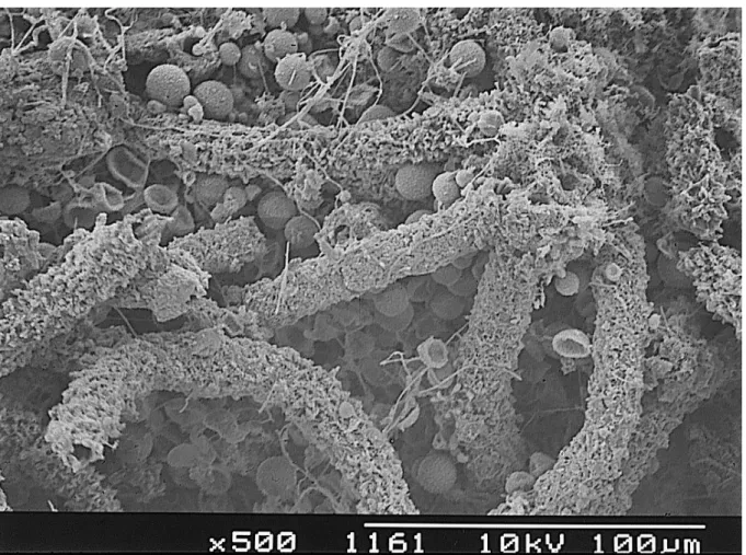
Scanning electron microscopy analysis ... 158
5.3.2.3. Statistical Analysis ... 160
5.4. Discussion... 162
5.4.1. Lithotypes characteristics ... 162
5.4.2. Stone bioreceptivity experiment... 165
5.5. Conclusions... 172
CHAPTER SIX Appraisal of endolithic growth and its biodeteriorating potential on limestones... 177
6.1. Introduction ... 177
6.2. Materials and methods ... 178
6.2.1. Investigated stone materials ... 178
6.2.2. Analysis of endolithic growth by microscopy techniques ... 179
6.2.2.1. Scanning electron microscopy ... 179
6.2.2.2. Confocal laser scanning microscopy ... 179
6.2.3. Biocalcarenite mineralogical and micromorphological characterisation... 180
6.2.3.1. X-ray diffraction... 180
6.2.3.2. SEM-EDS analysis ... 180
6.3. Results ... 181
6.3.1. Lithotypes colonisation by endolithic biofilms ... 181
6.3.2. Biocalcarenite mineralogical and micromorphological characteristics ... 188
6.3.2.1. X-ray diffraction... 188
6.3.2.2. SEM-EDS analysis ... 189
6.4. Discussion... 193
Concluding remarks ... 199 7.1. Conclusions ... 199 7.2. Further perspectives ... 205 RESUMO ... 209 REFERENCES ... 221 APPENDICES Appendix one... 251 Appendix two ... 253 Appendix three... 257
Fig.1.1. Geological map of Western Europe... 5
Fig. 1.2. SEM image showing green algae cells (arrows 1) adhered to Ançã limestone (arrow 2) and embedded in matrix of extracellular polymeric substances (arrow 3). ... 13
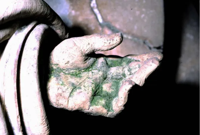
Fig. 1.3. Stone statue from Queluz National Palace (Portugal) showing green biofilms... 17
Fig. 1.4. SEM image of Scytomena julianum mat. The calcified sheaths are composed of triradiate calcite crystals. ... 20
Fig. 1.5. Confocal microscope image of filaments of Nostoc punctiforme in the vegetative growth state (biofilm sample come from Nerja Cave, Málaga, Spain). Pigment fluorescence is shown as red colour, polysaccharides labelled with Con-A-AlexaFluor 488 as green colour, and nucleic acids stained with Hoechst 33258 as blue colour. ... 29
Fig. 1.6. Polymerase chain reaction (PCR).. ... 31
Fig. 1.7. Principle of DGGE.. ... 32
Fig. 1.8. Molecular cloning using a plasmid vector. ... 34
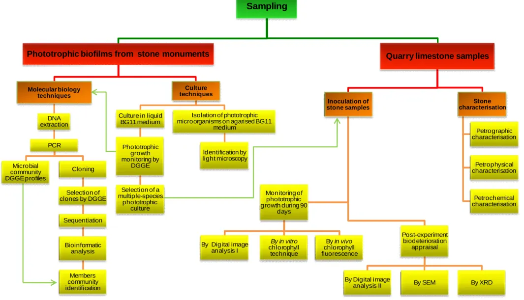
Fig. 1.9. Diagram of the experimental work performed to study the primary bioreceptivity of five different limestone types... 45
Fig. 2.1. Percentage of lithotypes present in the monuments, statues and historic buildings reported in this study. ... 50
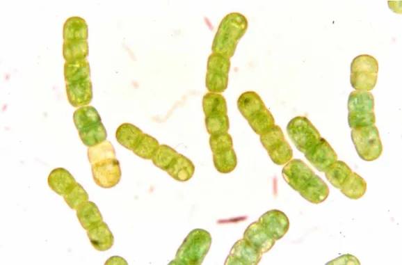
Fig. 2.2. Gloeocapsa sp. observed under optical microscope (1000x). ... 51
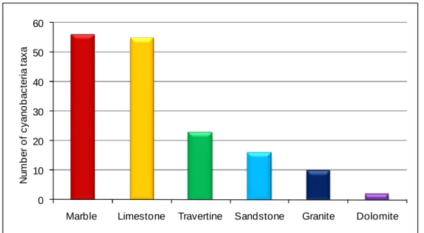
Fig. 2.3. Number of cyanobacteria taxa found on each lithotype... 52
Fig. 2.4. Klebsormidium flaccidum biofilm on a terracotta statue, Cathedral of Seville. ... 54
Fig. 2.5. Light microscope micrograph of trichomes of Borzia periklei, composed of 4-8 cells. ... 55
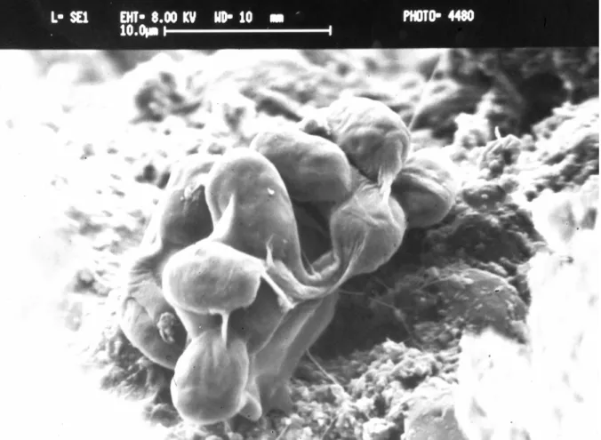
Fig. 2.6. SEM micrograph of a biofilm induced in vitro on sandstone. Gloeothece cells and sheath... 56
Fig. 2.7. Number of chlorophyte taxa found on each lithotype... 61
Fig. 3.1. View of the outside limestone monuments studied... 69
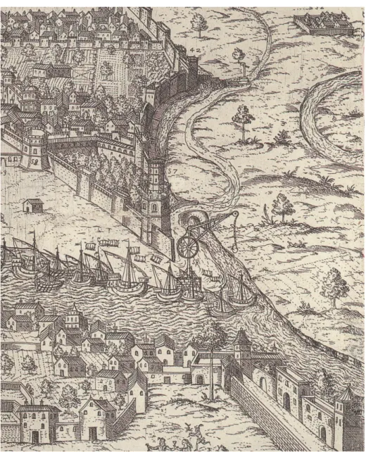
Fig. 3.2. 17th century engraving depicting the landing in Seville of San Cristobal stone from El Puerto de Santa María (Cadiz) with the help of a crane...71
cloning site. ... 77
Fig. 3.4. Biofilms cultivation in BG11 liquid medium. ... 79 Fig. 3.5. DGGE profiles of 16S rRNA gene from the natural green biofilm collected
from Orologio Tower, in Martano (natural sample), obtained after amplification using bacterial and cyanobacterial primers (Lanes “bact” and “cya”, respectively)... 86
Fig.3.6. DGGE profiles of 16S rRNA gene from the natural green biofilm collected
from Santa Clara-a-Velha Monastery (natural sample) and its liquid culture monitored during 90 days (Lanes 15, 30, 45, 60, 75 and 90, corresponding to the sampling days).. ... 87
Fig. 3.7. DGGE profiles of 16S rRNA gene from the natural green biofilm collected
from Ajuda National Palace (natural sample) and its liquid culture monitored during 90 days (Lanes 15, 30, 45, 60, 75 and 90, corresponding to the sampling days).... 88
Fig. 3.8. DGGE profiles of 16S rRNA gene from the natural green biofilm collected
from Cathedral of Seville (natural sample) and its liquid culture monitored during 90 days (Lanes 15, 30, 45, 60, 75 and 90, corresponding to the sampling days).. ... 89
Fig. 3.9. DGGE profiles of 16S rRNA gene from the natural green biofilm collected
from Cathedral of Granada (natural sample) and its liquid culture monitored during 90 days (Lanes 15, 30, 45, 60, 75 and 90, corresponding to the sampling days) ... ..90
Fig. 4.1. Scheme of the laboratory chamber used to assess the ability of phototrophic
communities to colonise stone samples: 1 – Phototrophic liquid culture; 2 – Silicon tubing; 3 – Automatic pumps; 4 – Sprinklers; 5 – Outlet valve device; 6 – Control unit; 7 – Stone samples. ... 104
Fig. 4.2. Abundance graphical representation of the different microbial groups
detected in the 16S rDNA clone libraries from Santa Clara-a-Velha Monastery (Coimbra, Portugal) green biofilm ... 107
Fig. 4.3. Abundance graphical representation of the green algae and fungi types
detected in the 18S rDNA clone libraries from Santa Clara-a-Velha Monastery (Coimbra, Portugal) green biofilm ... 107
Fig. 4.4c. Light microscope micrograph of Leptolyngbya sp. ... 108
Fig. 4.4d. Light microscope micrograph of Leptolyngbya sp. ... 108
Fig. 4.4e. Light microscope micrograph of Pleurocapsa sp. ... 109
Fig. 4.4f. Light microscope micrograph of Pleurocapsa sp... 109
Fig. 4.5. DGGE fingerprints using cyanobacterial-specific primers of the isolated phototrophic strains identified by 16S rDNA amplification, DGGE, construction of clone libraries and sequence analyses. Accession numbers and similarity (%) of the closest related database entries are given between brackets... 109
Fig. 4.6. Distribution pattern of the phototrophic biofilms developed on Ançã limestone samples during 90 days in a laboratory chamber. ... 110
Fig. 4.7. DGGE fingerprints using cyanobacterial-specific primers from samples of artificially colonising biofilms on Ançã limestone incubated in a laboratory chamber over three months (15, 30, 45, 60, 75, and 90 days). Lane I corresponds to the inoculum used to start the colonisation process of these stone samples. Lanes 15 to 90 represents the microbial communities’ profiles of this stone colonisation experiment at the indicated sampling time... 112
Fig. 5.1. One representative sample of each studied lithotype: CA (Ançã limestone), CL (Lioz limestone), SC (San Cristobal stone), PF (Escúzar stone) and PL (Lecce stone)... 119
Fig. 5.2. Scheme of the non-commercial incubation system: 1 – fluorescent lamp; 2 – Control unit; 3 – water pump; 4 – Silicon tubing for water circulation; 5 – Plastic grid; 6 – Stone samples. ... 130
Fig. 5.3. Spectrofluorometer (SPEX Fluorolog-3 FL3-22) fitted with a fibre-optic platform (Horiba Jobin Yvon F-3000) from Departamento de Química, Faculdade de Ciências e Tecnologia, Universidade Nova de Lisboa (Portugal). ... 132
Fig. 5.4. Multi-layer file of Ançã limestone samples (CA), elaborated by Adobe Photoshop© software. Adapted from Gomes (2008). ... 133
Fig. 5.5. One representative sample of each lithotype (CA – Ançã limestone, CL – Lioz limestone, SC – San Cristobal stone, PF – Escúzar stone, and PL – Lecce stone) selected for the stone bioreceptivity experiment. ... 138
Fig. 5.6. Photomicrograph of a thin section of CA lithotype (crossed polars). ... 139
Fig. 5.9. Photomicrograph of a thin section of PF lithotype (crossed polars). ... 139 Fig. 5.10. Photomicrograph of a thin section of PL lithotype (crossed polars). ... 139 Fig. 5.11. Open porosity of the studied lithotypes: CA (Ançã limestone), CL (Lioz
limestone), SC (San Cristobal stone), PF (Escúzar stone) and PL (Lecce stone). The values correspond to average ± SD for 27 samples of each lithotype... 140
Fig. 5.12. Representative kinetics of water absorption by capillarity of the studied
lithotypes: CA (Ançã limestone), CL (Lioz limestone) with enlarged inset, SC (San
Cristobal stone), PF (Escúzar stone) and PL (Lecce stone)... 141 Fig. 5.13. Visual impression (left image) and computed 3D surface topography (right
image) of one representative measured area of Ançã limestone (CA)... 144
Fig. 5.14. Visual impression (left image) and computed 3D surface topography (right
image) of one representative measured area of Lioz limestone (CL)... 144
Fig. 5.15. Visual impression (left image) and computed 3D surface topography (right
image) of one representative measured area of San Cristobal stone (SC). ... 144
Fig. 5.16. Visual impression (left image) and computed 3D surface topography (right
image) of one representative measured area of Escúzar stone (PF)...145
Fig. 5.17. Visual impression (left image) and computed 3D surface topography (right
image) of one representative measured area of Lecce stone (PL)... 145
Fig. 5.18. Upper view of one representative sample of each lithotype at the
inoculation time and after 30, 60 and 90 days of incubation... 148
Fig. 5.19. Lateral views of the SC and PF samples depicting green colour
appearance. ... 149
Fig. 5.20. Maximum concentration values of chla (µg.cm-3) present on the stone samples of CA, CL, SC, PF and PL lithotypes, measured by the in vitro chla quantification technique after 30, 60 and 90 days of incubation... 150
Fig. 5.21. Intensities of chla fluorescence obtained for each lithotype (excitation
wavelength: 430 nm): A) Fluorescence intensity values of chla at 684 nm measured immediately after inoculation (initial fluorescence), and after 30, 60 and 90 days of incubation. Each column corresponds to the mean value of an average of 15 measurements ± SD. B) Chla fluorescence spectra measured after 90 days of incubation. Each lithotype spectrum is an average of 15 spectra... 152
inoculation; B) After 45 days incubation; C) After 90 days incubation. The detected particles are then measured using ImageJ software... 154
Fig. 5.23. Sum of the stone surface areas covered by the biofilms, quantified by
digital image analysis... 154
Fig. 5.24. CA sample micrograph (left) and false-colour image obtained by Principal
Component Analysis (PC1, PC2, PC3) (right). The thin blue band with occasional occupations in the empty spaces is the photosynthetic biofilm. ... 156
Fig. 5.25. CL sample micrograph (left) and false-colour image obtained by Principal
Component Analysis (PC1, PC2, PC3) (right), showing the biofilm as a thin blue band at the top of the image. ... 156
Fig. 5.26. SC sample micrograph (left) and false-colour image obtained by Principal
Component Analysis (PC1, PC3, PC3) (right). The biofilm is presented as a blue band inside the stone sample. ... 157
Fig. 5.27. PF sample micrograph (left) and false-colour image obtained by Principal
Component Analysis (PC1, PC3, PC3) (right), enhancing the extent of the endolithic biofilm (blue band). ... 157
Fig. 5.28. PL sample micrograph (left) and false-colour image obtained by Principal
Component Analysis (PC1, PC3, PC3) (right). The biofilm is represented in dark red to black. ... 157
Fig. 5.29. Bands corresponding to the PC1, PC2 and PC3 of the original image of PL
used in this work. ... 158
Fig. 5.30. SEM images of transversally cut fresh samples of CA lithotype after 90
days-incubation: a) phototrophic biofilm in external view; b) and c) details of image 5.30a. White arrows indicate EPS; empty arrows show mineral substratum; black arrows indicate microbial cells. ... 159
Fig. 5.31. SEM images of transversally cut samples of SC lithotype after 90
days-incubation: a) phototrophic biofilm in external view; b) and c) details of image 5.31a. White arrows indicate EPS; empty arrows show mineral substratum; black arrows indicate microbial cells... 159
Fig. 5.32. SEM images of transversally cut samples of PF lithotype after 90
days-incubation: a) phototrophic biofilm in external view; b) and c) details of image 5.32a. White arrows indicate EPS; empty arrows show mineral substratum; black arrows indicate microbial cells... 159
incubation: a) phototrophic biofilm in external view; b) and c) details of image 5.33a. White arrows indicate EPS; empty arrows show mineral substratum; black arrows indicate microbial cells. ... 160
Fig. 5.34. Correlation among the variables capillarity coefficient (Capillarity), surface
roughness (Ra and Rz), open porosity (Porosity), water vapour permeability
(Permeability), concentration of chla (Chlorophyll) and chla fluorescence (Fluorescence), and the factors (components)... 161
Fig. 6.1. SEM-BSE image from PF lithotype depicting an interaction result between
the inoculated phototrophic microorganisms and the substratum. It is particularly noticeable the endolithic activity creating small cavities (white arrows) in the calcite crystals. ... 182
Fig. 6.2. SEM-BSE images of phototrophic microorganisms in PF samples
illustrating: A) epilithic microorganisms randomly scattered on the stone surface and the likely depths endolithic cells penetrating the stone through the accessible void spaces; B) detail from 6.2A showing compact cell aggregates of the endolithic colonisation; C) distribution map of Ca showing the dissolutions features of the calcite in the endolithic microhabitat analysed by EDS microanalytical Line-Scan analysis. ... 182
Fig. 6.3. SEM-BSE images from PF samples depicting: A) grains with cubic
crystalline habit forming concentric or dispersed aggregates on the stone surface; and B) in the interior; C) enlarged view from image 6.3B. ... 183
Fig. 6.4. SEM-BSE images from SC samples showing randomly scattered cells within
the pores of the calcarenite together with calcite grains... 184
Fig. 6.5. SEM-BSE images from SC samples showing: A) cubic crystalline
aggregates within the calcite grains; B) close view of the cubic crystalline aggregates. ... 184
Fig. 6.6. SEM-BSE images from PL samples showing: A) the epilithic microbial
colonisation, in which cells are distributed on the surface of the sample or very close to the surface; B) close view of the microbial cells showing disaggregation of the calcite grains. ... 185
Fig. 6.7. Three-channel maximum intensity projection of pigment fluorescence of
unicellular phototrophic microorganisms in SC sample. It consists of a two-dimensional compound image of x-y optical selected section of acquisition. ... 186
selected fragment of the PF sample. ... 187
Fig. 6.9. Three-dimensional extended focus projections in x-y, x-z and y-z views of a
selected fragment of the PL sample. ... 187
Fig. 6.10. A) SEM image of non resin-cast samples of PF lithotype showing a biofilm
deposition with Fe and Mn in its composition; B) EDS spectrum performed for this sample area. ... 190
Fig. 6.11. A) SEM image of a Mn oxide and clay minerals coating a calcareous (Ca)
fissure in the PF lithotype; b) EDS spectrum obtained in position 1 from image 6.11A. ... 191
Fig. 6.12. SEM image of colonised PF lithotype presenting needle-like shaped
goethite and very small iron-rich spherical particles, mostly likely ferrihydrite. ... 192
Fig. 6.13. SEM images of PF lithotype showing: A) calcite crystals coated by
iron-oxyhydroxides and a globular aggregate of calcite grains surrounded by an iron-film; B) one globular aggregate of calcite grains with iron-rich coating and other porous round particle, likely biogeneous... 192
Table 2.1. Investigated monuments, statues and historic buildings in European
countries from the Mediterranean Basin, and their bibliographic references. ... 57
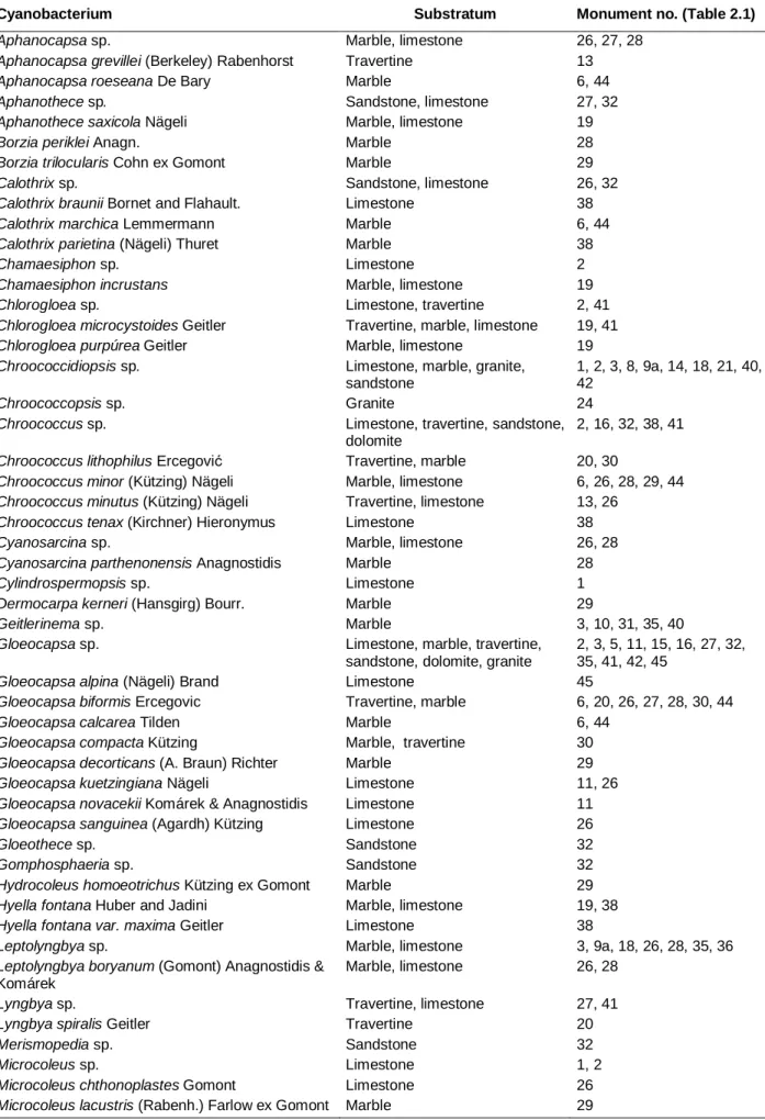
Table 2.2. Cyanobacteria reported on stone monuments, statues and historic
buildings in European countries from the Mediterranean Basin, on different substrata. ... 58
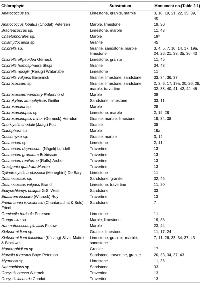
Table 2.3. Chlorophyta reported on stone monuments, statues and historic buildings
in European countries from the Mediterranean Basin, on different substrata. ... 62
Table 3.1. Sampling areas and biofilm descriptions. ... 73 Table 3.2. Primers used in this study. ... 75 Table 3.3. Occurrence of prokaryotic microorganisms in the green biofilms of the
studied monuments, detected by amplification using bacterial and cyanobacterial primers... 83
Table 4.1. Phylogenetic affiliations of eukaryotic microorganisms derived from the
natural green biofilm of Santa Clara-a-Velha Monastery (Coimbra, Portugal). ... 106
Table 5.1. Quarry, location and monument of the five studied lithotypes. ... 122 Table 5.2. Major physical and chemical characteristics of the five lithotypes, together
with the ANOVA results of open porosity (n = 27), capillarity water absorption coefficient (n = 27), water vapour permeability (n = 3), surface roughness (Ra and Rz)
(n = 3 for each parameter) and stone suspension pH (n = 3). ... 143
Table 5.3. Chemical analysis in weight percent of the five lithotypes. Values are
average ± SD (n = 3). ... 146
Table 5.4. ANOVA results obtained for the concentration of chla present on the
studied lithotypes after 30, 60 and 90 days of incubation. ... 151
Table 5.5. Intensity of chla fluorescence values measured at 684 nm together with
ANOVA results obtained for the five studied lithotypes after 30, 60 and 90 days of incubation. ... 153
C
HAPTER ONE
General introduction
CHAPTER ONE
General introduction
1.1. State of the art
1.1.1. Limestones as building materials
The Mediterranean Basin countries are often recognised as the cradle of some of the world’s most important cultural heritage in stone. The wide distribution of stone monuments and lithic works of art, and their cultural, artistic and religious importance emphasise the general need to safeguard this praiseworthy cultural heritage.
Stone materials have been used since the beginning of mankind for convenience, endurance and visual impact. Their selection for construction purposes has been invariably driven by questions of durability, availability, workability, cost and appearance. Despite the bewildering variety of stone types, carbonate rocks have been preferentially used as construction material in the architectural heritage of Mediterranean Basin region. Among them, limestones were prised for their attractive appearance, ease of quarrying, workability and exceedingly distribution across the Earth’s surface.
Limestones are sedimentary rocks that contain 50% or more of calcium carbonate (CaCO3) and possibly aragonite (CaCO3); the essential carbonate
minerals are calcite (CaCO3), aragonite (CaCO3) and dolomite (CaMg(CO3)2)
(Chilingar et al., 1967). Aragonite is a polymorph of calcite, unstable at normal surface temperatures and pressures, which may change into calcite. Dolomite is calcium magnesium carbonate intimately associated with calcite since it is in most cases a replacement of calcite or aragonite. On the basis of chemical composition, particularly, Ca/Mg (weight) ratios, Chilingar et al. (1967) proposed the classification of limestones into the following types: highly dolomitic limestone, when the range in
Ca/Mg ratio is 4.74-16; dolomitic limestone when Ca/Mg ratio is between 16-60; slightly dolomitic limestone, when the range in Ca/Mg ratio is 60-105, and calcitic limestone when the Ca/Mg ratio is superior than 105. Although some limestones are very pure consisting almost wholly of one or more carbonate minerals, they may contain other minerals. The most common include silica (commonly quartz sand), silicates and clay. If present in amounts greater than 10% the limestone rock may be termed as siliceous limestone or argillaceous limestone. A system based on grain size is widely used recognising three categories: calcilutite (grains <62 µm), calcarenite (62 µm to 2 mm) and calcirudite (>2 mm) (Grabau, 1904, 1913; Powers, 1962). Other classification systems of carbonate rocks are currently used, which are based on the textural components (Leighton and Pendexter, 1962; Folk, 1962) and depositional texture (Dunham, 1962; Embry and Klovan, 1971).
About 75% of the Earth’s surface is formed by sedimentary rocks, in which argillaceous rocks are the most abundant, occupying about 45% of the area; sandstones form about 30% and limestones about 28% (Dimes, 1998). Due to the availability of rocks and transport facilities, stonemasons trended to use the local stone materials for building construction. Thus, it is not surprising that memorials of our heritage are build of sedimentary rocks, being limestones and sandstones the predominant stone types used. Figure 1.1 depicts the geologic map of Western Europe, evidencing the presence of rocks from every age and almost every kind of rock. In the Southern Europe countries, especially in Spain and Italy, the broad distribution of tertiary sedimentary rocks explains the wide use of limestones in their architectural heritage. Great stages of geologic evolution are also setting in central Portugal, where extent layers of Cretaceous and Jurassic are quarried. In the north region of Lisbon, between Lameiras and Negrais, crops out one of the most traditional ornamental Portuguese stones, locally known as Lioz limestone (beige-white limestone). This rock is a Cretaceous fossiliferous limestone which owes its high value to its vivid colours and well known durability (Figueiredo et al., 2007). In the proximity of Coimbra (central Portugal), set in the northern branch of the Lusitanian Basin, layers dating from the Bajocian (Middle-Jurassic) are quarried for the Ançã limestone: an oolitic rock widely used in well known monuments in the region due to its workability and light colour.
In what concerns the geology of Spain, a remarkable stone diversity is observed. It includes one of the most complete Palaeozoic sedimentary successions in Europe. In the North and West of the Peninsula, siliceous formations are seated on hard and durable crystalline rocks (granites, schist and gneiss) forming acid soils and poor in carbonates. Sedimentary rocks based on limestones and marls (argillaceous limestones) are seated on the heavily weathered Meseta (central Spain) that form the major agricultural zones. It constitutes the oldest and most complex geologic formation of the Iberian Peninsula. Some of the most remarkable Spanish cultural heritage built in limestone is found in Andalusia, where huge amounts of upper Miocene formations, such as San Cristobal and Escúzar stones, were quarried for the construction of the cathedrals of Seville, Jerez, Granada and the walls of the Alhambra Palace, etc.
Great creations like magnificent buildings or beautiful temples can also be admired in Italy, which has a rich geology from erupting volcanoes, with all kind of volcanic rocks, to sedimentary rocks seated in the Salento Peninsula (South-eastern Italy). In the historic city of Lecce (Southern Italy) crops out the city's main export,
Lecce stone, because it is very soft and malleable, thus suitable for sculptures. This
Tortonian (Miocene epoch) rock formation is extended throughout the Salentine Peninsula, particularly in the regions of Lecce, Corigliano d'Otranto, Melpignano, Cursi and Jerseys.
Portugal, Spain and Italy contain some of the best exposed outcrop geology in Europe, which provide huge amounts of limestone materials for construction purposes. Cretaceous and Jurassic formation outcrops from Portugal, and Miocene formations from Spain and Italy, provide some of the materials more frequently used in several important buildings that have been constructed in these countries. Unfortunately, we are confronted with some problems concerning their preservation. Physical and chemical weathering both in monument or in natural geological outcrops are observed, which induce stone disaggregation and decomposition resulting from material loss (Smith, 2003). Physical weathering can be caused specifically by salt crystallisation and freeze-thaw processes, as well as hydric, thermal and wet-dry cycling. Chemical weathering can essentially be understood as stone disintegration resulting from reactions induced on mineral constituents of rock by water, carbon dioxide and oxygen. Whereas rock weathering is essential for
pedogenesis, the deterioration of cultural heritage artefacts in stone represents an irretrievable loss to the history of mankind.
Deterioration of monument stones is a complex process influenced by the intrinsic properties of the stone materials (geological time, mineralogical composition, porosity, etc.) and by extrinsic factors, such as climatic conditions, atmospheric pollution and biological colonisation. Other factors, sometimes acting synergistically, include damage induced by quarrying, transport and stone preparation procedures, microclimatic conditions (affected by local urban geometry and building design), adjacent materials used, such as inappropriate jointing material and metal elements, etc. Unfortunately, there are even more factors contributing to stone deterioration: man destructive actions, lack of maintenance, and inappropriate conservation works carried out in the building. These factors are determined by the economical situation and lack of the knowledge and experience in the conservation of monument stones.
Limestones have been intensely studied in conservation, largely because they have been used in buildings and statuary all over the world and because they are mineralogically quite simple (Sabbioni, 2003). Several researches have studied the deterioration processes occurring on limestone surfaces (Maurício and Figueiredo, 2000; Maurício et al., 2005; Dionísio, 2007; Figueiredo et al., 2007). In many dense limestones, the rate of deterioration may be gradual and, given climatic conditions, largely predictable. However, there are many commonly limestone types which do not decay gradually, but instead experience episodic and sometimes catastrophic breakdown, especially in polluted urban environments. The increase of atmospheric contamination undoubtedly accelerates stone deterioration, especially on limestones where loss is primarily related to dissolution of calcium carbonate induced by the solvent action of acid rainwater. Its penetration into the pores hastens the rate of stone deterioration tremendously, first because water is itself an effective deteriorating agent, dissolving, hydrating and hydrolysing minerals, and, secondly because it holds in solution substances (carbonaceous particles, sulphur compounds, soluble salts) responsible for leaching of surfaces, pH reduction, dark crusts, efflorescences and subflorescences (Bell, 1993b; Camuffo, 1995; Ordóñez et al., 1997; Papida et al., 2000). In addition, the absorption of ground water through capillary rise transport organic acids which may also dissolve the sparingly soluble calcium carbonate (Drever and Stillings, 1997).
The problem of understanding the deterioration of limestones is compounded by the large range of stone varieties, with different textural, mineralogical and physical characteristics, and by their varying weathering responses under different climatic and environmental conditions. The interactions between these numerous and synergistically acting factors lead to a dynamic and complex process of physical, chemical and biological deterioration. Laboratory studies have demonstrated the combined process of physical, chemical and biological factors on the deterioration of stone (Badalyan et al., 1996; Papida et al., 2000). However, little research has been focused on the problem of microorganisms as agents of stone deterioration (biodeterioration). This study represents a multidisciplinary work focused on the biodeterioration susceptibility of different limestone types widely used in Portuguese, Spanish and Italian monuments.
1.1.2. Biodeterioration of stone: definitions and history
Within the last few years it has been widely accepted that rocks, either in natural geological outcrops or in stone monuments, are common habitats for a wide variety of microorganisms, such as bacteria, cyanobacteria, actinomycetes, algae, fungi and lichens. Krumbien (1983, 1988) studied the interactions between these microorganisms and the lithic substrata, reporting that microorganisms play an important and substantial role in all rock alteration processes. Aesthetical, physical and chemical changes are induced on the lithic substrata, where the formation of coloured patinas are visibly noticed (Urzì et al., 1991; Gorbushina et al., 1993; May et al., 1993; Sterflinger and Krumbein, 1997). In some natural environments, chemical and physical transformations in the materials can be seen as a necessary and positive process. A very practical example is the essential biotransfer process of rocks for soil formation (pedogenesis). However, when a rock is used as building material, these transformations induced by stone colonising microflora in association with other environmental agents are clearly seen as a negative or destructive process, both from cultural and economical viewpoints. Therefore, the comparison of the concepts “biodegradation” and “biodeterioration” leads to a consideration of the manifold relations between organisms and materials and to a systematisation of these fields of science (Hueck, 2001).
The term biodeterioration was defined about 45 years ago by Hueck (1965) as “any undesirable change in the properties of a material caused by the vital activities
of living organisms”. The term biodegradation is also of common use as a
synonymous of biodeterioration. However, the definition of biodegradation with a negative, detrimental connotation is unjustified, according to Krumbein (1988). Biodegradation involves a positive or useful connotation in relation to ecology, waste management and environmental remediation. This concept is commonly used for the combined effects of erosion and weathering, i.e. the wearing down of the Earth’s surface by physical, chemical and biological action, essential for pedogenesis (Krumbein, 1988). In geological nomenclature, weathering is the decomposition process of rocks, minerals and soils through direct contact with the atmosphere, and is a necessary process in most cases.
For a detailed historic review of these and related terms it is recommended the following literature: Krumbein (1966, 1988), Krumbein et al. (2003) and Gorbushina and Krumbein (2005).
The main processes of stone biodeterioration have gone through interesting theories related to the biogenic attack. For a long time, microbial effects on stones were described as physical, chemical or pollution derived processes. When considering the physical and chemical damage of a given monument, only stone characteristics and abiotic factors (e.g. rain, wind, relative humidity, sunlight, temperature, pollution, etc.) were taken into consideration. The research carried out by Krumbein (1966, 1988) demonstrated the catalytic power of rock biota in the natural process of rock decay, and the role of microorganisms in the partially or completely deterioration of stone materials used in cultural heritage assets. After recognition of the biological origin on the alteration of stone, an increasing awareness on the manifold aspects of stone biodeterioration yielded. A considerable number of investigations have begun to elucidate the role of microorganisms in the deterioration of building stone (Griffin et al., 1991; Bock and Sand, 1993; May et al., 1993; Koestler et al., 1996; Wakefield and Jones, 1998; Saiz-Jimenez, 1995, 1999). However, initial researches considered biodeterioration as a secondary deterioration problem, occurring only after an advanced state of deterioration predetermined by physical and chemical parameters (Griffin et al., 1991). In other words, abiotic factors were thought to condition stone surfaces exposed outdoors through fissure formations and enrichment of inorganic and organic nutrients. When the surface of
the lithic substratum had undergone this process of alteration, living organisms colonised the surface area. Furthermore, the effects of stone biodeterioration were thought to induce only an aesthetic problem, typically seen as an unacceptable appearance of staining of the stone surfaces by biogenic pigments (Urzì et al., 1991). Recently, several analytical approaches demonstrated that even in the early stages of stone exposure, primary biodeteriorating effects can be clearly defined (Koestler et al., 1996; Gaylarde and Morton, 1999). These studies also demonstrated that microorganisms are involved in the physical and chemical deterioration of stones (Saiz-Jimenez, 1999; Dornieden et al., 2000; Warscheid and Braams, 2000), and the rate and extent of microbial colonisation are strongly influenced by environmental parameters, such as, water availability, climatic conditions, and by petrologic parameters (e.g. mineralogical composition, porosity, roughness).
From the above mentioned investigations, it is clear that the concept of stone biodeterioration is now seen in a more holistic notion in which the biological deterioration is controlled by synergism/antagonism relationships among the colonising species and environmental factors (Koestler et al., 1996; Wakefield and Jones, 1998; Warscheid and Braams, 2000). Thus, stone biodeterioration cannot be considered as an isolated phenomenon. It generally occurs with other physical, chemical, or physicochemical deterioration processes, being difficult to attribute damage specifically to a single cause. Moreover, although microorganisms colonise permanently various materials, their damaging activity is not constant, but rather periodic and defined by the variety of different conditions as a result of the habitat in which they dwell.
Detailed literature, summarised by Warscheid and Braams (2000), Krumbein (2004) and Gorbushina and Krumbein (2005), gives an idea about the early phases and the later opinions changing with new findings.
Stone monuments can be a paradigm of a complex environment in which microorganisms can be confronted as it encompasses heterogeneity and dynamic in time and space. This heterogeneity of most cultural heritage substrata is linked to the numerous interrelationships among various biological populations and between these populations and the substratum, as well as other processes occurring in the surrounding environment. Thus, the study of stone biodeterioration involves complex and multidisciplinary approaches.
Over the years, scientific approaches and conservation strategies of stone biodeterioration have evolved to reach a high level of sophistication into a still growing scientific community. Yearbooks and symposium publications (e.g. Hougthon and Eggins, 1988; Rossmoore, 1991; Saiz-Jimenez, 2003) have been useful sources, where biodeterioration, biofilms and bioreceptivity and, obviously strategies to avoid and eliminate biological colonisation are the central focus.
1.1.3. Colonisation of stone by phototrophic microorganisms
It is amply recognised that microbial communities are truly ubiquitous in aquatic and terrestrial ecosystems as well as on man-made materials, including cultural heritage assets exposed outdoors. A high microbial diversity in the form of bacteria, cyanobacteria, algae, fungi, as well as lichens can find a suitable habitat for their growth both on rock surfaces and on monument stones. Photoautotrophic microorganisms (green microalgae, cyanobacteria and lichens) are particularly damaging of stone surfaces due to their ability to survive in inhospitable environments. They can develop on stone surfaces without any presence of organic matter (Tiano, 1998). Thus, photoautotrophic microorganisms have the greatest ecological importance as pioneer organisms in the colonisation of stone materials (Bock and Sand, 1993; Gómez-Alarcón et al., 1995; Lamenti et al., 2000; Tomaselli et al., 2000 a,b; Bellinzoni et al., 2003). Several investigations demonstrated that different stone materials, such as limestones, granites, sandstones and marbles, can promote significant microbial growth under oligotrophic conditions (Guillitte and Dreesen, 1995; Tiano et al., 1995; Prieto and Silva, 2005; Miller et al., 2006). In fact, these microorganisms can grow using the mineral components of a stone and sunlight as energy source. Their phototrophic mode of life allows their exposition to visible solar radiation becoming the dominant primary producers of environments that may be considered extreme, such as hotsprings, deserts, alkali lakes, polar areas (Bell, 1993b; Friedmann, 1982; Büdel et al., 2004), as well as, cultural heritage assets such as vertical surfaces of monuments (Saiz-Jimenez et al., 1990; Ortega-Calvo et al., 1991a; Ariño et al., 1997; Tomaselli et al., 2000b; McNamara et al., 2006). These ecosystems may often be considered “extreme” because of their exposure to high temperature, sun radiation and drought for long periods of time and
also to wide temperature variations and high salt concentrations (Daffonchio et al., 2000).
Taxonomically, cyanobacteria are an evolutionarily coherent group of photoautotrophic microorganisms within the eubacteria (Wilmotte, 1994; Castenholz, 2001). This group constitutes the only phototrophic prokaryotes capable of carrying out oxygenevolving, plantlike photosynthesis, and shares a common ancestry with the chloroplasts of higher plants and green algae (Nelissen et al., 1995). They conduct photosynthesis on specialised infolded cytoplasmic thylakoid membranes, rather than in chloroplasts. They can be found in a single cell, colonies of unicellular organisms or organised in filaments. The pigments are spread in a membrane system at the periphery of the cells, which have a mucilaginous sheath, often highly pigmented (Graham and Wilcox, 2000). They contain chlorophyll a, carotenoids (yellow/orange pigments), phycocyanin (blue pigment) and phycoerythrin (red pigment).
Green microalgae or Chlorophyta, are a diverse group of oxygenic photoautotrophic microorganisms within the eukaryotes, containing therefore a nucleus enclosed within a membrane, and conduct photosynthesis within chloroplasts. They may be unicellular, colonial or multicellular but typically consist of a relatively small number of cells. Chlorophyll a and b and carotenoid pigments are present, which occur in plastids (Graham and Wilcox, 2000).
Cyanobacteria and microalgae develop on stone surfaces forming biofilms composed of a mono- or multilayer of cells embedded in a hydrated extracellular polymeric matrix which hold the cells together (Morton et al., 1998; Warscheid, 2000; Roldan et al., 2003). These glue-like extracellular polymeric substances (EPS) secreted by the cells enhance the attachment of the biofilm community, allowing the adhesion of microbial cells to the lithic substratum (Cecchi et al., 2000; Wimpenny et al., 2000). The attachment of microorganisms on stone surfaces is regulated biologically by the microbial cell structure and their surface charge; it is physically controlled by the stone surface, which partially determines the availability of water, nutrients, niche possibilities, and thus the survivability of the microorganisms. Preferentially, phototrophic microorganisms adhere to humid stone surfaces, on rough or porous surfaces, cracks and fissures, where water is retained and evaporation is slow due to protection against winds or direct sunshine. Inoculation is
cyanobacteria and algae can be brought in by wind and rain (Saiz-Jimenez, 1999; Alakomi et al., 2004). Subsequent formation of surface covering biofilms improves the living conditions for the phototrophic microflora. Their slimy surfaces favour the adherence of nutritive airborne particles (dust, pollen, spores, oil-and coal-fired carbonaceous particles) and microorganisms from the atmosphere (Saiz-Jimenez, 1995; Koestler et al., 1996; Warscheid, 2000).
The use of sophisticated techniques, consisting mainly of microscopy analyses, illustrates the composition, function and structure of biofilms. These biofilms are composed of population or communities of different microorganisms (microalgae, cyanobacteria, bacteria and fungi) immobilised on the stone surface (substratum) and frequently embedded in an organic polymer matrix formed by EPS (Fig. 1.2).
2
1
1 3
Fig. 1.2. SEM image showing green algae cells (arrows 1) adhered to Ançã limestone (arrow 2) and
EPS may vary in chemical and physical properties, but consist mainly of polysaccharides with fatty acids, proteins and enzymes (Krumbein and Urzì, 1991; Kawaguchi and Decho, 2002; Young et al., 2008). It may also contain exoenzymes and inorganic inclusions such as clay particles and minerals (Wilderer and Characklis, 1989). Confocal laser scanning microscopy (CLSM) studies revealed that EPS are organised into complex exopolymeric matrix, which consists of a heterogeneous distribution of cells and cellular aggregates with void spaces or water channels (Costerton et al., 1995; Kawaguchi and Decho, 2002). Zhang et al. (1998) pointed out that the major component in the biofilm matrix is water - up to 97%. Mazor et al. (1996) showed that addition of 0.5 mg of Microcoleus sp. EPS per gram of sand retained approximately 30% of the water-holding capacity of the sand after 24h of desiccation at 55ºC, while sand samples without EPS dried out completely. Direct visual observation, either with CLSM (Neu and Lawrence, 1999; Kawaguchi and Decho, 2002; Roldán et al., 2004), scanning electron microscopy (SEM) (Hernandéz-Mariné et al., 2004; De los Ríos and Ascaso, 2005) or Fluorescent In
Situ Hybridization (FISH) (Urzì and Albertano, 2001; Urzì et al., 2003), revealed the
high biodiversity and distribution of phototrophic biofilms on stone surfaces. The top layer of biofilms is typically dominated by phototrophic microorganisms, such as cyanobacteria, green algae and diatoms, while underlying layers are composed by anoxygenic bacteria, such as green and purple sulfur bacteria (Martinez-Alonso et al., 2005).
According to their distribution pattern, the microorganisms that inhabit the rocks have been classified as epilithic and endolithic (Golubic et al., 1981). Epilithic microorganisms grow on the external surface of the rock, whereas endolithic microorganisms live in the interior of rocks, penetrating some millimetres into the rock pore system. Golubic et al. (1981) proposed a terminology which divides endolithic organisms into three groups: chasmoendolithic (colonising fissures and cracks), cryptoendolithic (colonising structural cavities within porous rocks) and euendolithic (penetrating actively into the interior of rocks and forming tunnels). Specialised euendolithic cyanobacteria penetrate and colonise the interior of limestone and dolomite rocks as well as loose sand-size particles, shell hash and other skeletal fragments. Generally, endolithic colonisation is a successful strategy of survival when surface environmental conditions are adverse for life on stone. The endolithic microhabitat gives protection from intense solar radiation and desiccation, and it
provides mineral nutrients, moisture and growth surfaces (Walker et al., 2005). The protection provided by the rock leads to the abundance of endolithic microorganisms in extreme environments, as cold and hot deserts, semiarid lands and even polar regions (Friedmann, 1982; Bell, 1993b; Walker et al., 2005). Microscopy techniques have also shown that penetration of growing organisms into rock and the diffusion of their excreted products may arise to depths of several millimetres (Saiz-Jimenez, 1999; Koestler, 2000; Salvadori, 2000; Pohl and Schneider, 2002; Barberousse et al., 2006; Young et al., 2008). Pohl and Schneider (2002) applied computerised image analysis to detect and quantify the biomass and depth of penetration of endolithic microorganisms into carbonate rock surfaces. They observed that the investigated natural carbonate rocks were endolithically colonised by lichens, cyanobacteria, algae and fungi. Phototrophic microorganisms from intensely insolated dry sites retreated to depths of 150-250 µm below the rock surface and revealed “cushions” of EPS oriented toward the surface, which protect them against intense light and provide water retention. In addition, these authors demonstrated that as soon as the endolithic biofilm was established it exerted an overall protective effect on the carbonate rocks. Salvadori (2000) examined by SEM the endolithic communities inhabiting stone Italian monuments and observed that euendolithic cyanobacteria and fungi could easily penetrate the calcite crystals of marble.
It is important to stress that phototrophic biofilms occur on contact surfaces at the interface between the solid substratum and the atmosphere. Thus, they are constantly subjected to adverse environmental conditions, such as, intense solar radiation, desiccation, temperature, moisture fluctuations, lack of nutrients, etc. Hence, the metabolic activity of phototrophic biofilms centres on retention of large amounts of water into its structure, protecting the cells from desiccation, fluctuating environmental conditions, as well as biocides, ultraviolet radiation, and prolonging their vegetative life (Prakash et al., 2003; Gorbushina, 2007). This leads to species characteristic of very different habitats and ecological requirements. Due to the presence of EPS, the biofilm incorporates large amounts of water into its structure ensuring the maintenance of moisture by balancing changes in humidity and temperature, which permits cyanobacteria and algae to resist drought periods (Gómez-Alarcón et al., 1995; Saiz-Jimenez, 1999; Schumann et al., 2005).
Summarised literature on the history, composition, structure and function of biofilms is presented by Christensen (1989), Costerton et al. (1994, 1995), Sutherland (2001) and Donlan (2002).
A number of factors influence the settlement, growth, distribution and metabolic activity of phototrophic biofilms on stone monuments, including i) intrinsic stone properties, such as geological origin, chemical composition, pore space structure and surface conditions that strongly influence water availability, nutrients and niche possibilities; ii) environmental parameters (e.g. solar radiation, temperature, water regime, wind, atmospheric pollution, etc.) iii) specific microclimatic parameters (e.g. orientation, exposure to shadow, permanent capillary humidity, etc.); iv) building maintenance. Yet clearly, the diversity and abundance of green algae and cyanobacteria are dependent on the availability of water allowing microorganisms to form biofilms on virtually any surface (Gorbushina, 2007). Water availability is one of the most important factors since all organisms need water for their metabolim. Bellinzoni et al. (2003) reported that the water factor, and in particular rain, represented the main limiting factor for the growth of phototrophic microflora in travertine walls. The shading of trees in Northern exposed surfaces reduced water evaporation favouring microflora growth. Ortega-Calvo et al. (1992) studying green biofilms on building stones from Spain and Northern Europe, showed that green algae only became dominant after the improvement of water retention properties to stone surfaces by pioneering cyanobacteria. Nevertheless, the stone surface has also several characteristics that are important in the attachment and development of phototrophic microorganisms. The extent of microbial colonisation appears to increase as the surface roughness increases. This is because shear forces are diminished, and total surface area is higher on rougher surfaces (Morton et al., 1998; Donlan, 2002). Tomaselli et al. (2000b) showed that high values of porosity and surface roughness played a greater role than mineral composition in promoting microbial establishment. Hernández-Mariné et al. (2001) examined phototrophic biofilms coating the limestone surface of the Puigmoltó sinkhole located in the East cost of the Iberian Peninsula, demonstrating that biofilms developed according to the decreasing irradiance; in the upper part of the sinkhole a discontinuous light green biofilm was formed with large amounts of EPS, whereas below 5 m in depth the biofilm occurred in patches and very few organisms were able to grow along the gradient of conditions.