HFE mutations in patients with hereditary haemochromatosis
in Sweden
E . M . P. CA R D O S Oa , P. S TÅ Lb , K . H AG E Na , J. M . CA B E DAc , d , S. E S I Ne , M . D E S O U S Ad & R . H U LTC R A N T Za From the aDepartment of Gastroenterology and Hepatology, Karolinska Hospital, Stockholm; the bDepartment of Gastroenterology and Hepatology, Huddinge University Hospital, Huddinge; and the eMicrobiology and Tumourbiology Centre, Karolinska Institute, Stockholm; Sweden. cDepartment of Clinical Haematology, Santo António Hospital, and dDepartment of Molecular Pathology and Immunology, Abel Salazar Institute for Biomedical Sciences, Porto, Portugal
Abstract. Cardoso EMP, Stål P, Hagen K, Cabeda JM, Esin S, De Sousa M, Hultcrantz R (Karolinska Hospital, Stockholm; Huddinge University Hospital, Huddinge; and Karolinska Institute, Stockholm, Sweden; Santo António Hospital; Abel Salazar Institute for Biomedical Sciences, Porto, Portugal). HFE mutations in patients with hereditary haemochromatosis in Sweden. J Intern Med 1998; 243: 203–8.
Objective.To determine the frequency of mutations (C282Y and H63D) in a newly identified gene HFE in patients with hereditary haemochromatosis (HH) in Sweden.
Design.Molecular genetic analyses of the HFE gene (polymerase chain reaction (PCR) followed by enzyme restriction) were performed in genomic DNA from unrelated patients with a clinical diagnosis of HH and in healthy subjects.
Settings.Patients with HH treated with phlebotomies at Karolinska Hospital and Huddinge Hospital were analyzed.
Subjects. Eighty-seven unrelated patients with HH and 117 healthy controls.
Results.It was found that the HFE C282Y mutation occurs in 94.2% of chromosomes from patients with HH. Eighty patients (92.0%) were homo-zygous for the C282Y mutation and one was heterozygous. Three patients were heterozygous for both C282Y and H63D mutations. One patient was homozygous and one was heterozygous for the H63D mutation. One patient carried normal alleles. In healthy controls, the C282Y mutation occurred in nine subjects (7.7%), all of which were heterozygous. The H63D mutation was found in 28 control subjects, one of which was homo-zygous.
Conclusions. We found that the majority of patients with HH have the C282Y mutation in the HFE gene. The frequency of the H63D mu-tation was higher in controls than in patients with HH, although in chromosomes at risk the frequency of the H63D mutation was higher in patients.
Keywords: genotype, haemochromatosis, HFE, inherited disorders, iron.
Introduction
Hereditary haemochromatosis (HH) is considered to be one of the most common autosomal recessive dis-eases in white people [1]. HH is characterized by a defect in the still unknown mechanism(s) regulating iron absorption. As a result, patients with HH initially accumulate iron in the plasma transferrin pool and later in parenchymal organs, which may lead to organ damage, such as liver cirrhosis, diabetes, arthropathy, cardiac arrhythmias and hypogonadism.
Recently, a candidate gene for HH was cloned by Feder et al. [2]. The gene is located on chromosome 6, three megabases telomeric of the HLA region, and has an organization and nucleotide sequence similar to that of HLA class I genes. This gene is now called
HFE. The authors found two missense alterations in
the HFE gene. The most important mutation, which results in a cysteine–tyrosine substitution at amino acid 282 (C282Y), was present in 83% of the 178 patients tested. A second mutation, resulting in a his-tidine–aspartate substitution at position 63 (H63D),
E . M . P. CA R D O S O et al. 204
was reported to be increased in patients with HH het-erozygous for the C282Y mutation [2, 3].
Other studies in the USA [3, 4], Australia [5] and France [6] confirmed that the C282Y mutation is pre-sent in a high percentage of patients with HH. However, the frequency of mutation varies somewhat between these different reports. A group in Italy showed an even lower incidence (69%) of the C282Y mutation in chro-mosomes from patients with HH [7]. The objective of the present study was to determine the presence of these two mutations in patients with HH in Sweden.
Subjects and methods
SubjectsA total of 87 patients with HH were examined: 67 men with a mean age of 47 years (range 20–73 years) and 20 women with a mean age of 49 years (range 25–72 years). The diagnostic criteria used were high transferrin saturation (.60% in males and .50% in females) and increased ferritin levels . 300 mg L21(except in one 20-year-old patient) and
a liver biopsy with typical iron staining indicating pri-mary haemochromatosis according to Scheuer et al. [8]. All patients included in the study were, from a clinical, biochemical and histopathological point of view, homozygous [9]. Siblings to patients with HH in the study were excluded. Patients were treated weekly with phlebotomies of 450 mL (1 L of blood contains approximately 0.5 g of iron). Patients with other causes of iron overload, such as excessive intake of oral supplementation, were excluded and none had received more than two units of blood from transfu-sions. Alcohol consumption was less than 30 g per day in all patients except one, who consumed 60 g of alcohol per day. All patients were negative for anti-bodies to hepatitis C, tested using the recombinant immunoblot assay II (Chiron Corporation, Emeryville, CA, USA). Of the male patients with 5 g or less of iron stores (n5 11), one was a blood donor and one had ulcerative colitis. No explanation was available for the other subjects with little iron. DNA from 117 random healthy Swedish subjects was used as controls. The individual identity of these samples was not known.
Methods
Genomic DNA was isolated from blood using the QIAamp Blood Kit (QIAGEN, Germany). Two
por-tions of the HFE gene surrounding both mutapor-tions were amplified separately using the polymerase chain reaction (PCR) with HFE specific primers (for the C282Y mutation, forward primer: 59- GTG ACC TCT TCA GTG ACC and reverse primer: 59- AAT GAG GGG CTG ATC CAG; for the H63D mutation, forward primer: 59-ATG GGT GCC TCA GAG CAG and reverse primer: 59- AGT CCA GAA GTC AAC AGT). PCR was performed using the GeneAmp PCR system 9600 (Perkin-Elmer, Norwalk, CT, USA) in a final volume of 100 mL. The PCR mixture contained 1 3 buffer (10 mM Tris-HCl, 1.5 mM MgCl2, 50 mM KCl, pH 8.3),
0.2 mM of each dNTP, 0.3 mM of each primer, 2.5U of Taq DNA polymerase (Boehringer Mannheim, Germany) and 80–100 ng of DNA. PCR was carried out for 35 cycles (annealing at 628C and 508C for the C282Y and H63D mutation, respectively. The ampli-fied products (10 mL) were digested with Sna BI (BioLabs, New England, MA, USA) for the C282Y mutation or with Bcl I (BioLabs) for the H63D muta-tion, according to the manufacturer9s recommenda-tions. The digested products were then run on a 4% NuSieve 3:1 agarose (FMC BioProducts – Europe, Denmark) gel at 100 V for 1.5 h and photographed under UV light. The C282Y mutation creates a Sna BI restriction site. Thus, the 237-bp PCR reaction prod-uct digested with Sna BI shows two fragments of 197 and 40 bp in mutant DNA. In normal DNA, no Sna BI site is created and, therefore, only one fragment cor-responding to the 237-bp PCR product is visible in the gel (Fig. 1a). The H63D mutation removes the Bcl I site in the 210-bp PCR product, while in normal DNA Bcl I digestion results in two fragments of 129 and 81 bp (Fig. 1b).
Statistic analyses
The statistical significance of the data was deter-mined using the x2
-test. A P value of,0.05 was con-sidered significant.
Results
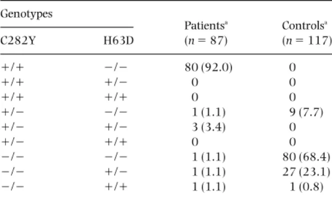
The HFE genotypes for the patients with HH and nor-mal subjects are summarized in Table 1. Of the patients, 92.0% were homozygous and 4.5% were heterozygous for the C282Y mutation. In three patients, wild-type alleles were present. No person homozygous for the C282Y mutation was found amongst the control subjects.
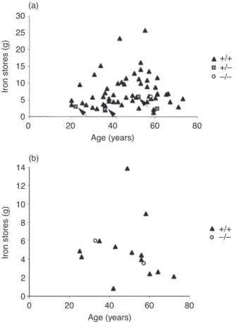
As can be seen in Fig. 2, heterozygous patients had lower iron stores in general than homozygous patients. Ten patients over 39 years old who were homozygous for the C282Y mutation had 5 g or less of iron stores. Of these, one patient was a blood donor and one had ulcerative colitis.
As shown in Table 2, the C282Y mutation fre-quency was higher in patients with HH than in con-trol subjects. The frequency of H63D was higher in the controls than in the patients with HH. However, because the C282Y mutation is in complete linkage disequilibrium with the H63D mutation, the only chromosomes at risk of the H63D mutation are those without the C282Y mutation. Thus, six of the 10 chromosomes at risk carried the H63D mutation, giving a frequency of 60% in patients with HH as compared with the controls where 29 of 225 chro-mosomes at risk carried the H63D mutation giving a frequency of 13% (P , 0.001).
The clinical data of patients with normal alleles for the C282Y are presented in Table 3. As can be seen they all showed many features typical of haemochro-matosis, both biochemical and clinical.
Discussion
The frequency of HH, which is a common genetic disease, differs in various parts of Europe and the
Fig. 1NuSieve gel electrophoresis of the polymerase chain reaction (PCR) fragments digested by enzyme restriction. (a) diagnosis of the C282Y mutation; (b) diagnosis of the H63D mutation. Band sizes (in bp) are shown on the lateral sides; The molecular weight marker (MWM) is the 100 bp ladder (Pharmacia Biotech, Sweden).
Table 1Analysis of mutations in the HFE gene in 87 patients with hereditary haemochromatosis and 117 normal subjects Genotypes Patientsa Controlsa C282Y H63D (n 5 87) (n 5 117) 1/1 2/2 80 (92.0) 00 1/1 1/2 00 00 1/1 1/1 00 00 1/2 2/2 01 (1.1) 09 (7.7) 1/2 1/2 03 (3.4) 00 1/2 1/1 00 00 2/2 2/2 01 (1.1) 80 (68.4) 2/2 1/2 01 (1.1) 27 (23.1) 2/2 1/1 01 (1.1) 01 (0.8) a
Number of subjects with the genotype. Parentheses denote percentage.
E . M . P. CA R D O S O et al. 206
USA [10–14]. Reports from Sweden have also shown variations in the prevalence of HH [15–20].
Two mutations in HFE have been described. The C282Y variant disrupts the interaction of the HFE chain with the b2-microglobulin and thereby
elimi-nates cell-surface presentation. The other mutation (H63D) does not interfere with the expression of the
HFE protein on the cell surface [21]. The role of the
H63D mutation in HH is still unclear.
In controls, we found no homozygous persons for the C282Y mutation, while 7.7% were heterozygous, giving an allelic frequency of 3.8% for the C282Y mutation. If this mutation was indeed alone respon-sible for HH, the prevalence for HH would be 0.16%. Our data correspond well with previously published frequencies on the prevalence of HH in the Stockholm area [19].
We found that C282Y mutation in approximately 94% of chromosomes from patients with HH, while only approximately 4% of chromosomes from the control population carried this mutation. It has been shown that the C282Y mutation of the HFE gene is present in a high percentage of white HH patients. However, the frequency varies between groups from different countries [2–7]. In Australia, the C282Y mutation was present in 100% of HH patients [5]. In contrast, only 69% of chromosomes from Italian patients with HH carried this mutation [7]. Our study confirms that the C282Y mutation is strongly associated with HH. One of the C282Y negative subjects was homozygous for the H63D
Table 2Mutation frequencies in the HFE gene in 87 patients with hereditary haemochromatosis and in 117 normal subjects
Patientsa Controlsa Mutation (n5 174) (n5 234) Pb C282Y 164 (94.2) 09 (3.8) ,0.001 H63D 006 (3.4) 29 (12.4) ,0.01 a
Number of chromosomes with mutation. Parentheses denote percentage.
b
x2
-test.
Table 3Clinical data on three out of 87 patients who did not carry the C282Y mutation
Genotypes Serum Serum Liver Related
Patient Age ALTa
Serum irona
transferrin ferritina
biopsy Removed Liver symptoms/
no. (years/sex) C282Y H63D (mkatal L21) (mmol mL21) saturationa
(%) (mg L21) iron gradeb
iron (g) histology relatives
1 33/F 2/2 2/2 0.36 40 89 0804 3 6.0 Fibrosis Arthritis 2 57/F 2/2 1/2 1.20 47 89 0746 3 3.6 Fibrosis Arthritis/ sibling with HH 3 58/M 2/2 1/1 1.26 44 93 1410 3 6.1 Fibrosis Diabetes a
Normal values: alanine transaminase (ALT): males ,0.8 mkatal L21, females ,0.6 mkatal L21; serum iron: 11–32 mmol mL21; serum
transferrin saturation: 10–60%; serum ferritin: 9–210 mg L21.
b
Hepatic iron overload grading according to Scheuer et al. [8].
80 30 0 0 Age (years) Iron stores (g) 20 25 15 5 10 20 40 60 +/+ +/Ð Ð/Ð (a) 80 14 0 0 Age (years) Iron stores (g) 10 12 8 4 6 20 40 60 +/+ Ð/Ð (b) 2
Fig 2Relationship between iron removed by phlebotomies and age at diagnosis in males (a) and in females with hereditary
haemochromatosis (HH) (b). s, negative for C282Y mutation; j, heterozygous for the mutation; m, homozygous patients. Patients in whom phlebotomy treatment had not been terminated are not represented. The arrows indicate heterozygosity for both C282Y and H63D mutations.
mutation. However, the importance of this homozy-gosity is not known, as one of the control subjects was also homozygous for this mutation. All patients in Table 3 had several clinical findings strongly indicating the diagnosis of HH, such as high transferrin saturation, associated symptoms, and in one patient, affected relatives. The fact that there are patients who do not have any of these two mutations suggests that additional mutations may be present either within the HLA region or on other, not yet detected, loci [2, 7]. In the present study we also found a similar frequency of the HFE C282Y mutation in Sweden, as compared with data from the USA, Australia and France, but a considerably higher frequency than that reported from Italy [7]. This also indicates that other factors than these mutations in the HFE gene may contribute to the expression of the disease. Our patients were all diagnosed with accepted criteria. Treatment showed that many had rather low iron levels. This has not been found in other studies. The explanation of this difference is not evident. One reason may be the often used health testing of people without symptoms of disease in Sweden, which recruits patients with only biochemical alterations, and probably a mild expression of the disease.
The role of the other mutation (H63D) in the phe-notypic expression of HH is unclear. In controls, the frequency of H63D mutation is higher than the C282Y mutation. If the H63D mutation had a criti-cal role in the development of HH, we would expect to find more homozygous patients for this mutation. However, amongst four patients heterozygous for the C282Y mutation, three were also heterozygous for the H63D mutation, compared with the controls where none of the individuals heterozygous for the C282Y mutation had the H63D mutation. This indi-cates that either this mutation is in linkage disequi-librium with other yet undiscovered mutations, or that the H63D itself is a deleterious mutation, which together with the C282Y mutation causes haemochromatosis. Because we found that of the 10 chromosomes at risk in the patients, six (60%) had the H63D mutation, this mutation may be important for the expression of the HH.
The finding of one HH patient negative for both mutations indicates that other not yet detected muta-tions may also be responsible for the phenotypic expression of the disease.
Acknowledgements
This study was supported by the Swedish Medical Research Council (no. 9127), the Swedish Society of Medicine, Ruth and Richard Julins Foundation and the Karolinska Institute. Miss Elsa Cardoso is a recipi-ent of a PRAXIS XXI Grant BD/5095/95.
The authors are very grateful to Dr Annika Lindblom, Department of Genetics, Karolinska Hospital, for providing the normal control DNA and to Ms Helena Åkerbrant for collecting patient DNA samples.
References
1 Crawford DHG, Powell LW, Halliday JW, Leggett BA. Factors influencing disease expression in haemochromatosis. Annu Rev Nutr 1996; 16: 139–60.
2 Feder JN, Gnirke A, Thomas W, Tsuchihashi Z, Ruddy DA, Basava A, et al. A novel MHC class I-like gene is mutated in patients with hereditary haemochromatosis. Nat Genet 1996;
13:399–408.
3 Beutler E, Gelbart T, West C, Lee P, Adams M, Blackstone R, et al. Mutation analysis in hereditary haemochromatosis. Blood Cells Mol Dis 1996; 22: 187–94.
4 Calandro L, Thorsen T, Barcellos L, Griggs J, Baer D, Sensabaugh GF. Mutation analysis in hereditary haemochro-matosis. Blood Cells Mol Dis 1996; 22: 194a–b.
5 Jazwinska EC, Cullen LM, Busfield F, Pyper WR, Webb SI, Powell LW. Haemochromatosis and HLA-H. Nat Genet 1996;
14:249–51.
6 Jouanolle AM, Gandon G, Jezequel P, Blayau M, Campion ML, Yaouang Y, et al. Haemochromatosis and HLA-H. Nat Genet 1996; 14: 251–2.
7 Carella M, D’Ambrosio L, Totaro A, Grifa A, Valentino MA, Piperno A, et al. Mutation analysis of HLA-H gene in Italian Haemochromatosis patients. Am J Hum Genet 1997; 60: 828–32.
8 Scheuer PJ, Williams R, Muir AR. Hepatic pathology in rela-tives of patients with haemochromatosis. J Pathol Bacteriol 1962; 84: 53–64.
9 Powell LW, Jazwinska E, Halliday JW. Primary iron overload. In: Brock JH, Halliday JW, Pippard MJ, Powell LW, eds. Iron Metabolism in Health & Disease. London: W. B. Saunders Company Ltd, 1994; 227–70.
10 Wiggers P, Dalhoj J, Kiaer H, Ring-Larsen H, Petersen PH, Blaabjerg O, Horder M. Screening for haemochromatosis: prevalence among Danish blood donors. J Intern Med 1991;
230:265–70.
11 Jonsson JJ, Johannesson GM, Sigfusson N, Magnusson B, Thjodleifsson B, Magnusson S. Prevalence of iron deficiency and iron overload in adult Icelandic population. J Clin Epidemiol 1991; 44: 1289–97.
12 Olsson KS, Marcell R, Ritter B, Olander B, Åkerblom A, Östergård H, Larsson O. Iron deficiency and iron overload in Swedish male adolescents. J Intern Med 1995; 237: 187–94. 13 Bell H, Thordal C, Raknerud N, Hansen T, Bosnes V, Halvorsen
R, et al. Prevalence of haemochromatosis among first-time and repeat blood donors in Norway. J Hepatol 1997; 26: 272–9.
E . M . P. CA R D O S O et al. 208
14 Edwards CQ, Griffen LM, Goldgar D, Drummond C, Skolnick MH, Kushner JP. Prevalence of haemochromatosis among 11,065 presumably healthy blood donors. N Engl J Med 1988;
318:1355–62.
15 Olsson KS, Heedman PA, Staugård F. Preclinical haemochro-matosis in a population on a high-iron-fortified diet. J Am Med Assoc 1978; 239: 1999–2000.
16 Olsson KS, Ritter B, Rosen U, Heedman PA, Staugård F. Prevalence of iron overload in Central Sweden. Acta Med Scand 1983; 213: 145–50.
17 Olsson KS, Eriksson K, Ritter B, Heedman PA. Screening for iron overload using transferrin saturation. Acta Med Scand 1984; 215: 105–12.
18 Lindmark B, Eriksson S. Regional differences in the idiopathic haemochromatosis gene frequency in Sweden. Acta Med Scand 1985; 218: 299–304.
19 Hallberg L, Björn-Rasmussen E, Jungner I. Prevalence of hereditary haemochromatosis in two Swedish urban areas. J Intern Med 1989; 225: 249–55.
20 Olsson KS. Prevalence of haemochromatosis in Scandinavia. In: Hallberg L, Asp NG, eds. Iron Nutrition in Health and Disease. London: John Libbey & Company Ltd, 1996; 273–7. 21 Feder JN, Tsuchihashi Z, Irrinki A, Lee VK, Mapa FA, Morikang
E, et al. The haemochromatosis founder mutation in HLA-H disrupts beta2-microglobulin interaction and cell surface
expression. J Biol Chem 1997; 272: 14025–8.
Received 10 June 1997; accepted 23 September 1997.
Correspondence: Dr Rolf Hultcrantz MD, Department of Gastroenterology and Hepatology, Karolinska Hospital, S-171 76