Case report
Atypical phenotype in two patients with
LAMA2
mutations
Joana Marques
a,⇑,1, Sofia T. Duarte
b,c,1, So´nia Costa
d, Sandra Jacinto
b,c, Jorge Oliveira
e,
Ma´rcia E. Oliveira
e, Rosa´rio Santos
e,f, Elsa Bronze-da-Rocha
f, Ana Rita Silvestre
g,
Eula´lia Calado
c, Teresinha Evangelista
b,gaServicßo de Neurologia, Instituto Portugueˆs de Oncologia de Lisboa, Francisco Gentil, Rua Professor Lima Basto, 1099-023 Lisboa, Portugal bInstituto de Medicina Molecular, Faculdade de Medicina da Universidade de Lisboa, Av. Professor Egas Moniz, 1649-028 Lisboa, Portugal cServicßo de Neurologia Pedia´trica, Hospital D. Estefaˆnia, Centro Hospitalar Lisboa Central, Rua Jacinta Marto, 1169-045 Lisboa, Portugal
dServicßo de Neurologia, Hospital Vila Franca de Xira, Estrada Nacional No. 1, Povos, 2600-009 Vila Franca de Xira, Portugal eUnidade de Gene´tica Molecular, Centro de Gene´tica Me´dica Dr. Jacinto Magalha˜es, Centro Hospitalar do Porto, Pracßa Pedro Nunes,
No. 88, 4099-028 Porto, Portugal
fDepartamento de Cieˆncias Biolo´gicas, Laborato´rio de Bioquı´mica, Faculdade de Farma´cia da Universidade do Porto, Rua de Jorge Viterbo Ferreira,
No. 228, 4050-313 Porto, Portugal
gLaborato´rio de Neuropatologia, Servicßo de Neurologia, Hospital de Santa Maria, CHLN, Av. Professor Egas Moniz, 1649-035 Lisboa, Portugal Received 18 September 2013; received in revised form 17 December 2013; accepted 9 January 2014
Abstract
Congenital muscular dystrophy type 1A is caused by mutations in theLAMA2 gene, which encodes thea2-chain of laminin. We report two patients with partial laminin-a2 deficiency and atypical phenotypes, one with almost exclusive central nervous system involvement (cognitive impairment and refractory epilepsy) and the second with marked cardiac dysfunction, rigid spine syndrome and limb-girdle weakness. Patients underwent clinical, histopathological, imaging and genetic studies. Both cases have two heterozygous LAMA2 variants sharing a potentially pathogenic missense mutation c.2461A>C (p.Thr821Pro) located in exon 18. Brain MRI was instrumental for the diagnosis, since muscular examination and motor achievements were normal in the first patient and there was a severe cardiac involvement in the second. The clinical phenotype of the patients is markedly different which could in part be explained by the different combination of mutations types (two missense versus a missense and a truncating mutation).
Ó2014 Elsevier B.V. All rights reserved.
Keywords: Laminina2-chain; Merosin; Cardiomyopathy; Epilepsy; Congenital muscular dystrophy 1A
1. Introduction
Mutations in LAMA2, encoding the a2-subunit of the extracellular matrix protein laminin, are normally associated with muscular dystrophy and white matter abnormalities of the central nervous system (CNS).
Classical congenital muscular dystrophy type 1A (MDC1A) is characterized by severe generalized muscle weakness, joint contractures and peripheral neuropathy
[1]. Primary partial laminin-a2 deficiency is associated with a less severe phenotype and with clinical and genetic heterogeneity[2–4].
In cardiac muscle, through its interactions with the
a2-chain of laminin 211, a-dystroglycan makes an important connection to the extracellular matrix, forming a link between the sarcolemma and the
http://dx.doi.org/10.1016/j.nmd.2014.01.004 0960-8966/Ó2014 Elsevier B.V. All rights reserved.
⇑Corresponding author. Tel.: +351 963113949; fax: +351 217229880.
E-mail address:[email protected](J. Marques). 1 Both authors contributed equally to this work.
www.elsevier.com/locate/nmd
ScienceDirect
extracellular matrix [5]. Symptomatic and subclinical cardiac involvement has been previously reported, notably in typical cases with absent laminin-a2 staining
[4,6,7]. Regarding partial defects, cardiac involvement
was documented only in one patient [8].
Previous studies indicate that laminin deficiency delays oligodendrocyte maturation through dysregulation of critical developmental signaling pathways[9]. Laminin-a2 binds to b1-integrin and is distributed punctately on cortical neuronal processes [10]. Cerebral white-matter changes are typical; cortical dysplasia and polymicrogyria are seldom seen. Most patients are cognitively normal, but epilepsy and mental retardation have been described[11].
We report two patients with atypical phenotypes for primary partial laminin-a2 deficiency. The first case is remarkable for the predominant CNS involvement with normal muscular strength and the second had an unusually severe cardiac involvement.
2. Case reports
2.1. Patient 1
This patient is the second child of healthy unrelated parents and he was born after an uneventful
pregnancy. Macrocephaly (3SD) was detected in the first year of life. Refractory epilepsy associated with progressive cognitive regression started by the age of 6, with atypical absences, myoclonic and atonic seizures. Physical exam revealed macrocephaly and bilateral divergent strabismus. Motor complaints were never reported. He had normal muscular strength, tone and deep tendon reflexes. His gait was normal, with preserved coordination, and he was able to run, climb stairs and ride his bike. Brain magnetic resonance imaging (MRI) showed an area of agyria in the occipital cortex. In T2-weighted images, extensive white matter abnormalities, swelling and widening of gyri, mainly frontal, were detected (Fig. 1A and B). Serial electroencephalograms (EEG) revealed bilateral multifocal paroxysmal activity, suggestive of symptomatic epilepsy (Fig. 1C). LAMA2 gene study was performed after extensive metabolic and genetic workup, given the occipital agyria and typical white matter abnormalities [12]. The highest creatine kinase (CK) value detected was 1589 UI/L. Electromyography (EMG), nerve conduction study and echocardiogram were unremarkable.
Epilepsy was initially controlled, but rapidly became refractory, with progressive cognitive impairment and
loss of expressive language. Motor function remains normal at age 21. Rufinamide significantly reduced the severity and frequency of atonic seizures.
2.2. Patient 2
This 55 year-old male patient was a single child of a healthy, non-consanguineous couple without family history of neuromuscular diseases. He presented delayed developmental motor milestones: he started walking after the age of 2 and always had difficulty running. By the age of 47 he sought medical attention, complaining of slowly progressive proximal muscle weakness and frequent falls, starting in his early forties. His neurological exam was notable for shoulder and pelvic girdle weakness, weak knee flexion and extension, and positive Gowers’ maneuver; muscular atrophy with scapular winging (Fig. 1H); lumbar hyperlordosis
(Fig. 1F) and dorsolumbar scoliosis. A rigid spine
syndrome was noted as well as multiple tendinous retractions (Fig. 1G). Brain MRI showed typical white matter abnormalities (Fig. 1D and E). CK values were 351 UI/L. EMG was consistent with a myopathic process. Echocardiogram showed impaired left ventricular contractility and mitral valve prolapse. ECG Holter was normal. A slowly but progressive worsening of his motor function was noted over the years; he remains ambulant with bilateral support. Cardiac function has clearly declined and he developed dilated cardiomyopathy with dysfunction of the left ventricle contractility (shortening fraction 18%), marked dilatation
of the left ventricle and congestive heart failure NYHA class II–III.
3. Muscle histology
Specimens were obtained by open left deltoid muscle biopsy, frozen and stained using standard techniques. Additionally, 6lm sections were immunolabeled with antibodies targeting the 300 kDa N-terminal fragment of laminin-a2 (NCL-merosin, Novocastra). Histological analysis in both cases revealed unspecific signs of muscular dystrophy (Fig. 2A–D). Immunohistochemistry labeling for dystrophin, sarcoglycans and dysferlin was normal, but a partial and irregular labeling for
laminin-a2 was documented in both specimens (Fig. 2E–G).
4. Molecular studies
4.1. Genomic DNA analysis
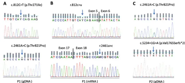
LAMA2 gene sequencing was performed according to Oliveira et al. [2], where all coding and adjacent intronic sequences were analyzed. Sequence variants were described according to the Human Genome Variation Society guidelines for mutation nomenclature (version 2.0) [13] and the reference sequence with accession NM_000426.3. Two heterozygous missense mutations were identified in patient 1, namely c.812C>T (p.Thr271Ile) in exon 5 and c.2461A>C (p.Thr821Pro) in exon 18 (Fig. 3). Family studies confirmed that these were allelic in the patient. Heterozygosity was also found in patient 2, who presented the missense mutation
Fig. 2. Histologic and molecular studies performed in both patients. (A) Patient 1, hematoxylin and eosin staining (10) discloses a mild increase in the
variability of fiber diameter; (B) patient 1, gomori trichrome staining (10); (C) patient 2, hematoxylin and eosin (10), revealing increased variability of
fiber diameter with round atrophic fibers, dispersed in the fascicles or in groups in the same area and frequent central nuclei; (D) patient 2, ATPase reaction (pH 9.4;10). Type 1 fibers predominated; (E) partial and irregular laminin-a2 labeling in patient 1 muscle specimen (20); (F) partial and
c.2461A>C (p.Thr821Pro), in common with patient 1, and the splicing mutation c.5234+1G>A (p.Val1765Serfs*21)
known to cause skipping of exon 36[2].
Population screening for these missense variants was carried out by direct sequencing of exon 5 in the case of c.812C>T and by high resolution melting curve analysis (hrMCA) of exon 18 in the case of c.2461A>C. Neither variants were detected in 300 control alleles, corroborating their pathogenic nature.
4.2. cDNA studies
Knowing that variants that are predictably missense can actually induce splicing defects[14], and since both variants are located in the vicinity of splice sites, we evaluated their possible effects on pre-mRNA processing. Transcript analysis was performed in a cryopreserved muscle specimen from patient 1, using primers designed specifically for the regions of interest. Results showed that neither appeared to have an effect on mRNA splicing (no exon skipping or cryptic splice site activation) and that bi-allelic expression was evidenced with both mutations detected in heterozygosity (Fig. 3).
4.3. Western Blotting
Laminin-a2, a-dystroglycan and b-dystroglycan expression in a muscle sample from patient 1 was assessed by Western Blotting (WB) analysis using monoclonal antibodies against the 80 kDa C-terminal segment of laminin-a2 (MAB1922, Chemicon, Millipore, Temecula, CA), a glycosylated epitope of a-dystroglycan (VIA4-1, Upstate, Millipore, Temecula, CA) and b -dystroglycan (NCL-b-DG, Novocastra, Leica Microsystems, Newcastle Upon Tyne, UK). The latter
was used as an internal control. The WB procedure was adapted from the protocol reported by Anderson et al.
[15]. For the purpose of densitometry, protein loading was normalized with the myosin band in the post-blotted gel. Immunoblot analysis revealed complete absence of merosin with the anti-80 kDa antibody. A 75% reduction was detected ina-dystroglycan expression by comparison with the control (Fig. 2, H), possibly due to technical issues related with the heterogeneous glycosylation pattern of the epitope recognized by the VIA4-1 antibody.
4.4. Bioinformatic analysis of missense variants
Besides the in vitro experiments described above we also assessed the pathogenicity of the missense variants using a diversity of bioinformatic tools and databases. As depicted in table S1 in supplementary data, all algorithms consistently suggested a deleterious effect for these mutations. In addition, the mutations c.2461A>C and c.812C>T were not present in SNP databanks including dbSNP, 1000 Genomes and Exome Variant Server.
5. Discussion
(p.Thr821Pro) in exon 18 and the splicing mutation c.5234+1G>A (p.Val1765Serfs*21) in intron 36, which
has been previously described[2]. The clinical phenotype of both patients is markedly different which could only in part be explained by the different allelic combination of mutations detected in each case. In the Leiden Muscular Dystrophy Pages (http://www.dmd.nl/LAMA2), we have found an entry for the c.2461A>C variant, but reported as being of unknown pathogenicity. Interestingly, this mutation was reported in this database in together with the c.5234+1G>A mutation, the same combination as in patient 2. However, the phenotypes of both patients seem to differ significantly (seizures, abnormal white matter, and cervical spine fusion as indicated in the Leiden database, without mention of a cardiac phenotype).
Reports of MDC1A (MIM: 607855) patients with severe cognitive impairment are very scarce and the severity of CNS involvement combined with absent muscular complaints, as described here for patient 1, is a new form of presentation of this disorder. In previous series of patients [7] there was no apparent correlation between mental retardation and severity of weakness, suggesting that different mechanisms contribute to muscular and CNS involvement. Rufinamide was shown to be effective in epileptic encephalopathies other than Lennox–Gastaut syndrome, particularly in patients with drop-attacks and (bi)frontal spike-wave discharges, matching our patient’s clinical features[16].
Cardiac involvement in laminin-a2 deficiency has been largely underreported. One study specifically addressed the cardiac involvement in children with laminin-a2 deficiency [6]. A cohort of 16 children with congenital muscular dystrophy was studied (6 with MDC1A) where two children with significant left ventricular dysfunction had complete laminin-a2 deficiency. In another series of 51 patients with MDC1A [4], 15 had cardiac assessment, of whom 5, with complete laminin-a2 deficiency, had cardiac abnormalities. Documentation on cardiac status was unavailable for the remaining patients. In the largest bibliographical review (248 published patients with abnormal immunohistochemical staining for laminin-a2) cardiac function was described in 20 [7]. Cardiac dysfunction was reported in 7 patients (7/20, 35%), of whom 4 were asymptomatic. Cardiac abnormalities varied, including right bundle branch block, dilated cardiomyopathy and “borderline changes in cardiac function”.
To the best of our knowledge, only one case of partial defect of laminin-a2 with cardiac involvement was previously reported [8]. Long-term evaluation led to a diagnosis of dilated cardiomyopathy with ventricular arrhythmias, requiring implantation of an intracardiac defibrillator. In this patient, LAMA2 gene analysis revealed two different mutations, a missense mutation in exon 29 (c.4405T>C, p.Cys1469Arg) and a nonsense mutation in exon 31 (c.4645C>T, p.Arg1549*).
We want to highlight the need for cardiac status assessment in this disorder, given the potential of severe cardiac involvement, even in patients with residual expression of the laminin-a2 chain.
This report widens the clinical presentation associated with genetic defects inLAMA2. These cases are probably under-diagnosed and seldom reported in the literature, especially when only subtle changes in laminin-a2 chain are detected in IHC studies. Considering the interplay role of laminin-a2 in the basal membrane and extracellular matrix, it is conceivable that mutations in this protein may only affect the interaction with other proteins maintaining at least partial interaction with a -dystroglycan, and leading to other phenotypes not directly related with muscle weakness.
Appendix A. Supplementary data
Supplementary data associated with this article can be found, in the online version, athttp://dx.doi.org/10.1016/ j.nmd.2014.01.004.
References
[1] Tome´ FM, Evangelista T, Leclerc A, et al. Congenital muscular dystrophy with merosin deficiency. C. R. Acad. Sci. III. 1994;317(4):351–7.
[2] Oliveira J, Santos R, Soares-Silva I, et al.LAMA2gene analysis in a cohort of 26 congenital muscular dystrophy patients. Clin. Genet. 2008;74(6):502–12.
[3] Tezak Z, Prandini P, Boscaro M, et al. Clinical and molecular study in congenital muscular dystrophy with partial laminin alpha 2 (LAMA2) deficiency. Hum. Mutat. 2003;21(2):103–11.
[4] Geranmayeh F, Clement E, Feng LH, et al. Genotype–phenotype correlation in a large population of muscular dystrophy patients with LAMA2 mutations. Neuromuscul. Disord. 2010;20(4):241–50. [5] Lapidos KA, Kakkar R, McNally EM. The dystrophin glycoprotein
complex: signaling strength and integrity for the sarcolemma. Circ. Res. 2004;94:1023–31.
[6] Spyrou N, Philpot J, Foale R, Camici PG, Muntoni F. Evidence of left ventricular dysfunction in children with merosin-deficient congenital muscle dystrophy. Am. Heart J. 1998;136:474–6. [7] Jones KJ, Morgan G, Johnston H, et al. The expanding phenotype of
Laminin alpha2 chain (merosin) abnormalities: case series and review. J. Med. Genet. 2001;38:649–57.
[8] Carboni N, Marrosu G, Porcu M, et al. Dilated cardiomyopathy with conduction defects in a patient with partial merosin deficiency due to mutations in the laminin-a2-chain gene: a chance association or a novel phenotype? Muscle Nerve 2011;44(5):826–8.
[9] Relucio J, Tzvetanova ID, Ao W, Lindquist S, Colognato H. Laminin alters fyn regulatory mechanisms and promotes oligodendrocyte development. J. Neurosci. 2009;29(38):11794–806.
[10] Schmid RS, Anton ES. Role of integrins in the development of the cerebral cortex. Cereb. Cortex 2003;13(3):219–24.
[11] Tsao CY, Mendell JR. Coexisting muscular dystrophies and epilepsy in children. J. Child Neurol. 2006;21:148–50.
[12] van der Knaap MS, Smit LM, Barth PG, et al. Magnetic resonance imaging in classification of congenital muscular dystrophies with brain abnormalities. Ann. Neurol. 1997;42(1):50–9.
[14] Santos R, Oliveira J, Vieira E, et al. Private dysferlin exon skipping mutation (c.5492G>A) with a founder effect reveals further alternative splicing involving exons 49–51. J. Hum. Genet. 2010;55(8):546–9, Epub. 2010 Jun 10.
[15] Anderson LVB. Multiplex western blot analysis of the muscular dystrophy proteins. In: Bushby KMD, Anderson LVB, editors.
Muscular dystrophy: methods and protocols. Walker JM, editors. Methods in molecular medicine series, vol. 43, III. Totowa, NJ: Humana Press; 2001. p. 369–86.