University of Lisbon Faculty of Medicine of Lisbon
Dynamics and function of nicotinic acetylcholine receptors in the nervous system
Catarina da Cunha Fernandes
Supervision by Professor Ana Maria Sebastião, PhD and Co-Supervision by Professor Darwin K. Berg, PhD
PhD in Biomedical Sciences Speciality of Neurosciences
Lisbon 2011
All the opinions expressed in this publication are those of the author and do not necessarily reflect the views of the University of Lisbon.
Todas as afirmações efectuadas no presente documento são de exclusiva responsabilidade do seu autor, não cabendo qualquer responsabilidade à Universidade de Lisboa pelos conteúdos apresentados.
A impressão desta dissertação foi aprovada pela
Comissão Coordenadora do Conselho Científico da
Faculdade de Medicina de Lisboa em reunião de 21 de
Junho de 2011.
The experimental work described in this thesis was performed at the Institute of Pharmacology and Neurosciences, Faculty of Medicine and Unit of Neurosciences, Institute of Molecular Medicine, under supervision of Professor Ana Maria Ferreira de Sousa Sebastião, PhD, and at the Division of Neurobiology, University of California San Diego, under the supervision of Professor Darwin K. Berg, PhD.
O trabalho experimental descrito nesta dissertação foi realizado no Instituto de Farmacologia e Neurociências, Faculdade de Medicina e Unidade de Neurociências, Instituto de Medicina Molecular, sob orientação da Professora Doutora Ana Maria Sebastião, e na Divisão de Neurobiologia, Universidade da Califórnia San Diego, sob orientação do Professor Doutor Darwin K. Berg.
List of originaL PubLications incLudedinthis thesis
I. Fernandes CC, Berg DK, Gómez-Varela D. (2010) Lateral mobility of nicotinic acetylcholine receptors on neurons is determined by receptor composition, local domain, and cell type. J Neurosci 30: 8841-51. (Chapter 3)
ii. Fernandes CC, Pinto-Duarte A, Ribeiro JA, Sebastião AM. (2008) Postsynaptic action of brain-derived neurotrophic factor attenuates alpha7 nicotinic acetylcholine receptor-mediated responses in hippocampal interneurons. J Neurosci 28:5611-8. (Chapter 4)
iii. Campbell NR, Fernandes CC, Halff AW, Berg DK. (2010) Endogenous signaling through alpha7-containing nicotinic receptors promotes maturation and integration of adult-born neurons in the hippocampus. J Neurosci 30: 8734-44. (Chapter 5)
other PubLicationsfromthe author:
iV. Diógenes MJ, Fernandes CC, Sebastião AM, Ribeiro JA. (2004) Activation of adenosine A2A receptor facilitates brain-derived neurotrophic factor modulation of synaptic transmission in hippocampal slices. J Neurosci 24:2905-13
V. Biber K, Pinto-Duarte A, Wittendorp MC, Dolga AM, Fernandes CC, Von Frijtag Drabbe Künzel J, Keijser JN, de Vries R, Ijzerman AP, Ribeiro JA, Eisel U, Sebastião AM, Boddeke HW. (2008) Interleukin-6 upregulates neuronal adenosine A1 receptors: implications for neuromodulation and neuroprotection. Neuropsychopharmacology 33:2237-50
VI. Neff RA 3rd, Gómez-Varela D, Fernandes CC, Berg DK. (2009) Postsynaptic scaffolds for nicotinic receptors on neurons. Acta Pharmacol Sin 30:694-701.
VII. Campbell NR, Fernandes CC, John D, Lozada AF, Berg DK. (2011) Nicotinic control of adult-born neuron fate. Biochem Pharmacol, in press.
To my parents. To António.
Aos meus pais. Ao António.
Acknowledgements
These studies were carried out in the Institute of Pharmacology and Neurosciences, Institute of Molecular Medicine, Faculty of Medicine, University of Lisboa, during the years 2004-2007, as well as in Division of Biology, University of California San Diego during 2007-2011.
At the end, I have the feeling that this is not my dissertation: this is teamwork. I would like to deeply thank all that contributed for making this project possible - Obrigada!
I want to express my deepest gratitude to my advisor Professor Ana Maria Sebastião, Ph.D, who gave me the opportunity to work under her supervision and offered me invaluable guidance, support, advice, confidence, and time for this project. I will be forever grateful for her kind mentorship.
I would also like to acknowledge my advisor and mentor Professor Darwin Berg, Ph.D, for his unshaken devotion to this project and science in general. Thank you for receiving me in your lab and for sharing your energy, enthusiasm and passion for science. The joy of science clearly lives within him. Or may be I should say that the joy of life clearly lives within him instead. Thank you for being an inspiration.
Furthermore, I wish to thank Professor Joaquim Alexandre Ribeiro, M.D., Ph.D., Professor Alexandre de Mendonça M. D., Ph.D, and Professor Nicholas Spitzer, Ph. D, for their constructive criticism.
I would like to thank those who directly contributed to the experimental work in this thesis, David Gómez-Varella, António Pinto Duarte and Nolan Campbell. David taught me how to record and analyse single-particle tracking experiments and he also did the imaging for the immunostaining experiments. Thank you for helping me in every single step of this project. António collaborated with me in patch-clamp experiments shown in chapter 4. Thank you for your persistence and all the great ideas that you had for our project. Nolan did the stereotaxic injections, the BrdU injections, morphological
and survival analyses shown in chapter 5. Thank you for inviting me to work with you in this project. It was always a pleasure to work with you, even when experiments did not want to collaborate with us.
I would like to thank my colleagues in my early years at Laboratorio de Neurociências, back to the year of 2003. Joana Coelho, Inês Pereira, Ana Rita Costenla, Bruno Teixeira da Silva and Nuno Canas. We are no longer colleagues, but we kept our friendship alive. Later, at the Instituto de Farmacologia e Neurociências, many thanks to all my colleagues, specially Bruno Fontinha, Sandra Vaz, Natália Assaife-Lopes, Vasco Sousa, Paula Pousinha, Maria José Diógenes and Sylvie Duflot. Special thanks to Alexandra Botelho for helping me with administrative issues.
To the past and current members of Berg Lab: Rob Neff, Xulong Wang, Natasha Guonko, Jeff Schoellerman, Andrew Halff, Adrian Lozada, Giordano Lippi, Danielle John, Kerri Massey and Jingjing Duan for helping, supporting, and laughing with me. Special acknowledgements to Xiao Yun Wang for her unconditional help in the lab.
To all my friends in San Diego. These last four years have been a wonderful time to me thanks to all of you. Your friendship made me feel at home when I was not so sure where home was anymore. Thank you for your generosity and for inspiring me in so many different ways. Special thanks to my dear friend Conny Plank for being such an amazing and special person without even realizing it or making any effort.
The support of my very dear friends has been invaluable to me. This is especially so for Rita Baptista, Alda Mendes, Margarida Suárez, Inês Correia, Inês Moura Martins and Mauro Figueira: you are all warmly thanked for the great times and for keeping me in touch with the real world. I barely remember my life before Rita. You have been the best friend in the past 25 years and it has been a pleasure growing up with you. Thank you for inspiring me every single day and for showing me how to pursuit happiness. Aldinha, you have always been an inspiration. Thank you for showing me how to fight for what (we think) is right. “Guida”, “Inês Pequenina” e “Inês de Setúbal”, my dearest friends from FCUL –Thank you so much for your sincere friendship, for supporting me
and taking care of me. Mauro, I missed you so much in San Diego. Our dinners, the movies, the (crazy!) dancing and the concerts... Thank you for making our friendship so simple and special.
All my relatives, especially my uncles, aunts and cousins. My grandparents Armindo, Celeste and Amélia - I thank you for your love and support. Ao meu Avô Armindo, quero agradecer por me ensinar que na vida é sempre possivel recomeçar. À minha avó Celeste agradeço por me ensinar que as coisas simples são também as mais belas. À minha avó Amélia, agradeço por ser a minha irmã mais velha. Todas as brincadeiras e as férias de Verão intermináveis, os gelados e bolas de berlim. O melhor café com leite do mundo. A tua alegria, força, coragem e determinação.
To my parents, Amorim and Maria José Fernandes, who gave me the happiest childhood one can have. You have always been with me, even in these last years, when we were 9169 km apart. Best gift I ever had was your unconditional love and I can’t imagine a better gift. Thank you for showing me all the good things in life, especially when life does not seem to have a lot of good things, and that laughing is the best thing I can do when going through a tough time.
Finally, my deepest acknowledgments are dedicated to António Pinto Duarte. Thank you for loving me. For supporting me in all possible ways and for showing me that dreams are tangible, even in the darkest moments of life. For our daily life, all the adventures and travels. For all the surprises. For shaping me up a better person. For making me laugh like no one else does. For making everything special.
This project was financially supported by the Fundação para a Ciência e Tecnologia (POCI 2010/FSE) and Fundação Calouste Gulbenkian.
O coração, se pudesse pensar, pararia.
Could it think, the heart would stop beating.
tabLeof contents
1. generaL introduction 1
1.1 Scope of the thesis 3
1.2 Chapter overview 3
1.3 Neuronal transmission and synaptic strength 5
1.3.1 Neuronal communication: evolution of the nervous system 5 1.3.2 From the neuromuscular junction to the brain 6 1.3.3 Synaptic transmission in central synapses – Hippocampus as a model system 7 1.3.4 Synaptic plasticity - basis for learning and memory? 10 1.3.4.1 Mechanisms subjacent to modifications on synaptic strength in the CNS 11 1.3.4.2 Trafficking of neurotransmitter receptors – exocytosis, lateral diffusion 13
and endocytosis
1.3.4.3 Molecular determinants of lateral diffusion in the cell membrane – 15 - lipids, scaffolds and cytoskeleton
1.3.4.4 Trafficking of neurotransmitter receptors that underlie synaptic plasticity 18 – the example of long-term potentiation in CA1 hippocampal region
1.3.5 New neurons in old brains – adult neurogenesis in the hippocampus 19
1.4 Nicotinic acetylcholine receptors 23
1.4.1 Nicotinic acetylcholine receptors: subunits and subtypes in the nervous system 25 1.4.2 Structure of nAChR subunits – Implications for function 27
1.4.3 Postranslational modifications of nAChRs 29
1.4.4 Trafficking of nAChRs – putting nAChRs in the cell surface 30 1.4.4.1 Chaperones and scaffolds as intrinsic factors that regulate the trafficking of nAChRs 30
1.4.5 Signaling cascades mediated by nAChRs 33
1.4.6 Function of nAChR in the nervous system 34
1.4.6.1 Cholinergic signaling in the hippocampus 35 1.4.6.2 Cholinergic signaling in pathology - the cholinergic hypothesis of Alzheimer’s disease 40 1.5 Neurotrophins as regulators of synaptic strength 41
1.5.1 Brain Derived Neurotrophic Factor 41
1.5.1.2 Signaling mediated by TrkB receptors 43 1.5.1.3 A role for cAMP in gating TrkB-mediated signaling 45
1.6 Rationale and objectives 47
2. materiaL and methods 49
2.1 Ethics 51
2.2 Methods for Chapter 3 51
2.2.1 Single Particle Tracking: imaging the lateral diffusion of membrane molecules 51
with Quantum Dots
2.2.1.1 Primary neuronal culture and transfection 51
2.2.1.2 Pharmacological manipulations 54
2.2.1.3 Synapse labeling and receptor tagging 54
2.2.1.4 Acquisition of SPT movies 55
2.2.1.5 Tracking procedure and trajectory decomposition 56
2.2.1.6 Quantification of diffusion properties 56
2.2.2. Immunostaining of Fixed Cells 57
2.2.2.1 Fluorescence image acquisition and quantitative analysis 58
2.2.3 Statistical analyses 58
2.3 Methods for Chapters 4 and 5 58
2.3.1 Patch Clamp technique in tissue slices 58
2.3.1.1 Hippocampal slices preparation 60
2.3.1.2 Patch Clamp recordings 61
2.3.1.3 Statistical analysis 64
2.3.2 Identification of Adult-Born Neurons in the Granule Cell Layer 65
2.3.2.1 Stereotaxic viral injection 65
2.3.2.2 BrdU labelling 65
2.3.2.3 Imaging and quantification 65
2.4 Chemicals 66
3. LateraL mobiLityof nicotinic recePtorson PeriPheraL neuronsis determinedby
recePtor comPosition, LocaL domainand ceLL tyPe 69
3.2 Results 72 3.2.1 Expression of nAChRs on neurites of CG neurons in culture 72
3.2.2 Lateral mobility of nAChRs on neurons 73
3.2.3 Cytoskeletal determinants of nAChRs mobility 77 3.2.4 Effects of cholesterol depletion on receptor mobility 79 3.2.5 Constraint of nicotinic receptors by the PDZ-containing postsynaptic scaffolds 82 3.2.6 Mobility of a7-nAChRs on chick sympathetic ganglion neurons 85
3.3 Discussion 87
3.3.1 Mechanisms that control the lateral diffusion of a7- and a3*-nAChRs 87 3.3.2 Context of nAChRs mobility with respect of other transmitter receptors 89 3.3.3 nAChRs – a heterogeneous population of receptors? 91 3.3.4 Functional consequences of nAChR lateral diffusion 91
4. PostsynaPtic actionof brain-deriVed neurotroPhic factor attenuatesa7
nicotinic acetyLchoLine recePtor-mediated resPonsesin hiPPocamPaL interneurons 93
4.1 Introduction 95
4.2 Results 96
4.2.1 BDNF induces a rapid depression of a7-nAChR-mediated currents 96 4.2.2 Inhibition of TrkB receptors impairs BDNF-induced suppression of a7-nAChR-mediated
currents 99
4.2.3 BDNF action on a7-nAChRs process requires the PLCg/PKC Pathway and Ca2+ ions as a
cofactor 102
4.2.4 The attenuation of a7-nAChRs function by BDNF involves the actin cytoskeleton 104
4.3 Discussion 105
4.3.1 Regulation of a7-nAChRs by BDNF 105
4.3.2 Involvement of PLC/PKC pathway on the regulation of a7-nAChR 107 4.3.3 Short- and long-term actions of BDNF on a7-nAChRs 108 4.3.4 Regulation of a7-nAChRs and its impact on hippocampal circuitry 109 5. endogenous signaLLingthrougha7-containing nicotinic acetyLchoLine
recePtors Promotes maturationand integrationof aduLtborn neuronsinthe
5.1 Introduction 113
5.2 Results 114
5.2.1 Nature of GABA-induced activation signaling in adult-born neurons depends
on a7-nAChRs 114
5.2.2 GABAergic PSCs retain immature temporal characteristics in the absence of
a7-nAChR-mediated signaling 118
5.2.3 Adult-born a7KO neurons have reduced dendritic arbors and receive less
synaptic activity compared with their age-matched WT counterparts 119 5.2.4 Reduced survival of adult-born a7KO neurons through the critical period 121
5.3 discussion 121
5.3.1 a7-nAChRs set the tempo for the maturation of GABAergic signaling in
adult-born neurons 122
5.3.2 Compromised integration of adult-born in the absence of a7-nAChR-mediated
signaling 124
5.3.3 Neurogenesis and cholinergic signaling in the hippocampus 125
6. generaL discussionand future PersPectiVes 125
6.1 Dynamic regulation of nAChRs in the nervous system 131 6.2 Role of a7-nAChRs in the CNS – much more to know
then what is known 137
6.3 Therapeutic relevance of nAChRs in pathology 141
6.4 Perspective 146
Figure Index
Figure 1.1 – A simplified diagram of the hippocampus in the brain and
an hippocampal slice. 8
Figure 1.2 – The neural circuitry in the rodent hippocampus. 9
Figure 1.3 – Receptor exchanges between synaptic, extrasynaptic and intracellular
compartments. 15
Figure 1.4 – Adult hippocampal neurogenesis. 21
Figure 1.5 – Distribution of nAChRs in the brain. 24
Figure 1.6 - Model of the a7-nAChR. 27
Figure 1.7 - Neurotrophin signaling pathways via TrkB receptor. 44
Figure 2.1 - Images showing the number of QDs. 55
Figure 2.2 - Images showing the number of QDs. 55
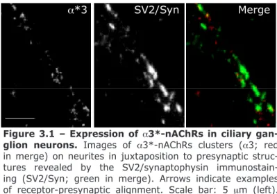
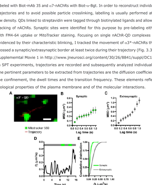
Figure 3.1 – Expression of a3*-nAChRs in ciliary ganglion neurons. 73 Figure 3.2 – Expression of a7-nAChRs in ciliary ganglion neurons. 73 Figure 3.3 – Different mobilities of surface a3*-nAChRs in synaptic and
extrasynaptic spaces. 74
Figure 3.5 – Comparison of lateral diffusion values for nAChRs using different
synaptic markers. 76
Figure 3.6 – Lateral diffusion of nAChRs in the presence of the vehicle DMSO. 77
Figure 3.7 – Different cytoskeletal regulation for a3*-nAChR and a7-nAChR lateral
diffusion. 78
Figure 3.8 – Selective effects of cholesterol depletion. QD-nAChR trajectories were examined in extrasynaptic and synaptic space for control conditions (Ctr) or after
cholesterol depletion with either COase or MbCD. 80
Figure 3.9 – Expression of GFP via transfection as a negative control does
not change nAChR diffusion properties. 83
Figure 3.10 – Regulation of nAChR mobility by PDZ-containing proteins. 84
Figure 3.11 – Effect of vehicle (DMSO) on the lateral diffusion of
α7-nAChRs on sympathetic neurites. 85
Figure 3.12 – Lateral diffusion of surface a7-nAChRs in synaptic
and extrasynaptic space on sympathetic ganglion neurites. 86
Figure 4.1 – Patch clamp recordings of a7 nAChR-mediated currents. 97
Figure 4.2 – BDNF inhibits a7 nAChR-mediated currents in CA1 hippocampal
interneurons. 98
Figure 4.3 – BDNF-induced depression of a7 nAChR-mediated currents
Figure 4.4 – A2A receptors do not influence per se the amplitude or
the kinetics of a7 receptor-mediated currents. 100
Figure 4.5 – The inhibitory action of BDNF on a7 nAChR-mediated
responses does not involve the activity of Src kinases-family. 101
Figure 4.6 – Signaling pathways involved in BDNF-induced inhibition
of a7 nAChRs-mediated currents. 103
Figure 4.7 – F-actin disruption prevents the acute action of
BDNF on a7-nAChRs. 104
Figure 5.1 – Recording of GABA-mediated PSCs in adult-born neurons. 115
Figure 5.2 – Delayed maturation of the chloride gradient in adultborn a7KO neurons extends the period of depolarizing GABAergic responses. 116
Figure 5.3 – Inactivating the a7-nAChR gene prolongs an immature
pattern of chloride transporters in the hippocampus. 117
Figure 5.4 – In the absence of a7-nAChRs, GABAergic PSCs display immature
kinetics. 118
Figure 5.5 – Adultborn neurons lacking a7-nAChRs receive less
synaptic inputs than normal. 120
Figure 5.6 – Absence of a7-nAChRs decreases the chance of survival for
adultborn neurons during the critical period. 121
Figure 6.1 - Illustration of the different cellular mechanisms constraining
Figure 6.2 – BDNF inhibits a7-nAChR-mediated currents in CA1
hippocampal interneurons. 136
Figure 6.3 - Potential mechanisms underlying a7-nAChRs-mediated
abbreViations
a7KO a7-nAChR knockout animal
ACh acetylcholine
aCSF artificial cerebrospinal fluid
AMPA a-amino-3- hydroxy-5-methylisoxazole-4-propionic acid
AP action potential
APC adenomatous polyposis coli
BDNF brain-derived neurotrophic factor
Biot biotinylated
BrdU 5-bromo-2-deoxyuridine
a-Bgt a-bungarotoxin
Ca2+ calcium
CaMKII calcium/calmodulin-dependent protein kinase II cAMP 3’-5’-cyclic adenosine monophosphate
CG ciliary ganglion
Ch choline
ChAT choline acetyltransferase
Cl- chloride
CNS central nervous system
COase cholesterol oxidase
CREB cAMP response element binding protein CRIPT cysteine-rich interactor of PD 23
Di diffusion coefficients
DAG diacylglycerol
DG dentate gyrus
E embryonic day
ECl chloride equilibrium potential
EB1 end binding protein 1
EC entorhinal cortex
ECD extracellular domain
ERK extracellular signal-regulated kinase F-actin filamentous actin
FRAP fluorescence recovery after photobleaching GABA g-aminobutyric acid
GCL granule cell layer
GDP giant depolarizing potentials
GFP green fluorescent protein
Gly glycine
GK guanylate kinase
HF hippocampal formation
IP3 inositol 1,4,5 triphosphate K252a tyrosine kinase inhibitor
KB ketone body
KO Knock-out
LTM long-term memory
LTP long-term potentiation
MAGUKs membrane associated guanylate kinases MAPK mitogen-activated protein kinase
MbCD methyl-β-cyclodextrin
mEPSC miniature excitatory postsynaptic current MMLV Moloney’s Murine Leukemia Virus
MSD mean square displacement
MW Mann–Whitney U
NA Numerical aperture
nAChR nicotinic acetylcholine receptor
nDBB nucleus of the diagonal band of Broca
NMDA N-methyl-D-aspartate
NGF nerve-growth factor
NPC neuronal progenitor cells
NMJ neuromuscular junction NT-3 neurotrophin-3 NT-4/5 neurotrophin-4/5 P postnatal day p75NTR pan-neurotrophin receptor PBS phosphate-buffered saline
PDZ post synaptic density protein (PSD95)/Drosophila disc large tumor suppressor (DlgA)/ zonula occludens-1 protein (zo-1)
PI3K phosphatidylinositol-3-OH kinase
PKA protein kinase A
PKB protein kinase B
PKC protein kinase C
PLCg phospholipase C, g subunit
PNS peripheral nervous system
PSC postsynaptic current
PSD postsynaptic density
QDs quantum dots
RFP red fluorescent protein
RNAi RNA interference
ROI region of interest
SFK Src-family tyrosine kinase
SGZ subgranular zone
SH3 Src-homology 3
Shc Src homology 2/-collagen-related protein SSCs spontaneous synaptic currents
STM short-term memory
SPT single particle tracking
Sub subiculum
SV2 synaptic vesicle protein 2
t test Student’s t test
TM transmembrane
trkA/B/C tropomyosin-related kinase A/B/C VGCC voltage-gated calcium channels
VTA ventral tegmental area
abstract
Nicotinic acetylcholine receptors (nAChRs) are broadly distributed in the nervous system. Due to the sub-cellular location and high diversity of nAChRs, they are thought to play a key role in setting the synaptic strength between neurons.
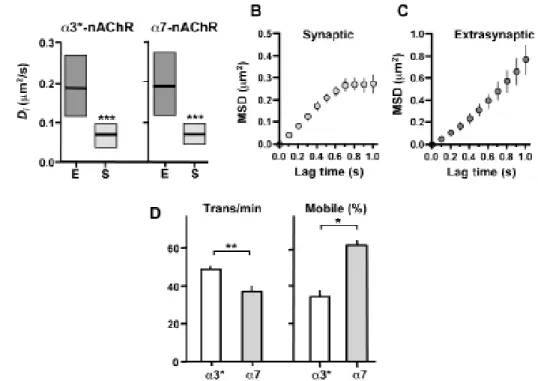
The present work aimed to study the mechanisms that regulate the fate of neuronal nAChRs on the cell membrane and clarify their role in the central nervous system. I used single-particle tracking to follow surface nAChRs on neurons. Both heteropentameric a3-containing receptors (a3*-nAChRs) and homopentameric a7-containing receptors (a7-nAChRs) access synapses by lateral diffusion, where they moved slower and showed a constrained behavior. The nature of synaptic restraints was receptor-dependent, since the disruption of either lipid rafts or PDZ scaffolds rendered half of the stationary a3*-nAChRs to be mobile without changing the proportion of mobile a7-nAChRs.
I next investigated the acute action of brain-derived neurotrophic factor (BDNF) on the function of a7-nAChRs. Patch-clamp experiments showed that BDNF rapidly decreased a7-nAChRs-mediated responses in hippocampal interneurons. This effect was dependent on the activation of TrkB receptors, occured through the phospholipase C/protein kinase C pathway and involved the actin cytoskeleton.
Finally, the role of a7-nAChRs on the adult neurogenesis was investigated. Stereotaxic retroviral injection into the dentate gyrus of wild-type and a7-knockout (a7KO) mice was used to label and birthdate adult-born neurons for morphological and electrophysiological measures; BrdU injections were used to quantify cell survival. In a7KO mice, adult-born neurons developed with truncated dendritic arbors and experienced a prolonged period of immature GABAergic signaling. Under these conditions, neurons received fewer synaptic inputs and were more prone to die during the critical period when adult-born neurons are normally integrated into networks.
Overall, the findings here reported support a regulatory role for a7-nAChRs in the nervous system, pointing out an important role of these receptors on synaptic transmission and plasticity in the brain.
sumário
Os receptores colinérgicos do tipo nicotínico (nAChRs) são vastamente expressos no sistema nervoso. Na região do hipocampo, em particular, os mecanismos desencadeados por nAChRs influenciam processos de atenção, memória, aprendizagem, e constituem importantes alvos terapêuticos para doenças do fórum neurológico, neurodegenerativo e psiquiátrico.
Atendendo à sua distribuição sub-celular e à elevada diversidade de subunidades expressas no sistema nervoso central, estes receptores são geralmente associados a fenómenos de regulação da força sináptica entre neurónios. Ainda mais, quando activados, os nAChRs permitem a entrada directa e/ou indirecta de cálcio (Ca2+),
elevando consequentemente a concentração citoplasmática deste ião. O aumento da concentração de Ca2+ citoplasmático, mesmo que transitória, activa várias e
diferentes cascatas de sinalização que podem, inclusivamente, alterar a expressão génica. Por outro lado, se a entrada de Ca2+ for excessiva, são activados fenómenos de
excitoxicidade neuronal que eventualmente podem culminar em morte celular. Assim, a localização e função dos nAChRs são de extrema importância e consequentemente alvo de mecanismos de regulação nos neurónios.
O trabalho descrito nesta dissertação teve como objectivo estudar os mecanismos intracelulares que regulam o destino de nAChRs na membrana celular, assim como mecanismos que regulam a sua função, e por fim clarificar o papel dos nAChRs no sistema nervoso central.
Para estudar a localização e o tráfego dinâmico dos nAChRs em neurónios, recorri à técnica de de single-particle tracking SPT com quantum dots. Dado que nesta técnica são estudadas partículas individuais, é possível distinguir diferentes tipos de nAChRs atendendo à mobilidade. Este estudo foi efectuado em culturas de neurónios de gânglios ciliares, que expressam dois subtipos de nAChRs: heteropentaméricos contendo a subunidade a3 (a3*-nAChRs) e homopentaméricos contendo a subunidade a7 (a7-nAChRs). Os a3*-nAChRs medeiam a transmissão sináptica nos gânglios ciliares, enquanto que os a7-nAChRs têm um papel regulador. Quanto à dinâmica dos nAChRs, identifiquei uma população imóvel e uma outra móvel, tanto para os a3*-nAChRs como para os a7*-nAChRs. Curiosamente, no caso dos a3*-nAChRs, cerca de 70%
da população apresentava-se imóvel, enquanto apenas cerca de 30% dos a7-nAChRs se apresentava imóvel. Ambos os subtipos de nAChRs apresentavam movimento do tipo Browniano quando se deslocavam em áreas extrasinápticas e do tipo restrito quando em áreas sinápticas. Ambos os subtipos possuíam constantes de difusão superiores nas áreas extrasinápticas do que nas sinápticas. Distiguiu-se ainda, tanto para os a3*-nAChRs, como para os a7-nAChRs, uma subpopulação móvel, que transita entre domínios sinápticos e extrasinápticos através de difusão lateral. Os valores de constantes de difusão foram semelhantes para os a3*- e a7-nAChRs. Verificou-se, contudo, que a natureza das restrições sinápticas é diferente para os dois tipos de receptores. Por exemplo, perturbações ao nível das jangadas lipídicas (“lipid rafts”) ou das proteínas do citoesqueleto contendo domínios PDZ aumentam a proporção de a3*-nAChRs móveis para cerca de 70%, sem contudo alterarem a proporção de a7-a3*-nAChRs móveis. Curiosamente, os mecanismos que regulam a difusão dos nAChRs depende do ambiente celular. Por exemplo, o colesterol é importante para a difusão dos a7-nAChRs em neurónios de gânglios ciliares, mas não em neurónios de gânglios lombares. A acção aguda da neurotrofina brain-derived neurotrophic factor (BDNF) na função dos a7-nAChRs foi também investigada. O BDNF é uma molécula que existe em grande abundância no hipocampo e que geralmente é libertada durante períodos de grande actividade neuronal. O BDNF foi inicialmente descrito como uma molécula de efeitos relativamente lentos, que ocorriam na escala de horas ou mesmo dias. Mais recentemte, foram descritas acções rápidas do BDNF em vários receptores ionotrópicos, o que me motivou estudar uma hipotética acção desta molécula sobre os a7-nAChRs. Este estudo foi efectuado em fatias de hipocampo de rato (3-4 semanas) preparadas agudamente. Utilizou-se a técnica de patch clamp para registar correntes iónicas geradas pela aplicação de agonistas dos a7-nAChRs. A amplitude máxima destas correntes foi usada como uma medida da activação dos a7-nAChRs. A aplicação exógena de BDNF inibiu rapidamente as respostas mediadas pelos a7-nAChRs expressos nos interneurónios do
stratum radiatum da área CA1 do hipocampo. Este efeito é dependente dos receptores
TrkB para o BDNF, ocorre através das vias da fosfolipase C/cinase C de proteínas e requer a activação de receptores de adenosina do tipo A2A . A inibição dos a7-nAChRs não era dependente, contudo, da acção de cinases da família das Src. Demonstrou-se ainda que a regulação dos a7-nAChRs pelo par BDNF/receptor TrkB depende do
citoesqueleto de actina e está comprometida na ausência de Ca2+ extra- e intra-celular.
Por fim, investigou-se o papel desempenhado pelos a7-nAChRs na neurogénese adulta. Nos mamíferos, a neurogénese no giro dentado mantem-se activa durante o adulto e parece ser essencial para o funcionamento do hipocampo. As células progenitoras dos neurónios expressam a7-nAChRs, o que despertou o interesse em investigar o papel destes receptores no desenvolvimento e maturação das células precursoras de neurónios. Injecções com 5-bromo-2-desoxiuridina (BrdU) permitiram observar que os neurónios dos animais knockout para o gene dos a7-nAChRs (a7KOs) são mais vulneráveis durante a fase de integração na rede neuronal que os neurónio de animais controlo. Esta fase crítica de morte neruonal ocorre entre a segunda e quarta semana após o início do processo de diferenciação e é determinante para a integração de neurónios na rede. De seguida, investigou-se o ritmo de desenvolvimento/maturação dos neurónios, na presença e ausência de a7-nAChRs. A injecção extereotáxica de retrovírus no giro dentado de animais adultos normais (controlos) e em a7KO foi utilizada para marcar e datar neurónios gerados no adulto, permitindo uma posterior análise morfológica (número de ramificações e comprimento das dendrites) e electrofisiológica (potencial de membrana, potencial de inversão do cloro, cinética das correntes ácido g-aminobutírico (GABA)-érgicas, frequência e amplitude das correntes sinápticas espontâneas) destes neurónios. A análise das propriedades dos neurónios foi feita três semanas depois da injecção, quando os neurónios controlo apresentavam parâmetros morfológicos e electrofisiológicos característicos de neurónios maturos. Ainda mais, durante esta idade, os neurónios estão a atravessar o período crítico de integração no circuito. Verificou-se que, nos a7KOs, os neurónios gerados no adulto apresentavam-se menos diferenciados do que nos animais controlo, quer a nível morfológico, quer a nível electrofisiológico. Neste caso, os neurónios possuíam árvores dendríticas truncadas e menos complexas. Apresentavam também um prolongamento do período em que o neurotransmissor GABA actua como despolarizante. De facto, as próprias correntes GABAérgicas mediadas pelos receptores GABAA apresentavam uma cinética característica de estadios imaturos. Em comparação com os neurónios controlo, apresentavam ainda uma menor frequência de correntes sinápticas espontâneas, que por sua vez possuíam também uma menor amplitude.
o tráfego dinâmico dos nAChRs na membrana celular. Demonstrei, também, que estes mecanismos dependem da constituição dos receptores, do domínio subcelular e do tipo de célula. Verifiquei ainda que os a7-nAChRs são um dos alvos das acções rápidas do BDNF. Esta regulação poderá ser importante na regulação da transmissão e plasticidade sináptica no cérebro. Por fim, identificou-se um papel determinante dos a7-nAChRs na sobrevivência e no ritmo de desenvolvimento, maturação neuronal e integração dos neurónios durante a neurogénese no adulto. Em suma, os resultados apresentados nesta tese apontam para um papel fundamental dos nAChRs no hipocampo e são propostos vários factores intra e extracelulares que regulam o tráfego e função destes receptores nos neurónios.
Palavras-chave: Receptores colinérgicos nicotínicos; difusão lateral; brain-derived neurotrophic factor; neurogénese no adulto; hipocampo
Chapter 1
chapter 1
General IntroductIon
1.1 Scopeofthe theSIS
The research presented in this thesis is aimed to clarify of the mechanisms that regulate the functional expression of neuronal nicotinic acetylcholine receptors (nAChRs) on the cell surface and elucidate about the role played by these receptors in the central nervous system (CNS).
1.2 chapter overvIew
Due to the broad distribution and diversity of nAChRs in the nervous system, as well as to the characteristics of nAChR-dependent signaling, these receptors are thought to play a key role in setting the synaptic strength between neurons. In the section 1.3 of Chapter 1 some of the cellular and molecular mechanisms currently known to regulate synaptic strength will be revised. In section 1.4, nAChRs and nAChRs-mediated signaling will be described, stressing out the reasons why these receptors are in a favorable position to act as a neuromodulators of synaptic transmission in the CNS. In section 1.5, there is a general description of the neurotrophin brain-derived neurotrophic factor (BDNF), including some intracellular pathways activated by BDNF to induce rapid modifications in neurotransmitter receptors function. Finally, the “Objectives and Rationale” are summarized in section 1.6.
Chapter 2 is about the Materials and Methods used in this thesis. In this chapter,
the techniques used will be explained in detail, pointing out their advantages and limitations. Live imaging of quantum dots (QDs) and immunostaining of fixed cells were used to study the trafficking of a3*- and a7-nAChRs. Patch-clamp experiments were done to 1) evaluate the acute action of BDNF on a7-nAChR function and 2) investigate the impact of a7-nAChRs in the development and integration of adult-born neurons
in the hippocampus. Stereotaxic retroviral injection into the dentate gyrus was used to label and birthdate adult-born neurons for morphological and electrophysiological measures; BrdU injections were used to quantify cell survival.
Chapter 3 is the first chapter for the Results section. Real-time imaging with QDs was
used to study the lateral diffusion of neuronal a3*- and a7-nAChRs. These experiments were executed in dissociated chick ciliary ganglion neurons. We started by characterizing the mobility of a3*- and a7-nAChRs in this system. Both receptors could access synaptic domains by lateral diffusion, displaying Brownian motion in extrasynaptic space and being constrained and move more slowly in synaptic space. The nature of their movement restraints was, however, different for a3*- and a7-nAChRs; lipid rafts, PDZ-containing scaffolds, microtubules, and actin filaments differentially affected their mobility. We found that control of nAChR lateral mobility, therefore, is determined by mechanisms that are domain-specific, receptor subtype-dependent, and cell-type constrained. The outcome is a system that could tailor nicotinic signaling capabilities to specific needs of individual locations.
Chapter 4 will focus on the acute action of the neurotrophin BDNF on the function
of a7-nAChRs. Patch clamp experiments were performed in fresh hippocampal slices taken from young rats. Acetylcholine- or choline-evoked currents were recorded in CA1 interneurons and were used as a measure of a7-nAChR function. BDNF rapidly reduced the amplitude of a7-nAChR mediated currents when applied in the perfusion solution. This effect was dependent on phospholipase C/protein kinase C signaling pathway and required Ca2+ as a cofactor. The present findings disclose a7-nAChR as a novel target
for rapid actions of BDNF that might play important roles on synaptic transmission and plasticity in the brain.
Chapter 5 will elucidate about the functional relevance of a7-nAChRs in the adult
hippocampal formation (HF). In these experiments, adult mice were stereotaxically injected with Moloney murine leukemia virus-green fluorescence protein (MMLV-GFP) into the dentate gyrus to label and birthdate adultborn neurons in vivo; BrdU injections were used to quantify cell survival. In a7-nAChRs knockout mice, we observed a
reduced survival, delayed maturation and deficient integration of adult-born neurons in the network. This evidence points to a critical role of a7-nAChR in the fate of newborn dentate granule neurons.
This thesis will end with Chapter 6. A synopsis of all major findings is given. Furthermore, the possible consequences and implications of the new insights gained in the present study will be given and future perspectives for the study of nAChRs in the nervous system will be discussed.
Chapter 7 includes all the references used in this dissertation.
1.3 neuronal tranSmISSIonand SynaptIc StrenGth
1.3.1 Neuronal communication: evolution of the nervous system
The survival of an organism relies on the ability that cells have to communicate between each other and generate a global and effective response facing the changes that occur in the environment. When one looks at the evolutionary scale, the more evolved an organism is, the more complex is its nervous system and the more sophisticated is its behavior. But despite the large behavioral differences found in behavior among species, comparative studies of both vertebrates and invertebrates have revealed that brains evolved rather conservative compared with other morphological structures. Likewise, vertebrates, from lampreys to humans, are strikingly similar in the overall brain organization. In the last century, electrophysiological, pharmacological and molecular studies have provided a global understanding of the fundamental mechanisms of neuronal communication, which were also found to be highly conserved across a range of animal species. Despite it, changes in neural connections, neurotransmitters and membrane properties have occurred frequently in evolution. Thus, the brains of animals are a combination of small novelties that appear against a background of conserved features.
and maintain its homeostasis. Consequently, the network developed in a way to sense perturbations upon the endogenous and exogenous systems, appropriately respond to these perturbations. Moreover, neural networks have the ability to form a memory of any particular episode in a way that allows them to respond more efficiently on the next time the same episode occurs. The capacity of the neural activity generated by an experience to modify neural circuit function and thereby modify subsequent thoughts, feelings, and behavior is generally called neuronal plasticity. Information storage and plasticity require that synapses carry out two opposing tasks: maintaining stable long-term synaptic connections while at the same time remaining plastic and allowing for rapid changes in synaptic strength. A major challenge has been to understand how the large array of synaptic proteins that govern these opposing processes are regulated to selectively establish, maintain, and modify the strength of synapses.
1.3.2 From the neuromuscular junction to the brain
As an emergent structure, some of the properties of the nervous system can be explained by the low-level properties of units in the context of their interactions. It is now consensual that the synaptic function is the basic and universal property of neural circuits. The simplest type of synapse that one can find in nature is the neuromuscular junction (NMJ). In contrast to synapses in the CNS, the NMJ connects two different cell types, the neuronal cells and muscle cells. By the simplicity of the NMJ and due to its easy technical accessibility, it become one of the most important model systems in synapse research and was the base for the molecular principles of neurotransmission. Essentially, the function of the NMJ, as any chemical synapse, consists in the conversion of an electric signal conducted by the presynaptic cell to a chemical signal, which can be perceived by the target cell.
Though NMJ and neuron-neuron synapse share the basic mechanisms of synaptic transmission, there are clear differences between both types of synapses. In both cases, the electrical signal [or action potential (AP)] induces depolarization of the presynaptic terminal membrane to the extent of opening voltage-gated calcium channels (VGCC). The transient increase of the local concentration of cytoplasmic Ca2+ induces
consequently, stimulates the release neurotransmitters from the active zone into the synaptic cleft. The neurotransmitters selectively bind to neurotransmitter receptors located in the postsynaptic density (PSD) of the neighboring cell, opening their internal pores and allowing the influx of ions. This inward current eventually generates a new AP, leading to the propagation of the electric signal, which spreads within the muscle cell.
While in the NMJ, neurotransmitter vesicles are always loaded with acetylcholine, in neuron-neuron synapses, the type of the neurotransmitter released can vary, depending on several factors (e.g. cell type, developmental stage of the cell). The endplate of the NMJ comprises only two types of nAChRs, while the neuronal PSD can express panoply of neurotransmitter receptors, which play either excitatory or inhibitory actions.
The molecular/structural differences found between NMJ and neuron-neuron synapses can be easily understood if one looks at their jobs assignments. The synaptic contact at the muscle end plate is optimized to propagate and amplify a single AP in the muscle with maximal reliability. In neuron-neuron synapses, the focus lies less on the transmission of a single AP but rather on integration of multiple signals within the neuronal network and on the complexity of the network itself. In the CNS, a single neuron gets input from several thousands of other neurons. In contrast to the simple architecture of motor neurons, the complex organization of neuronal networks demands regulatory mechanisms orchestrating excitatory actions.
1.3.3 Synaptic transmission in central synapses – Hippocampus as a model system
Synaptic transmission is the basis of most nervous system function, including controlling body parts, memory, learning and cognition. Complex and highly regulated steps take place at both pre- and post-synaptic components to guarantee normal neuronal communication. The hippocampal formation (Fig 1.1; HF) is probably one of the brain regions more extensively studied in the CNS due to its central role learning and memory. Furthermore, the anatomical structure of HF allows one to cut thin slices out of the HF in a way that preserves all of the major connections. For these reasons, HF has been
the primary model for transmission in central synapses.
The rodent HF is a C-shaped structure that is situated in the caudal part of the brain. Three distinct subregions can be distinguished: the dentate gyrus (DG), the hippocampus proper (consisting of CA3, CA2 and CA1) and the subiculum (Sub) (Amaral and Lavenex, 2007; Burwell and Witter, 2002). The cortex that forms the HF has a three-layered appearance (for a review, see Förster et al., 2006). The first layer is a deep layer, comprising a mixture of afferent and efferent fibres and interneurons. In the DG, this layer is called the hilus, whereas in the CA regions it is referred to as the stratum oriens. Superficial to this polymorph layer is the cell layer, which is composed of principal cells and interneurons. In the DG this layer is called the granule layer, whereas in the CA regions and the subiculum it is referred to as the pyramidal cell layer (stratum pyramidale). The most superficial layer is referred to as the molecular layer (the stratum moleculare) in the DG and the subiculum. In the CA region, the molecular layer is subdivided into a number of sublayers. In CA3, three sublayers are distinguished: the stratum
lucidum, which receives input from the DG; the stratum radiatum, comprising the apical
dendrites of the neurons located in the stratum pyramidale; and, most superficially, the stratum lacunosum-moleculare, comprising the apical tufts of the apical dendrites. The lamination in CA2 and CA1 is similar, with the exception that the stratum lucidum is missing in CA1.
The principal neurons of the different HF subfields are interconnected via the excitatory trisynaptic circuit (Fig 1.2; Witter and Amaral, 2004; van Strien et al., 2009). According to the canonical model, the first step of the trisynaptic HF pathway is formed by a unidirectional projection from the DG to CA3: the mossy fibres. The Schaffer collaterals, which originate in CA3 and project to CA1, are the next step in the polysynaptic loop. Finally, CA1 send their projections to the Sub and deep layers of entorhinal cortex (EC), while Sub cells send their projections mainly to EC.
Hippocampal formation
Hippocampal slice Figure 1.1 – A simplified diagram of the hip-pocampus in the brain and an hippocampal slice.
The net flow of information in the HF is strongly modulated by the action of the local-circuit inhibitory interneurons, whose cell bodies are distributed throughout all layers of the HF. Although interneurons are poorly represented in the HF and comprise ~10-15% of the total neuronal population, they exert a powerful control on network excitability and information processing in the brain since a single inhibitory nervous cell may contact 1000-3000 pyramidal cells via extensive arborization (Li et al., 1992; Buhl et al., 1994a). For this reason, interneurons can phase the output of principal cells giving rise to a coherent oscillatory activity (Klausberger et al., 2003; Klausberger et al., 2004; Klausberger and Somogyi, 2008), which has been implicated in encoding, consolidation and retrieval of information in the hippocampus (for review, see Freund and Buzsaki, 1996). Because of their central role in pacing, timing and synchronizing neural circuits in both spatial and temporal domains, knowledge on the mechanisms that control and/or modulate interneuronal function will be crucial to understand hippocampal computation.
Different types of interneurons appear to perform specific and diverse functions in the hippocampus (Freund and Buzsaki, 1996; Klausberger and Somogyi, 2008). For example, some interneurons potently inhibit pyramidal cells by acting directly on their
CA1
CA3
EC
DG
Schaffer collaterals Mossy fibers Perforant pathFigure 1.2 – The neural circuitry in the rodent hippocampus. An illustration of the hippo-campal circuitry. The traditional excitatory trisynaptic pathway (entorhinal cortex (EC)–dentate gyrus (DG)–CA3–CA1–EC) is depicted by solid arrows. The axons of layer II neurons in the en-torhinal cortex project to the dentate gyrus through the perforant pathway (PP). The DG sends projections to the pyramidal cells in CA3 through mossy fibres. CA3 pyramidal neurons relay the information to CA1 pyramidal neurons through Schaffer collaterals. CA1 pyramidal neurons send back-projections into deep-layer neurons of the EC. The DG cells also project to the mossy cells in the hilus and hilar interneurons, which send excitatory and inhibitory projections, respectively, back to the granule cells
.
cell bodies or axon hillocks (Gulyás et al., 1993; Buhl et al., 1994b; McBain et al., 1994; Sik et al., 1995; Miles et al., 1996), whereas others inhibit pyramidal cell activity at their dendrites (Han et al., 1993; Gulyas et al., 1993a,b; Sik et al., 1995; Miles et al., 1996). Another group of interneurons specifically inhibit other interneurons (Acsady et al., 1996; Gulyas and Freund, 1996; Hajos et al., 1996; Blasco-Ibanez et al., 1998). Some interneurons also appear to show long-range projections that cross the area border and are involved in the coordination of spike timing across sub-areas (Sik et al., 1994, 1995). The large variety of inhibitory cells in the hippocampus explains why it has been so complicated to find common properties that would allow grouping them in different subtypes. Most if not all hippocampal interneurons produce the neurotransmitter g-aminobutiric acid (GABA). Choline acetyltransferase (ChAT), the synthesizing enzyme of acetylcholine, is localized in a small number of interneurons in the CA1 region of the hippocampus and the dentate gyrus of the rat (Frotscher et al., 1986), but even these cells are also likely to contain GABA. According to Somogyi and Klausberger, there are at least 16 different types of interneurons just in the CA1 hippocampal area. It is currently thought that this high heterogeneity of interneurons is essential for shaping different patterns of activity in the neural network. Even from a minimalist point of view, at least 10 types of distinct interneurons innervate a single pyramidal cell and actually it is not known yet if all pyramidal cells are uniformly innervated. In addition, the type of inhibition played by a single interneuron in the network can change over time depending on the afferents that activate it (Croce et al., 2010). Finally, the intrinsic passive and active electrical properties of interneurons, their synaptic kinetics and the subcellular domains of the target neurons on which they make GABA-releasing synapses are also important for the impact that they can exert in the network (Morin et al., 1996).
1.3.4 Synaptic plasticity – basis for learning and memory?
The activity-dependent modifications of the efficacy of synaptic transmission (or synaptic strength) at preexisting synapses are generically referred as synaptic plasticity. A key concept is that synaptic strength is bidirectionally modifiable by different patterns of activity, this meaning that synapses can be potentiated or depressed depending on
the input. It has been proposed that synaptic plasticity plays a central role in the early development of neural circuitry and in the capacity to incorporate transient experiences into persistent memory traces (although the molecular foundations subjacent to these two processes might differ due to different requirements).
It is widely accepted that synaptic plasticity requires structural changes that occur too quickly to be accounted for by nuclear or even dendritic protein synthesis (Kasai et al., 2010). Later, these changes must be stabilized or consolidated in order for memory to persist (Harris et al. 2002). The temporary reversible changes are referred as short-term synaptic plasticity [or short-short-term memory (STM)], while the persistent changes are called (long-term synaptic plasticity [or long-term memory (LTM)]. Numerous forms of STM, lasting on the order of milliseconds to several minutes, have been observed at virtually every synapse examined in organisms ranging from simple invertebrates to mammals (Zucker and Regehr, 2002). These are thought to play important roles in short-term adaptations to sensory inputs, transient changes in behavioral states, and short-lasting forms of memory. Repetitive activation of excitatory synapses in the hippocampus can cause a potentiation of synaptic strength that could last for hours or even days (Bliss and Gardner-Medwin, 1973; Bliss and Lomo, 1973). This phenomenon, termed long-term potentiation (LTP), has been the object of intense investigation because it is widely believed that it provides an important key to understanding some of the cellular and molecular mechanisms by which memories are formed (Martin et al, 2000; Pastalkova et al, 2006; Whitlock et al, 2006).
1.3.4.1 Mechanisms subjacent to modifications on synaptic strength in the CNS
To understand the mechanisms that underlie synaptic plasticity, one should look in detail at synapses since they are considered the first level of organization beyond the molecule. Moreover, the molecular mechanisms that orchestrate or mediate synaptic structural changes often play a role in synaptic plasticity and memory.
The regulation of synaptic strength at central synapses is dependent on many factors (Sudhof and Malenka, 2008). At the presynaptic level, two principal points of regulating
transmitter release exist: (1) the peak Ca2+ concentration produced by an action
potential, i.e., the conversion of an action potential to a Ca2+ current (Katz, 1969); and
(2) the release probability per given Ca2+ concentration, i.e., the conversion of a Ca2+
signal to exocytosis (Perin et al., 1990). Intracellular messengers and extracellular modulators released by neurons and glia can influence the effectiveness of an action potential to evoke transmitter release by affecting at least one of the regulation points described. Moreover, the effectiveness of release can undergo sustained activity-dependent changes over time. For example, signaling back to the presynaptic terminal from the postsynaptic neuron has short-term effects on transmitter release and can even induce a number of forms of long-lasting synaptic plasticity (Fitzsimonds and Poo, 1998).
Besides the complex presynaptic machinery guaranteeing tuned neurotransmitter release, the PSD determines neurotransmitter response and responsiveness. Recent advances in high-resolution electron microscope tomography coupled with specific antibody labeling have allowed visualizing the anatomical structures of PSDs directly (Chen et al., 2008). The majority of these studies have been done at glutamatergic synapses. The first layer of a PSD mainly contains membrane receptors, ion channels and transmembrane cell-adhesion molecules, with N-methyl D-aspartate (NMDA) receptors at the centre and a-amino-3- hydroxy-5-methylisoxazole-4-propionic acid (AMPA) receptors at the periphery. The second layer is enriched with scaffold (or adaptor) proteins, which are closely coupled to the membrane receptors and ion channels and are arranged perpendicular to the PSD membrane. The third layer is comprised of scaffolds proteins that bind to other scaffold proteins and are arranged in parallel to the PSD membrane. The proteins in this third layer are further connected to the actin cytoskeleton. All of these membrane receptors and scaffold proteins form a protein network to which other cytoplasmic proteins and enzymes can bind.
The PSD is responsible for adjusting type, number, localization and properties of neurotransmitter receptors. In the CNS, the majority of fast excitatory input is mediated by glutamatergic AMPA, kainate, and NMDA receptors, while inhibitory transmission is governed almost entirely by glycine (Gly) receptors and GABAA receptors. The main
structure of these receptors is conserved, supporting the hypothesis of a common evolutionary origin (Chiu et al., 1999). However, in contrast to the structural similarities, there are pronounced functional differences among ligand-gated ion channels; the receptors are sensitive to different ligands and exhibit different ion selectivity. AMPAR and NMDAR are activated by glutamate and permeable for cations, inducing excitation in a neuron. GABAAR and GlyR are activated by GABA and glycine respectively, playing inhibitory roles since they allow chloride (Cl-) ions to enter the cell and, consequently
hyperpolarizing the membrane.
1.3.4.2 Trafficking of neurotransmitter receptors – exocytosis, lateral diffusion and endocytosis
Each type of receptor has its own complementary accessory proteins and regulatory elements, but there are some interesting commonalities between excitatory and inhibitory receptors of the CNS. Because neurons are highly arborized cells, newly synthesized receptors have to travel long distance along neuritis to reach the most distal synapses. This can be achieved either by vesicular transport from internal pools as well as by lateral diffusion on the cell membrane (Cognet et al., 2006; Newpher and Ehlers, 2008). These mechanisms of receptor delivery to synapses were first described for acetylcholine receptors in the NMJ (Axelrod et al., 1976; Anderson and Cohen, 1977) and have since been found to occur similarly for glutamate (Borgdorff and Choquet, 2002), GABA (Pooler and McIlhinney, 2007) and glycine (Meier et al., 2001) receptors in the CNS. In fact, receptor trafficking in the CNS has been particularly well documented for glutamate AMPA receptors, but it was shown to occur for most of receptor types. The overall number of receptors is adjusted by the rate of protein synthesis, rate of internalization and integration from and to the membrane, respectively. Receptors are synthesized at the endoplasmatic reticulum and processed at the Golgi apparatus. Insertion sites seem to be distinct for different receptors types. In all cases, and once in the cell membrane, receptors are stabilized and anchored through different mechanisms, involving transient specific binding to scaffold molecules, steric repulsive interactions with other transmembrane molecules such as other receptors, and interactions with synaptic adhesion molecules (Heine et al., 2008a) and submembrane
cytoskeletal fences (Allison et al., 1998).
The relatively high concentration of neurotransmitter receptors within the postsynaptic specialization and the interactions between these receptors and intracellular scaffold proteins both led to the predominant view that synaptic receptors are tightly fixed within the synapse. However, evidence for dynamic receptor populations and receptor exchange at synapses has long existed (Axelrod et al., 1976; Anderson and Cohen, 1978). More recently, single-particle tracking of glutamate receptors in the postsynaptic membrane has demonstrated that glutamate AMPARs rapidly alternate between periods of Brownian-like lateral mobility, often at extrasynaptic sites, and periods of confinement, mostly at synapses (Borgdorff and Choquet, 2002). The Brownian movement depends on thermal agitation of molecules (in this case, lipids shaking on proteins) and is characterized by a linear relationship between the surface explored versus time. The confinement at synapses results of molecular crowding and the presence of receptor binding sites at the synaptic scaffolds, which promote the concentration of receptors at the PSD.
It was recently shown that learning drives AMPA-type glutamate receptors into the synapse of postsynaptic neurons (Rumpel et al., 2005; Whitlock et al., 2006); if AMPA receptor synaptic incorporation is blocked, memory will also be reduced (Rumpel et al., 2005). Since learning and memory critically depend on the trafficking of receptors in the membrane, it is important to study the mechanisms that contribute for synaptic incorporation of receptors. The control of receptor diffusion into an out of the synapse is central for determinant of synaptic strength. The increase in synaptic receptors due to lateral diffusion is now thought to result from a complex set of events involving receptor-scaffolding protein unbinding, untethering of receptors from the cytoskeleton following depolymerization or a change in transmembrane adhesion molecules.
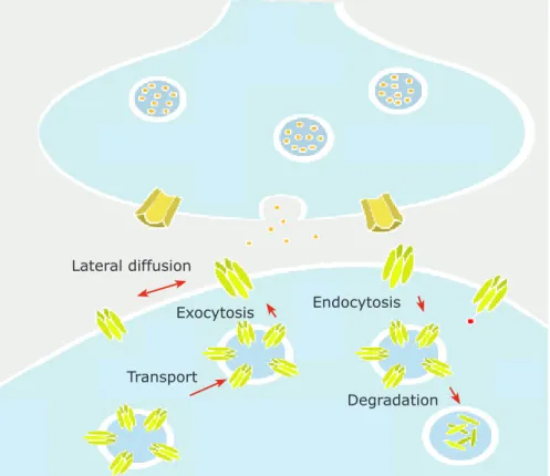
Trafficking within the surface membrane has recently emerged as a key step for regulating synaptic responses. Exocytosis, endocytosis and lateral trafficking have been highlighted as a key process in receptor renewal and concentration at synapses, accounting for the construction and plasticity of synapses in the membrane (Fig 1.3). Recent studies indicate that AMPAR endocytosis occurs in endocytic zones positioned
in the vicinity of the PSD (Blanpied et al., 2002; Petralia et al., 2003; Racz et al. 2004; Lu et al., 2007). Receptors are not simply degraded upon internalization, as previously thought (Gardner and Fambrough, 1979). Instead, a significant number of receptors, rather than being metabolized upon internalization, recycle back into the postsynaptic membrane. Interestingly, both endocytic zones and local receptor recycling are required to maintain a mobile pool of receptors at synapses (Petrini et al., 2009).
1.3.4.3 Molecular determinants of lateral diffusion in the cell membrane – lipids, scaffolds and cytoskeleton
Membrane compartmentalization explains, in part, the heterogeneous diffusion of receptors in the cell surface. The cohesive forces that assemble and maintain different microdomains in the cell membrane include lipid–lipid, protein–protein, and protein–lipid interactions, as well as sub- and supramembrane effectors (cytoskeletal, extracellular
Exocytosis
Transport
Degradation Endocytosis Lateral diffusion
Figure 1.3 – Receptor exchanges between synaptic, extrasynaptic and intracellular compartments. The extrasynaptic receptors can access synapses by lateral diffusion. The surface receptors are exchanged with the intracellular pool by insertion and internalization. The intracellular pool also features receptor synthesis, transport, recycling and degradation.
matrix) (Anderson and Jacobson K, 2002; Kusumi et al. 2004; Kwik et al., 2003; Murase et al., 2004; Nicolau et al., 2006; Lenne et al., 2006).
Biological membranes contain hundreds of lipids with different properties and can undergo dynamic changes in composition. The phospholipids form the major lipid part of biological membranes and are composed of two fatty acids plus a phosphate attached to a glycerol. Their hydrophilic head and hydrophobic tail constitute the basis for the formation of self-assembled phospholipid bilayers or micelles in aqueous environments. The great variety of phospholipid molecular species, the differences in their molecular shapes, physical properties, and their asymmetric distribution in the membrane bilayer possibly contribute to the formation of membrane microdomains (Shaikh et al., 2001). Cholesterol is another essential lipidic component of membranes that influences membrane fluidity, membrane protein trafficking, and consequently regulates neurotransmission and receptor trafficking (Allen et al., 2007; Renner et al., 2009). When introduced into lipid bilayers, cholesterol intercalates between the hydrocarbon parts of the other lipids, filling in the flickering spaces between the acyl chains. Because of its rigid planar structure, cholesterol increases the order of the neighboring acyl chains, making the membranes laterally more condensed and more densely packed. Thus, the physicochemical properties of the membrane are altered; in particular, permeability is decreased and mechanical strength and rigidity increased (Needham et al., 1988; Feigenson and Buboltz, 2001). Additionally, cholesterol can also appear concentrated in microdomains or rafts, which are composed by cholesterol and sphingolipids in the exoplasmic leaflet of the bilipid layer and cholesterol and glycerophospholipids in the endoplasmic leaflet. The lipid rafts can measure several tens of nanometers in diameter and behave as “moving platforms” (Simons and Ikonen, 1997; Pralle et al., 2000). One of the most important properties of lipid rafts is that they can include or exclude proteins to variable extents based on how well they fit within this organized lipid environment (Simons and Ikonen, 1997;). Proteins with raft affinity include glycosylphosphatidylinositol-anchored proteins, doubly acylatedproteins, such as Src-family kinases or the a-subunits of heterotrimeric G proteins, cholesterol-linked and palmitoylated proteins, and transmembrane proteins, particularly palmitoylated ones (Levental et al., 2010). Thus, lipid rafts serve as platforms that organize different
signaling components into dynamic modules and control their subcellular sorting and efficient function.
Cholesterol plays a crucial role in the generation of ordered domains in the plasma membrane that laterally segregate certain proteins, thus reducing their rate of lateral diffusion and, by virtue of this, increasing clustering and consequently signaling strength (Edidin, 2003; Hancock and Parton, 2005; and Hancock, 2006; Lingwood and Simons, 2010). For these reason, lipid rafts determine the functional properties of membrane-resident proteins like ion channels and transmitter receptors (Allen et al., 2007).
A vast number of components have been identified linking directly or indirectly AMPA and NMDA receptors in the PSD (Newpher and Ehlers, 2007). The number of scaffold proteins in the PSD exceeds the number of receptors by a big margin, ensuring plenty of ‘slots’ for the various binding partners. The members of the membrane associated guanylate kinases (MAGUKs) family are the most abundant scaffolds in excitatory synapses and are the best-described scaffolds of glutamate receptors (Elias and Nicoll, 2007). This family includes PSD95, PSD93, SAP102 and SAP97. A common feature of all these proteins is that they share the same organization with three N-terminal
post synaptic density protein (PSD95)/Drosophila disc large tumor suppressor (DlgA)/ zonula occludens-1 protein (zo-1) (PDZ) domains, a Src-homology 3 (SH3)
domain and a C-terminal catalytically inactive guanylate kinase (GK) domain (Cho et al., 1992; Feng and Zhang, 2009). These domains interact with a relatively weak binding affinity to the small peptide fragments situated at the very carboxyl tail of the scaffolds’ targets, thereby ensuring the dynamic range of synaptic responses. Despite the similarities in domain structure, PSD95-like MAGUKs (PSD-MAGUKs) are distinct in their N-terminal amino acid sequences, which could account for the selectivity of their interactions. For example, PSD-95 itself binds directly to the intracellular C-terminal of NMDA receptors (Kornau et al., 1995), and together with other associated PSD-95 molecules, links numerous components in an elaborate postsynaptic scaffold. On the other hand, PSD95 requires a TARP link to bind AMPARs (Hashimoto et al., 1999; Chen et al., 2000), as well to other components important for signal transduction such as calcium/calmodulin-dependent protein kinase II (CaMKII). SAP102 and PSD-93 are related members of the PSD-95 family and perform similar functions at glutamatergic
synapses depending on the developmental stage and location of the synapse (Elias et al., 2006). The fourth member of the family, SAP97, plays a different role, facilitating AMPA receptor trafficking to the surface membrane, for example (Nakagawa et al., 2004).
The cytoskeleton provides the stability for receptor anchoring and controls the “apparent viscosity” of mammalian plasma membranes (Gulley and Reese, 1981; Kusumi et al., 2005). A well-accepted hypothesis is that the cytoskeleton cortex hinders protein movements by controlling (1) the avidity of the postsynaptic scaffold for receptors; (2) creating fences below the membrane or by anchoring transmembrane molecules, which then act as obstacles to lateral diffusion (Kusumi and Sako, 1996; Saxton and Jacobson, 1997) and likely controlling (3) adhesion molecules, which create permeable barriers at the edge of excitatory and inhibitory synapses (Triller and Choquet, 2003). The cytoskeleton actions above described depend on both actin (Juliano, 2002; Yamagata et al., 2003; Bamji, 2005) and microtubules (Barth et al., 1997; Schoenwaelder and Burridge, 1999). Filamentous actin (F-actin) is the main cytoskeletal component and forms a large cortical meshwork that is concentrated beneath excitatory and inhibitory postsynaptic membranes, just below the postsynaptic scaffold (Dillon and Goda, 2005). Microfilaments are important for maintaining synaptic integrity and function and their depolymerization can disrupt the signaling of activated neurotransmitter receptors (Charrier et al., 2006). The cytoskeleton, together with the subsynaptic proteins, might constitute submembranous diffusion fences; adhesion proteins are probably passive obstacles that hinder diffusion, whereas scaffolding molecules probably restrict diffusion through specific interactions with given receptors (Choquet and Triller, 2003).
1.3.4.4 Trafficking of neurotransmitter receptors that underlie synaptic plasticity – the example of long-term potentiation in CA1 hippocampal region
The most extensively studied and therefore prototypic form of synaptic plasticity is LTP observed in the CA1 region of the hippocampus. In LTP, activation of AMPARs by presynaptically release glutamate must depolarize the membrane to an extension that allows relieving the voltage-dependent block of the NMDAR by magnesium (Mg2+)