Improving Outcomes with Multifocal
Intraocular Lenses
Filomena Ribeiro1, Tiago Ferreira1, Bernardo Feijóo2, Rita Dinis da Gama2, Cláudia Gonçalves3, Joana Couceiro3, Ana Souza e Silva3
1Hospital da Luz Refractive Department 2Hospital da Luz OCT Department 3Hospital da Luz Glaucoma Department
Background
With increasing life expectation, cataract surgery became one of the most frequent surgeries in developed countries. With the growing numbers and improving results came increased patient expectations. The introduction in the 80s of multifocal intraocular lenses (MIOL) offered the possibility of spectacle independence after cataract surgery or refractive lens exchange. Although the results with these intraocular lenses (IOLs) improved dramatically over the last decades, some of the problems inherent to the design of the lenses remain, namely reduced contrast sensitivity, especially in mesopic conditions and unwanted photic phenomena.
The preoperative evaluation of these patients is essential in screening retinal and optical nerve pathologies that may imply limitations on postoperative satisfaction.
Also, we cannot neglect the postoperative follow up of these patients, so it is vitally important to know the effects of optical diffractive multifocal lens on the interpretation of the results of evaluation by optical coherence tomography (OCT) and automated perimetry.
ImprovIng outcomes wIth
preoperatIve oct
OCT produces real-time, non-contact, high resolution, cross-sectional images of the retina, enabling the identifi-cation of the alterations in its morphology.1,2 OCT imaging may also be used to quantitatively measure structures such as retinal thickness or retinal nerve fiber layer thickness.1 Cata-ract influences both OCT image quality and retinal thickness measurements. However, even in the presence of cataract, OCT scans of individual patients remain reliable for clinical interpretation of gross retinal pathology (defined as signal strength ≥ 6/10), meaning that a foveal contour is often dis-cernable, whereas detail on intraretinal structures might be
lost.3,4 Furthermore, OCT has been shown more effective than indirect ophthalmoscopy or stereoscopic fundus photography in detecting maculopathy (such as idiopathic epiretinal mem-brane, age-related macular degeneration and ischemic atro-phy) in the preoperative examination of patients undergoing cataract surgery. Klein et al. studied 149 patients scheduled for cataract surgery and implantation of a MIOL or toric IOL whose clinical history and examination had excluded macu-lar pathology. In this group spectral-domain OCT identified macular abnormalities in 13,2% of scans.5 Similarly, In our experience, OCT is sometimes responsible for excluding some candidates to MIOLs, as illustrated in Figure 1.
Fig. 1 | 56 year-old woman being evaluated for cataract surgery whose fundoscopy and retinography (a) showed no rema-rkable findings. The macular OCT (b), however, revealed vitreomacular traction with loss of the normal foveal con-tour – a possible contraindication for MIOL implantation.
a
For reasons stated above, OCT has become a fundamen-tal evaluation tool in candidates for MIOL implantation, safeguarding the patient’s and surgeon’s interests. In short term follow-up studies, photic phenomena are the main cause of MIOL explantation.6 It is likely that, if studies with longer follow-up were conducted, retinal pathology might become an important cause of MIOL explantation and patient dissatisfaction, once again overstressing the role of OCT in this context. And, we must also consider that, as OCT rapidly becomes the standard in the evaluation and identification of vitreoretinal pathology, the application of this instrument to eyes with a MIOL will increase as patients age and the risk of vitreoretinal disorders, namely macular degeneration and preretinal membranes, also increases.
optIcal coherence tomography aFter mIol ImplantatIon
Multifocal intraocular lenses have been associated with quality of vision issues, particularly in low lightning conditions. But it is less well known the effect these IOLs can have on retinal imaging and measurements from devi-ces such as OCT. There is a report of reduced OCT signal strength with refractive multifocal contact lenses.7 A pre-vious study (Inoue et al.) described wavy artifacts in the image on the line-scanning ophthalmoscope of the spec-tral domain-OCT (Cirrus 4000 HD-OCT) in patients with diffractive MIOLs. However, despite these artifacts seen on OCT line-scanning ophthalmoscopic images, the OCT and fundoscopic images in eyes with a MIOL were comparable to those in eyes with a monofocal IOL.8
There are also two other studies9,10 which evaluated the impact of MIOLs on the accuracy of retinal OCT mea-surements through comparison with a control group with monofocal IOL. Skiadaresi et al. evaluated OCT measure-ments following implantation of LENTIS Mplus (Oculentis GmbH, Berlin, Germany), a refractive MIOL. They used
Topcon 3D OCT-1000 (Topcon, Oakland, USA) and found neither image artifacts nor alteration in macular thickness or volume measurements.10 Dias-Santos et al. accessed the accuracy of retinal OCT in patients with a diffractive MIOL: Acrysof ReSTOR SA60D3 (Alcon Laboratories, Fort Worth, USA) or Tecnis ZM900 (Abbott Medical Optics) using OCT Heidelberg Spectralis (Heidelberg Engineering, Heidelberg, Germany). They found a statistically signifi-cant decrease in the OCT image quality in the diffractive IOL group, but the measurements in the macular area were not affected by the optical design of diffractive IOLs.9
Our group also studied 30 eyes of 16 patients implan-ted with MIOL: TECNIS Symfony (17 eyes) or trifocal Finevision (13 eyes), which were compared with a control group with 12 eyes of 8 patients who underwent uneventful phacoemulsification with implantation of a monofocal IOL: TECNIS 1-Piece Aspheric IOL (Abbott Medical Optics) or Acrysof Aspheric IOL (Alcon Laboratories). The demogra-phic data of the patients is presented in Table 1.
We included only eyes without significant ocular comor-bidities (namely posterior capsule opacification, glaucoma, corneal or vitreoretinal pathology). The Cirrus-HD OCT 4000 was used to perform macular imaging at least 1 month postoperatively in all eyes. Acquisition was made with the macular cube 512x128 scan. This mode acquires scans at a length of 6.0x6.0 mm and with a resolution of 128 lines of 512 A-scans per line, with the fixation on the macula. Central thickness and macular volume were recorded. We also calculated the mean thickness in the 3 mm and 6 mm concentric circles of the automatic map (average of the all the quadrants in each circle). The signal strength was obtai-ned for all eyes. Statistical comparisons between groups for macular thickness, macular volume and OCT signal strength were assessed with Mann-Whitney U test, with a p-value of 0.05 being considered as statistically significant. Our results are summarized in Table 2.
There were no statistically significant differences in any measured or calculated values for macular thickness and table 1 | patient demographic data and implanted Iol model.
multifocal Iol group monofocal Iol group
sex (m/F) 4/12 2/6
age (y) 63 (range 50-80) 71 (range 58-84)
spherical equivalent (mean ± sd) (d) 0,88 ± 2,59 0,86 ± 2,88
macular volume between the two groups. However, median OCT signal strength was significantly higher (p=0,002) in the monofocal IOL group (9,50) compared with the MIOL group (9,00), indicating a better image quality in this group. Nevertheless, in the MIOL group mean signal strength was still over 6 – the minimum quality score recommended by the manufacturer. However, our results should be interpre-ted with caution since our monofocal IOL group had a small number of patients and we evaluated different models of IOL in each group.
In conclusion, the optical design of MIOLs may affect OCT imaging, however the available data show that it does not seem to compromise the role of this important tool in the diagnosis and follow-up of vitreoretinal disorders.
mIols In glaucoma patIents. Is It possIBle?
When approaching a patient with cataract and glaucoma who desires spectacle independence, the severity of the disease must be considered. As discussed above, with any premium surgery, an evaluation of the retina and optic nerve with OCT is increasingly more common, being a valuable tool not only to quantify glaucomatous damage but also to detect macular disease and predict outcomes. Preexisting visual field defects that might influence the function of a MIOL should also be considered.
Some of the new technologies included in premium IOLs are of particular importance in glaucomatous patients.
Considering that pupilar response in glaucoma patients can be altered, and as we know that pupil size can influence some types of premium IOLs performances, it is advisable to choose a premium IOL model independent of pupil’s size (ie. diffractive versus refractive multifocal IOLs). Aspheric IOLs compensate for the positive spherical aberration of the cornea and have been shown to improve mesopic and sco-topic contrast sensibility after cataract surgery, as well as to decrease the incidence of unwanted photic phenomena. It has been demonstrated that glaucoma reduces contrast sensitivity independent of visual acuity. This reduction affects primarily mesopic levels and is correlated not only with visual field loss but also quality of life. As glaucoma decreases contrast sensitivity, the choice of aspheric IOLs may be even more important in these patients. In addition, this technology may be combined with toricity to correct preexisting astigmatism which might reduce visual function after cataract surgery with a MIOL. Since corneal astigma-tism induction is greater with trabeculectomy than with other glaucoma surgeries, care must be taken with using toric IOLs when combining trabeculectomy with cataract surgery.11,12
A newer IOL technology is the TECNIS Symfony extended depth of focus IOL. This IOL not only corrects spherical but also chromatic aberration of the eye, resul-ting in an increased depth of focus which, as claimed by the manufacturer, improves intermediate vision and results in higher spectacle independence with less photic pheno-mena. Furthermore, the study of the visual performance of this IOL shows a superior contrast sensitivity function table 2 | medians of central macular thickness, mean macular thickness in 3 mm and 6 mm circles, macular volume, oct
sig-nal strength and statistical significance between the two IOL groups.
multifocal Iol group monofocal Iol group P-value Central macular thickness (μm)
median (min, max); IQr (221.00,298.00); 23.25269.50 (222.00-283.00); 14.00264.00 0.263
mean macular thickness 3 mm circle (μm) median (min, max); IQr
330.13
(286.75, 348.50); 31.13 (286.25,344.75);24.44318.13 0.146
mean macular thickness 6 mm circle (μm) median (min, max); IQr
282.50
(253.00,309.50); 27.94 (246.00,297.25); 22.56275.75 0.263
macular volume (mm3)
median (min, max); IQr (9.20-11.10);1.0310.40 (9.10,10.90);0.8010.00 0.240
oct signal strength median
in comparison with other MIOLs.13 Given these specific characteristics and outcomes, this IOL may be an option in patients with early to moderate glaucoma desiring some level of spectacle independence.
In summary, although concomitant cataract and glau-coma may represent, nowadays, a relative contraindication for implanting premium IOLs, a careful patient evaluation and selection can reveal potential candidates for MIOL implantation. For patients with ocular hypertension, glau-coma suspects and early stable glauglau-coma, any of the pre-mium IOLs (multifocal, extended depth of focus) can be an option. In moderate or severe glaucoma, the surgeon must consider several factors, including preexisting visual defi-cits, characteristics of the IOL to be implanted and the effect of surgery in the future evaluation and follow-up of glau-coma. In the particular case of glaucoma patients with pseu-doexfoliation it is debatable, even those with stable disease, given the risk of intraoperative complications and late pos-toperative IOL decentration when there is zonular weak-ness. So should be excluded progressing patients and those with severe glaucoma or pseudoexfoliative glaucoma.14
Functional and structural evaluation of glaucoma patient should be repeated soon after the eye has recovered following cataract surgery, because, as already mentioned, all the investigations can be compromised by crystalline opacification, and a new baseline can be established.
Cataract surgery can improve the quality of vision in patients with glaucoma, although lost contrast sensitivity and visual field defects remain unchanged. Unfortunately, there is little published data of the outcomes of cataract sur-gery with MIOLs in patients with glaucoma, most of the evi-dence being from anecdotal experience. One article reported outcomes of cataract surgery with MIOLs including patients with glaucoma and found similar outcomes to monofocal IOLs, except for improved near visual acuity.15 For these reasons, the use of a MIOL in these patients must be approa-ched with caution, through a careful informed consent pro-cess reviewing the benefits and drawbacks of this kind of lenses, especially as the severity of glaucoma increases.
postoperatIve automated perImetry wIth mIol
Glaucoma is an insidious chronic eye disease that results in retinal sensitivity loss. This loss may be evaluated through automated perimetry, which is a fundamental tool for glau-coma diagnosis and follow-up. Since both glauglau-coma and MIOLs may reduce contrast sensitivity, it is possible to theo-rize that MIOLs may affect proper glaucoma assessment.
Cataract also decreases the sensitivity of diagnostic tests that document glaucoma progression. There have been many studies regarding cataract extraction impact on visual field in glaucoma patients, but none of them was centered on MIOLs. Those studies have demonstrated that visual field parameters, namely mean deviation (MD) and pattern standard deviation (PSD) changed after cataract extraction. MD was shown to improve after cataract surgery in the majority of studies. Concerning PSD values, the data are not so consistent; some studies have shown no change in PSD postoperatively, while others have demonstrated dete-rioration of PSD.16
Patients with diffractive MIOL have clinically relevant reduction of the visual sensitivity as assessed with stan-dard automated perimetry size III and size V. The reduc-tion seems to be related to the multifocal design of the IOL rather than to pseudophakia.17 This reduction interfe-res with the assessment of common eye diseases such as glaucoma.
Farid et al. regarded the effect of MIOL on nonspeci-fic reduction of MD upon Humphrey standard achromatic perimetry (SAP) 10-2 testing with Swedish Interactive Threshold Algorithm (SITA) standard thresholds.18
A subtle contrast sensitivity (CS) change might already alter central 10-2 visual field performance. Pierre et al. noted that the luminance contrast values of patients with yellow-tinted IOLs were significantly lower than those of patients with clear IOLs, in a series of 25 patients.19 And Vingolo EM et al. compared the visual acuity and CS in eyes with the Acrysof ReSTOR multifocal intrao-cular lens and eyes with the monofocal Acrysof SA60AT IOL. The MIOL provided lower contrast sensitivity than monofocal IOL.20
There is stronger evidence of reduced retinal sensitivity in subjects with MIOLs compared to subjects with mono-focal IOLs, however most of the studies were performed in healthy subjects, not in patients with glaucoma.
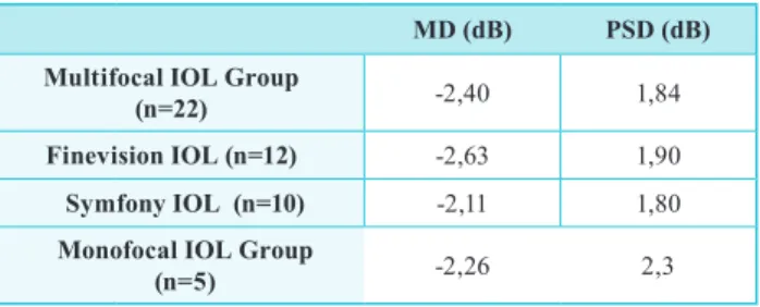
We studied a small group of healthy eyes with MIOL implantation (n=22), 10 eyes had TECNIS Symfony and 12 eyes had trifocal Finevision IOL. The results, in terms of MD and PSD, are shown in Table 3.
We found a mean MD of -2,4 dB, being lower for the Finevision Group (-2,63 dB) when compared to the Symfony group (-2,11 dB). This difference was not statisti-cally significant (p>0,05). We had a small monofocal IOL implanted group, with 5 eyes. As the literature reports, our MD value for monofocal IOLs was higher than the mean MD for multifocal. Even though Symfony eyes had higher MD than monofocal eyes. The small number of eyes inclu-ded does not allow definite conclusions.
conclusIons
A comprehensive preoperative assessment is manda-tory when considering the implantation of a MIOL. OCT has become an essential tool in this task, allowing a tho-rough macular analysis and identifying potential vitreoma-cular abnormalities that may compromise the surgical out-come. It should be part of the evaluation of every candidate to a MIOL.
The implantation of premium IOLs in patients with cataract and concurrent glaucoma is still controversial. The progressing nature of the disease, the defects in visual function that can be induced and the presence of anatomic characteristics that can compromise the surgical outcome complicate the decision of using premium IOLs in glau-coma patients. There are, however a subgroup of glauglau-coma patients in which the use of these IOLs can be considered: ocular hypertension, glaucoma suspects and early stable glaucoma.
The influence this IOL technology can have on postope-rative follow-up and evaluation of our patients is still uncer-tain. Although for OCT imaging the available data suggest some compromise of image quality, the retinal measure-ments made do not seem to be affected by the diffractive optics of the IOLs. For automated perimetry, the limited published data trends towards a reduced retinal sensitivity in SAP that seems related to the multifocal design of the IOLs. The lack of large randomized trials of premium IOLs use in patients with glaucoma does not allow to establish the impact these IOLs design can have on glaucoma diag-nosis or progression assessment. Nevertheless, it is proba-bly advisable to set a new perimetric baseline in patients with MIOLs with (suspect) glaucoma and preferably in all patients with MIOLs to guarantee a correct interpretation of any future abnormality.
Our goal should be to meet our patient’s expectations, without adversely influencing future disease diagnosis, monitoring and possible treatments.
With these considerations in mind, we will be able to take better advantage of this technology and the increased number of patients it brings to our practices for many years to come. Further studies are, however, necessary to achieve better outcomes.
reFerences
1. Hee MR. Interpretation of the optical coherence tomo-graphy image. In: Schuman JS, Puliafito C, Fujimoto JG, Duker JS, eds. Optical Coherence Tomography of Ocu-lar Diseases. :3-20.
2. Nordmann JP. Optical Coherence Tomography & Optic Nerve. Paris: Laboratoire Théa; 2014.
3. van Velthoven MEJ, van der Linden MH, de Smet MD, Faber DJ, Verbraak FD. Influence of cataract on optical coherence tomography image quality and retinal thickness. British Journal of Ophthalmology. 2006;90(10):1259-1262. doi:10.1136/bjo.2004.097022. 4. Garcia-Medina JJ, Del Rio-Vellosillo M,
Zanon--Moreno V, et al. Does Posterior Capsule Opaci-fication Affect the Results of Diagnostic Techno-logies to Evaluate the Retina and the Optic Disc? Biomed Res Int. 2015;2015(3):813242-813248. doi:10.1155/2015/813242.
table 3 | standard automated perimetry results: mean deviation (md) and pattern standard deviation (psd) values for each Iol group
md (dB) psd (dB)
multifocal Iol group
(n=22) -2,40 1,84
Finevision Iol (n=12) -2,63 1,90
symfony Iol (n=10) -2,11 1,80
monofocal Iol group
(n=5) -2,26 2,3
keypoints
• OCT identified macular abnormalities in 13,2% in
patients scheduled for cataract surgery and implantation of a MIOL whose clinical history and examination had excluded macular pathology.
• Optical design of MIOLs may affect OCT imaging,
however the available data show that it does not seem to compromise the role of this important tool in the diagno-sis and follow-up of vitreoretinal disorders.
• Although concomitant cataract and glaucoma may
represent, nowadays, a relative contraindication for implanting premium IOLs, a careful patient evaluation and selection can reveal potential candidates for MIOL implantation.
• It is probably advisable to set a new perimetric baseline
in patients with MIOLs with (suspect) glaucoma and preferably in all patients with MIOLs to guarantee a correct interpretation of any future abnormality.
• The limited published data trends towards a reduced
retinal sensitivity in standard achromatic perimetry that seems related to the multifocal design of the IOLs.
5. Klein BR, Brown EN, Casden RS. Preoperative macu-lar spectral-domain optical coherence tomography in patients considering advanced-technology intraocu-lar lenses for cataract surgery. Journal of Cataract & Refractive Surgery. 2016;42(4):537-541. doi:10.1016/j. jcrs.2016.01.036.
6. Foroozan R. Non-organic Visual Loss in Patients with Multifocal Intraocular Lenses. J Ophthalmic Vis Res. 2012;7(4):305-309.
7. Posner M, Naroo SA, Nithyanandarajah G, Trivedy M, Sharma A. Multifocal contact lenses and posterior pole imaging. Cont Lens Anterior Eye. 2010;33(3):151. doi:10.1016/j.clae.2010.02.005.
8. Inoue M, Bissen-Miyajima H, Yoshino M, Suzuki T. Wavy horizontal artifacts on optical coherence tomo-graphy line-scanning images caused by diffractive mul-tifocal intraocular lenses. Journal of Cataract & Refrac-tive Surgery. 2009;35(7):1239-1243. doi:10.1016/j. jcrs.2009.04.016.
9. Dias-Santos A, Costa L, Lemos V, et al. The impact of multifocal intraocular lens in retinal imaging with opti-cal coherence tomography. International ophthalmology. 2015;35(1):43-47. doi:10.1007/s10792-014-0016-8. 10. Skiadaresi E, McAlinden C, Ravalico G, Moore J.
Optical coherence tomography measurements with the LENTIS Mplus multifocal intraocular lens. Graefe’s Arch Clin Exp Ophthalmo. 2012;250(9):1395-1398. doi:10.1007/s00417-011-1901-8.
11. Paletta Guedes R-A, Paletta Guedes V-M, Aptel F. [Multifocal, toric, and aspheric intraocular lenses for glaucoma patients]. Journal francais d’ophtalmologie. 2011;34(6):387-391. doi:10.1016/j.jfo.2011.02.003. 12. Martínez Palmer A, Gómez Faiña P, España Albelda
A, Comas Serrano M, Nahra Saad D, Castilla Céspedes M. Visual function with bilateral implantation of mono-focal and multimono-focal intraocular lenses: a prospective, randomized, controlled clinical trial. J Refract Surg. 2008;24(3):257-264.
13. Ribeiro F, Salgado Borges J, González-Méijome JM, Escandón-García S. Challenges in evaluating features of new premium intraocular lenses. Journal of Emmetropia. 2016;7(1):5-6.
14. Tan H-Y, Wu S-C. Refractive error with optimum intraocular lens power calculation after glaucoma filte-ring surgery. Journal of Cataract & Refractive Surgery. 2004;30(12):2595-2597. doi:10.1016/j.jcrs.2004.05.016. 15. Iancu R, Corbu C. Premium intraocular lenses use
in patients with cataract and concurrent glaucoma: a review. Maedica (Buchar). 2013;8(3):290-296.
16. Koucheki B, Nouri-Mahdavi K, Patel G, Gaasterland D, Caprioli J. Visual field changes after cataract extraction: the AGIS experience. AJOPHT. 2004;138(6):1022-1028. doi:10.1016/j.ajo.2004.08.006.
17. Aychoua N, Junoy Montolio FG, Jansonius NM. Influence of multifocal intraocular lenses on stan-dard automated perimetry test results. JAMA Ophthalmol. 2013;131(4):481-485. doi:10.1001/ jamaophthalmol.2013.2368.
18. Farid M, Chak G, Garg S, Steinert RF. Reduction in mean deviation values in automated perimetry in eyes with multifocal compared to monofocal intraocular lens implants. Am J Ophthalmol. 2014;158(2):227–231.e1. doi:10.1016/j.ajo.2014.04.017.
19. Pierre A, Wittich W, Faubert J, Overbury O. Lumi-nance contrast with clear and yellow-tinted intraocu-lar lenses. Journal of Cataract & Refractive Surgery. 2007;33(7):1248-1252. doi:10.1016/j.jcrs.2007.03.024. 20. Vingolo EM, Grenga P, Iacobelli L, Grenga R. Visual
acuity and contrast sensitivity: AcrySof ReSTOR apo-dized diffractive versus AcrySof SA60AT monofo-cal intraocular lenses. Journal of Cataract & Refrac-tive Surgery. 2007;33(7):1244-1247. doi:10.1016/j. jcrs.2007.03.052.
Os autores não têm conflitos de interesse a declarar.
Trabalho não publicado cedendo os direitos de autor à Sociedade Portu-guesa de Oftalmologia.
contacto
Filomena Ribeiro, MD,PhD,FEBO
Head of ophthalmology Department at Hospital da Luz e-mail: [email protected]