Research Article
Antimicrobial, Antioxidant, Anti-Inflammatory,
and Cytotoxic Activities of Propolis from the Stingless Bee
Tetragonisca fiebrigi
(Jataí)
Jaqueline Ferreira Campos,
1Uilson Pereira dos Santos,
1Paola dos Santos da Rocha,
1Marcio José Damião,
1José Benedito Perrella Balestieri,
1Claudia Andrea Lima Cardoso,
2Edgar Julian Paredes-Gamero,
3Leticia Miranda Estevinho,
4Kely de Picoli Souza,
1and
Edson Lucas dos Santos
11School of Environmental and Biological Science, Federal University of Grande Dourados, Rodovia Dourados-Itahum,
Km 12, 79804-970 Dourados, MS, Brazil
2Course of Chemistry, State University of Mato Grosso do Sul, Rodovia Dourados-Itahum, Km 12, 79804-970 Dourados, MS, Brazil
3Department of Biochemistry, Federal University of S˜ao Paulo, Rua Pedro de Toledo 669, 04039-032 S˜ao Paulo, SP, Brazil
4CIMO/ESA, Department of Biology and Biotechnology, Agricultural College of Braganc¸a, Polytechnic Institute of Braganc¸a,
Campus Santa Apol´onia, 5301-855 Braganc¸a, Portugal
Correspondence should be addressed to Edson Lucas dos Santos; [email protected] Received 15 April 2015; Revised 3 June 2015; Accepted 3 June 2015
Academic Editor: Vincenzo De Feo
Copyright © 2015 Jaqueline Ferreira Campos et al. This is an open access article distributed under the Creative Commons Attribution License, which permits unrestricted use, distribution, and reproduction in any medium, provided the original work is properly cited.
Propolis from stingless beesTetragonisca fiebrigifound in Brazil is used in folk medicine by their nutritional and therapeutic properties. However, there are no scientific records evidencing such properties. The present study was designed to investigate the chemical composition and the biological properties of propolis fromT. fiebrigi. For this, the chemical composition of the ethanol extract of propolis (EEP) was determined by GC-MS and presented phenolic compounds, alcohol, and terpenes as its major class compounds. The antimicrobial activity was accessed in gram-positive and gram-negative bacteria and in fungi, isolated from different biological fluids and reference strains. The EEP was active against all microorganisms and showed antioxidant activity by scavenging free radicals, inhibiting hemolysis and lipid peroxidation in human erythrocytes incubated with an oxidizing agent. The anti-inflammatory potential of the EEP was confirmed by inhibition of the hyaluronidase enzyme. The cytotoxic activity was concentration-dependent against K562 cells, with a predominance of death by necrosis. Taken together, these results show that propolis fromT. fiebrigihas important therapeutic activities, which suggest its potential application in the pharmaceutical industry, as well as in health foods, beverages, and nutritional supplements.
1. Introduction
More than 200 species of stingless bees have been described in Brazil [1]. Among these species, Tetragonisca fiebrigi, popularly known in Brazil as Jata´ıor abelha-ouro (golden bee), covers a large part of the Brazilian territory, including the midwest, south, and southeast regions, as well as countries such as Argentina, Bolivia, and Paraguay [2,3]. Recently, the taxonomic classification of this species has been revised. The species was previously described as Tetragonisca angustula
fiebrigisubspecies along withT. a. angustula; however, molec-ular markers studies showed them to be distinct species [4,5]. Among the species of stingless bees,T. fiebrigiis known as a good producer of honey and propolis, which are highly prized in folk medicine for their nutritional and therapeutic properties. Despite the popular use of these products, only the antibacterial and antioxidant properties of the honey produced by this species have been described in the literature [6,7], and there are no scientific records of the therapeutic properties attributed to the propolis produced by these bees.
Propolis is a resinous substance produced by bees through the mixture of jaw secretions and the exudate collected from plant materials [8, 9]. This resin is used in the construction, maintenance, and asepsis of the nest [10]. There are records that ancient civilizations, such as the Incas, Greeks, Egyptians, and Romans, used propolis for its therapeutic properties, being described as antiseptic, healing, and antipyretic [11]. Additionally, this natural product has also been used in the preparation of drinks and foods for human nutrition, aiming at the improvement in health and disease prevention [12].
Indeed, recent studies expanded the knowledge on the therapeutic applications of propolis from different species of stingless bees, describing their antimicrobial [13, 14], antioxidant [15,16], and antitumor [12,17] properties.
These medicinal properties are directly related to the chemical composition. Studies of propolis from stingless bees describe the presence of phenolic acids, aromatic acids, terpenes, and carbohydrates [1,16,18]. The bee species and the botanical source from which the bees collect the resin are among the factors responsible for the physical, chemical, and biological characteristics of the propolis [12,15].
Therefore, this study describes for the first time the chemical composition and antimicrobial, antioxidant, anti-inflammatory, and cytotoxic properties of propolis produced by the stingless beeT. fiebrigi.
2. Materials and Methods
2.1. Preparation of the Ethanol Extract of Propolis (EEP).
Propolis samples fromT. fiebrigi bees (see Supplementary Material available online at http://dx.doi.org/10.1155/2015/ 296186) were collected from the state of Mato Grosso do Sul, in the midwest region of Brazil. Three colonies were identified, and six samples of propolis were collected with approximately 2.14 g each. The ethanol extract of propolis (EEP) was prepared with 4.5 mL of 80% ethanol per 1 g of propolis, kept in a water bath at 70∘C in a sealed container until total dissolution, and subsequently filtered to obtain the EEP [19].
2.2. Chemical Analysis. One milligram of dry extract was reacted with 50𝜇L pyridine + 100𝜇L bis(trimethylsilyl) triflu-oroacetamide (BSTFA) including 1% trimethylchlorosilane (TMCS) in a sealed glass tube for 30 min at 100∘C to prepare samples for gas chromatography [20]. The procedure was performed in triplicate. Samples were injected and analyzed by gas chromatography-mass spectrometry (MS). The procedure was performed in triplicate. The GC-MS analysis was performed on a gas chromatograph (GC-17A, Shimadzu, Kyoto, Japan) equipped with a mass spec-trometer detector (QP 5050a), using DB-5 (J&W, 5% phenyl-dimethylpolysiloxane), fused-silica capillary column (30 m in length× 0.25 mm i.d., and 0.25𝜇m film thickness), under the following conditions: carrier gas of helium (99.999% and flow rate 1.0 mL min−1), 1𝜇L injection volume, split ratio (1 : 10), initial oven temperature set at 85∘C, and heating from 85∘ to 315∘C at 4∘C min−1. The injector temperature
was 280∘C, and the quadrupole detector temperature was 315∘C. The MS scan parameters included an electron-impact ionization voltage of 70 eV, mass range of 45–600m/z, and scan interval of 0.5 s. Temperature-programmed retention indices [21] were calculated using mixtures of linear alkane (C8–C30) as external references. The identifications were completed by comparing the mass spectra obtained in the QP5050 libraries. In some cases, when identified spectra were not found, only the structural type of the corresponding component was proposed on the basis of its mass-spectral fragmentation. When possible, reference compounds were cochromatographed to confirm GC-retention times.
2.3. Antimicrobial Activity. In the present study microor-ganisms isolated from biological fluids were used, collected in the Hospital Centre, and identified in the Microbiology Laboratory of Escola Superior Agraria de Braganca (ESA), but also reference strains were obtained from the authorised distributor of ATCC (LGC Standards S.L.U., Barcelona). The microorganisms were Staphylococcus aureus ATCC 43300,
S. aureusESA 654,Staphylococcus epidermidisATCC 12228,
S. epidermidisESA 675,Enterococcus faecalisATCC 43300,
E. faecalis ESA 553, Klebsiella pneumonia ATCC 4352, K. pneumoniaeESA 154,Pseudomonas aeruginosaATCC 15442,
P. aeruginosa ESA 22, Proteus mirabilis ATCC 43300, P. mirabilisESA 37,Candida glabrataATCC 90030,C. glabrata
ESA 123, Candida albicans ATCC 90028, and C. albicans
ESA 345. The isolates were stored in Muller-Hinton medium plus 20% glycerol at −70∘C, before experimental use. The inoculum for the assays was prepared by diluting cell mass in 0.85% NaCl solution, adjusted to 0.5 in MacFarland scale, confirmed by spectrophotometric reading at 580 nm for bacteria and 640 nm for yeasts. Cell suspensions were finally diluted to 104CFU/mL in order to use them in the activity assays. Antimicrobial tests were carried out as previously described by Silva et al. [22], using Nutrient Broth (NB) or Yeasts Peptone Dextrose (YPD) on microplate (96 wells). Propolis extracts were diluted in dimethyl sulfoxide (DMSO) and transferred into the first well and serial dilutions were performed. The inoculum was added to all wells and the plates were incubated at 37∘C for 24 h (bacteria) and 25∘C for 48 h (yeast). Amphotericin B and gentamicin were used as controls. In each experiment a positive control (inoculated medium) and a negative control (medium) and DMSO control (DMSO with inoculated medium) were introduced. Antimicrobial activity was detected by adding 20𝜇L of 0.5% TTC solution (2,3,5-triphenyl-2H-tetrazolium chloride). The minimum inhibitory concentration (MIC) was defined as the lowest concentration of propolis extract that inhibited visible growth, as indicated by the TCC staining (dead cells are not stained by TTC).
performed in triplicate (𝑛 = 3) and results are expressed as mg/mL.
2.4. Antioxidant Activity
2.4.1. ABTS Radical Scavenging Assay. Free radical scav-enging capacity for EEP was studied as described by Re et al. [23], through the evaluation of the free radical scavenging effect on 2,2 -azinobis-(3-ethylbenzothiazoline-6-sulfonic acid) (ABTS) radical. The stock solutions included 7 mM ABTS solution and 140 mM potassium persulphate solution. The ABTS∙+ radical was then prepared by mixing the two stock solutions (5 mL of ABTS solution and 88𝜇L potassium persulphate solution) and left for 12–16 h at room temperature in the dark. The solution was then diluted by mixing 1 mL ABTS∙+radical with 50 mL ethanol absolute to obtain an absorbance of 0.70 nm±0.05 units at 734 nm using a spectrophotometer. Then, 20𝜇L of EEP (0.1–500𝜇g/mL) was mixed with 1980𝜇L of the ABTS∙+ radical and the absorbance was taken at 734 nm after 6 min using a spec-trophotometer. Ascorbic acid and butylated hydroxytoluene (BHT) were used as positive controls. Two independent experiments were performed in triplicate. The percentage of inhibition of the ABTS radical was calculated according to the following equation, where Abscontrolis the absorbance of
ABTS∙+radical without the tested sample:
% inhibition of ABTS= ((Abscontrol−Abssample) Abscontrol
)
× 100.
(1)
2.4.2. Antioxidant Assay Using a Human Erythrocyte Model
(1) Preparation of Erythrocyte Suspensions. Following approval by the Research Ethics Committee (Comitˆe de ´Etica em Pesquisa; CEP) of the University Center of Grande Dourados (Centro Universit´ario da Grande Dourados; UNIGRAN), Brazil (CEP process number 123/12), 10 mL of peripheral blood was collected from healthy donors into sodium citrate-containing tubes and was subsequently centrifuged at 1500 rpm for 10 min. After centrifugation, the blood plasma and leukocyte layers were discarded, and the erythrocytes were washed 3 times with saline solution and centrifuged. Finally, 10% erythrocyte suspension was prepared in saline, to obtain a final 2.5% after the treatments.
(2) Hemolytic Activity and Oxidative Hemolysis Inhibition. The protective effect of the propolis extract was evaluated according to the method described by Campos et al. [16], with minor modifications. The assays were conducted with erythrocyte suspensions. The erythrocytes were preincubated at 37∘C for 30 min in the presence of different concentrations of EEP or ascorbic acid (25–125𝜇g/mL). Then, 50 mM 2,2 -azobis-(2-amidinopropane) dihydrochloride (AAPH) solu-tion was added. A sample of 0.2% ethanol was used as a negative control. Total hemolysis was induced by incubating erythrocytes with distilled water. Basal hemolysis caused
by EEP was assessed by incubating erythrocytes with the extract without the presence of AAPH, and the control was assessed in erythrocytes incubated only with 0.9% NaCl. This mixture was incubated at 37∘C for 240 min, with periodical stirring. Hemolysis was determined after every 60 min of sample incubation for a total of 240 min; specifically, aliquots were removed, diluted in saline, and centrifuged at 1500 rpm for 10 min, after which the absorbance of the supernatant was read spectrophotometrically at 540 nm. The percentage hemolysis was measured with the formula𝐴/𝐵 × 100, where (𝐴) is the sample absorbance and (𝐵) is the total hemolysis. Two independent experiments were performed in duplicate.
(3) Inhibitory Efficiency against Lipid Peroxidation. The pro-tective effect of EEP against lipid peroxidation was evaluated according to the method described by Campos et al. [16], with minor modifications. Erythrocytes were preincubated at 37∘C for 30 min with different concentrations of EEP or ascorbic acid (25–125𝜇g/mL). A sample of 0.2% ethanol was used as a negative control. Next, 50 mM AAPH was added to the erythrocyte solution, which was then incubated at 37∘C with periodical stirring. Dosage of malondialdehyde (MDA), a byproduct derived from lipid peroxidation, was determined after every 60 min of sample incubation for a total of 240 min. The samples were centrifuged at 1500 rpm for 10 min, and 500𝜇L aliquots of the supernatant were transferred to tubes with 1 mL of 10 nmol thiobarbituric acid (TBA). As a standard control, 500𝜇L of 20 mM MDA solution was added to 1 mL of TBA. The samples were incubated at 96∘C for 45 min. The samples were then cooled, 4 mL of n-butyl alcohol was added, and the samples were centrifuged at 3000 rpm for 5 min. The sample supernatants were removed, and the absorbance was read at 532 nm. Two independent experiments were performed in duplicate. MDA levels in the samples were expressed in nmol/mL, obtained with the following formula:
MDA=Absorbancesample
× ( 20 × 220.32
AbsorbanceMDAstandard
) . (2)
1
2 34 5
6 7
8
9 10 12
13 1415 11
35 30 25 20 15 10 5
10 15 20 25 30 35 40 45 50 55 60 65 70 75 80 Retention time (min)
Ab
u
n
d
an
ce
Figure 1: GC-MS profile of EEP fromT. fiebrigi.
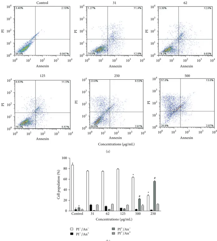
2.6. Cytotoxic Activity and Cell Death Profile. The K562 erythroleukemia cell line was grown in suspension in RPMI 1640 media (Cultilab, Campinas, Sao Paulo, Brazil) sup-plemented with 10% fetal bovine serum (FBS; Cultilab), 100 U/mL of penicillin, and 100𝜇g/mL of streptomycin in a humidified atmosphere at 37∘C in 5% CO2. The cytotoxic activity and cell death profile were evaluated according to the method described by Paredes-Gamero et al. [24], with minor modifications. Cells were seeded into 96-well plates (105cells/mL) and cultured in medium with 10% FBS in the absence or presence of EEP (31–500𝜇g/mL) for 24 h. After this period, the K562 cells were washed with PBS and resuspended in annexin-labeling buffer (0.01 M HEPES, pH 7.4, 0.14 M NaCl, and 2.5 mM CaCl2). The suspensions were stained with annexin-FITC and propidium iodide (PI) (Becton Dickinson, Franklin Lakes, NJ, USA), according to the manufacturer’s instructions. The cells were incubated at room temperature for 15 min. Ten thousand events were collected per sample, and the analyses were performed on a FACSCalibur flow cytometer (Becton Dickinson) with CellQuest software (Becton Dickinson). All the tests were performed in duplicate.
2.7. Statistical Analyses. The data are shown as the mean± standard error of the mean (SEM) and were analyzed for statistical significant differences between the groups, using Student’s𝑡-test, comparing the treatment with the control, using the Prism 5 GraphPad Software. The results were considered significant when𝑃 < 0.05.
3. Results
3.1. Chemical Composition. The identification of the propo-lis components was based on mass spectra interpretation, retention index, and basis data. The major compounds were benzoic and kaurenoic acids in relative area (Figure1). The EEP components were identified and listed in Table1.
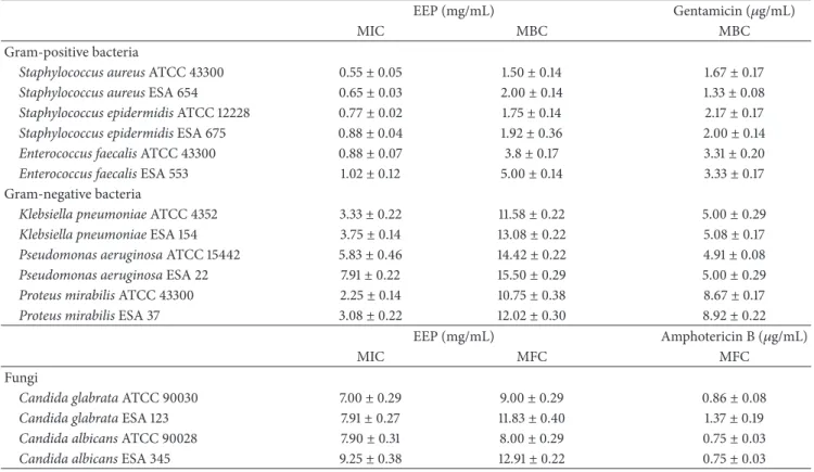
3.2. Antimicrobial Activity. The EEP showed antimicrobial activity against all microorganisms evaluated (Table2). The propolis fromT. fiebrigipresented greater activity against the gram-positive bacteria than against gram-negative bacteria.
The inhibition observed followed the sequence:S. aureus> S. epidermidis>E. faecalis>P. mirabilis>K. pneumonia> P. aeruginosa. Apart from this inhibitory activity, the EEP had bactericidal effects against all the organisms under study, ranging from1.50±0.14mg/mL, for the reference strain ofS. aureus, to15.50 ± 0.29mg/mL, for the isolatedP. aeruginosa. Regarding the fungicidal activity, it was observed both in the reference stains and in those isolated from biological fluids.
3.3. ABTS Radical Scavenging Assay. Considering the pres-ence of potentially antioxidant substances, anin vitro evalu-ation of the ABTS∙+ radical scavenging activity of EEP was performed at different concentrations. The 50% inhibitory concentration (IC50) and the maximal ABTS∙+ radical scav-enging activity of EEP and the controls used are shown in Table3. The IC50of EEP was approximately five times higher than that of synthetic antioxidant BHT.
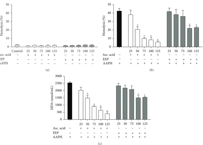
3.4. Oxidative Hemolysis Inhibition Assay. The EEP was also evaluated for its hemolytic activity and ability to protect erythrocytes against AAPH-induced hemolysis. When ery-throcytes were incubated only with ascorbic acid or EEP, no hemolysis was observed at the times and concentrations tested (Figure 2(a)), indicating that, at the concentrations evaluated, the extract is not toxic to this cellular model. Next, the ability to protect against AAPH-induced cell lysis of human erythrocytes was evaluated. The EEP showed antihe-molytic activity at all experimental period in a concentration and time-dependent manner (data not shown). After a 240-min incubation of erythrocytes with EEP, a reduction of46 ±
3.6% of hemolysis was observed in the highest concentration evaluated (Figure2(b)).
3.5. Efficiency of EEP on the Inhibition of AAPH-Induced Lipid Peroxidation. The ability of the EEP to protect against AAPH-induced lipid peroxidation of human erythrocytes was evaluated through the dosage of MDA. The EEP showed reduced MDA levels at all experimental period in a con-centration and time-dependent manner (data not shown). After a 240-min incubation of erythrocytes with EEP, a reduction of39.5±2.4% of MDA was observed in the highest concentration evaluated (Figure2(c)).
3.6. Anti-Inflammatory Activity. It was observed that the EEP inhibited the hyaluronidase enzyme in a concentration-dependent manner. The highest concentration of extract evaluated (75 mg/mL) was able to inhibit43.06 ± 3.06% of enzymatic activity (Figure3).
Table 1: Compounds identified in EEP fromT. fiebrigiby GC-MS.
Peak Retention time (min) Molecular mass Compound % TIC∗
1 13.10 122 Benzoic acid 9.2
2 15.31 286 Cinnamyl caffeate 1.5
3 21.04 270 Benzyl caffeate 1.5
4 23.76 148 Cinnamic acid 3.6
5 23.88 150 Hydrocinnamic acid 1.3
6 24.51 178 Hydrocinnamic acid ethyl ester 0.6
7 40.86 164 p-Coumaric acid 0.4
8 47.03 232 3-Phenyl-p-coumaric acid 2.8
9 50.67 180 Fructose 0.6
10 56.12 180 Glucose 1.5
11 59.94 302 Kaurenoic acid 11.8
12 63.25 152 4-Methoxybenzoic acid 3.0
13 68.59 286 Retinol 8.1
14 76.89 386 Cholesterol 12.4
15 79.39 430 Tocopherol 7.4
∗TIC: total ion current. The ion current generated depends on the characteristics of the compounds.
Table 2: Minimum inhibitory concentration (MIC), minimum bactericidal concentration (MBC), and minimum fungicidal concentration (MFC) for same microorganisms gram-positive bacteria, gram-negative bacteria, and the fungi.
EEP (mg/mL) Gentamicin (𝜇g/mL)
MIC MBC MBC
Gram-positive bacteria
Staphylococcus aureusATCC 43300 0.55±0.05 1.50±0.14 1.67±0.17
Staphylococcus aureusESA 654 0.65±0.03 2.00±0.14 1.33±0.08
Staphylococcus epidermidisATCC 12228 0.77±0.02 1.75±0.14 2.17±0.17
Staphylococcus epidermidisESA 675 0.88±0.04 1.92±0.36 2.00±0.14
Enterococcus faecalisATCC 43300 0.88±0.07 3.8±0.17 3.31±0.20
Enterococcus faecalisESA 553 1.02±0.12 5.00±0.14 3.33±0.17
Gram-negative bacteria
Klebsiella pneumoniaeATCC 4352 3.33±0.22 11.58±0.22 5.00±0.29
Klebsiella pneumoniaeESA 154 3.75±0.14 13.08±0.22 5.08±0.17
Pseudomonas aeruginosaATCC 15442 5.83±0.46 14.42±0.22 4.91±0.08
Pseudomonas aeruginosaESA 22 7.91±0.22 15.50±0.29 5.00±0.29
Proteus mirabilisATCC 43300 2.25±0.14 10.75±0.38 8.67±0.17
Proteus mirabilisESA 37 3.08±0.22 12.02±0.30 8.92±0.22
EEP (mg/mL) Amphotericin B (𝜇g/mL)
MIC MFC MFC
Fungi
Candida glabrataATCC 90030 7.00±0.29 9.00±0.29 0.86±0.08
Candida glabrataESA 123 7.91±0.27 11.83±0.40 1.37±0.19
Candida albicansATCC 90028 7.90±0.31 8.00±0.29 0.75±0.03
Candida albicansESA 345 9.25±0.38 12.91±0.22 0.75±0.03
Values are shown as means±SEM (𝑛 = 3).
indicating a concentration-dependent activity (Figures4(a) and4(b)).
4. Discussion
Propolis is a natural product that has a complex chem-ical composition. Substances in propolis composition are
potentially responsible for its biological activity and may act synergistically and in an isolated manner. In this study, phenolic compounds, aromatic acids, alcohols, terpenes, and sugars were observed in the EEP from T. fiebrigi. These compounds have been identified in other studies of propolis of stingless bees found in Brazil [18,25].
0 10 20 30 40 50
Asc. acid 25 50 75 100125
EEP AAPH
25 50 75 100125
Control H emo lysis (%) − − − − − − − − − − − − − − − − − − + + + + + − − − + − −+ + + + (a) 0 10 20 30 40 50
Asc. acid + + + + +
EEP
AAPH + + + + + +
25 50 75 100 125
25 50 75 100 125
+ + + + + + + + + + * * * * * * H emo lysis (%) − − − − − − − − − − − − (b) 0 500 1000 1500 2000 2500 3000 Asc. acid
25 50 75 100125
EEP AAPH
25 50 75 100125
* * * * * * MD A (nmo l/mL) + + − + + − + + − + + − + + + + + + + + + + + + + − − − − − − − − (c)
Figure 2: Human erythrocytes incubated at 240 min with ascorbic acid and EEP ofT. fiebrigi(25–125𝜇g/mL). (a) Hemolytic activity of treatments without the presence of AAPH and erythrocytes incubated only with 0.9% NaCl as control. (b) Hemolysis assessment after addition of AAPH in erythrocytes incubated with ascorbic acid and EEP. (c) Concentration of malondialdehyde (MDA) in nmol/mL after adding the oxidizing agent in erythrocytes incubated with different concentrations of treatments. * represents statistically significant results (𝑃 < 0.05) when the treated group was compared to the AAPH group (erythrocytes incubated only with oxidizing agent).
Table 3: IC50and maximum activity of ABTS radical scavenging of standard antioxidants and of EEP.
Sample IC50(𝜇g/mL)
Maximum inhibition
% 𝜇g/mL
Ascorbic acid 1.3±0.2 99.5±0.2 5
BHT 22.8±4.2 85.2±5.5 50
EEP 119.6±20.5 86.5±2.8 500
Values are means±SEM (𝑛 = 2).
the emergence of drug-resistant microorganisms appears as a serious global concern. In this context, it is important to find other substances effective against pathogens resistant to the conventional therapies. Natural products, such as propolis, appear as a viable alternative, either by taking advantage of the synergistic effects of all the compounds present or by investing in specific isolated compounds.
Indeed, different studies have shown a positive cor-relation between phenolic compounds and terpenes with antimicrobial action [26,27].
Phenolic compounds cinnamic acid, p-coumaric acid, and the diterpene kaurenoic acid identified in EEP from
0.20 0.39 0.78 1.56 3.13 6.25 12.5 25.0 50.0 75.0 0 10 20 30 40 50 Concentrations (mg/mL) In hib it io n (%)
Figure 3: Anti-inflammatory activity for inhibition of the activity of hyaluronidase by the EEP in different concentrations.
Control 31 62
125 250 500
104
103
102
101
100
104
103
102
101
100
104
103
102
101
100
104
103
102
101
100 104
103
102
101
100 104
103
102
101
100
104 103 102 101
100 100 101 102 103 104 100 101 102 103 104
104 103 102 101 100 104
103 102 101 100 104
103 102 101 100
PI PI PI
Concentrations (𝜇g/mL)
PI
PI
PI
Annexin Annexin Annexin
Annexin Annexin Annexin
(a)
*
* #
Control 31 62 125 500 250
#
Ce
ll
p
o
p
u
la
ti
o
n
(
%
)
Concentrations (𝜇g/mL)
100
80
60
40
20
0
PI−/An−
PI−/An+
PI+/An−
PI+/An+
(b)
Figure 4: Cytotoxic action of EEP fromT. fiebrigiagainst the K562 erythroleukemia cell line. (a) Representative diagrams obtained by flow cytometry of cells stained with annexin V-FITC/PI: the lower left quadrant (PI−/An−) represents the viable cells; the lower right quadrant (PI−/An+) represents the apoptotic cells; the upper left quadrant (PI+/An−) represents cells in necrosis; and the upper right quadrant (PI+/An+) represents cells in secondary necrosis. (b) Frequency of cell death, obtained from the corresponding diagrams of tested concentrations.*𝑃 < 0.05treated group versus control viable cells.#𝑃 < 0.05treated group versus control necrosis.
The phenolic compounds are described as important antibacterial agents that inhibit bacteria by promoting dam-age to the cell membrane and by inhibiting the synthe-sis of nucleic acids and the energy metabolism of these microorganisms. In addition, these compounds interfere with
virulence factors of various bacteria, including enzymes, toxins, and other signaling molecules [28].
that these microorganisms are responsible for common noso-comial infections in urinary tract, respiratory pneumonia, surgical site wound, gastrointestinal, and skin infections [22]. Propolis acts by inhibiting the growth and prolifer-ation of bacteria [29]; however, gram-positive bacteria are more sensitive to the action of propolis than gram-negative bacteria, which may be due to structural differences of the cell wall [14, 25]. Mirzoeva et al. [30] suggested that the antimicrobial activity of propolis is species-dependent and that its constituents are able to promote an increase in membrane permeability and inhibit bacterial motility.
Among the antimicrobial activities of propolis, the EEP showed fungicidal properties against C. glabrata and C. albicans. Kujumgiev et al. [31] correlated the antifungal effect of Brazilian propolis with the presence of diterpene acids, triterpenes, aromatic acids, and phenolic compounds, which were also identified in propolis fromT. fiebrigi.
Gallucci et al. [32] showed that many phenolic com-pounds inhibit fungal growth by interacting with the plasma or mitochondrial membrane of the fungus. de Castro et al. [33] reported that the mechanisms by which propolis pro-motes the death of fungi include vacuolar acidification and changes in the mitochondrial electron-transport chain, resulting in death by apoptosis; in turn, prolonged exposure results in secondary necrosis.
Recently, de Castro et al. [34] reported that propolis inhibits the morphological transition from yeast to hyphae, thus promoting the control of cell growth and differentiation, and induces cell death through the metacaspase pathway. Ramsdale [35] noted that many antifungal compounds pro-mote cell death mediated by the apoptotic pathway and can be metacaspase dependent or independent. In addition, anti-fungal compounds stimulate the production of reactive oxy-gen species or interfere with calcium/calmodulin/calcineurin signaling. The discovery of new antifungal agents, especially those that promote cell death by different pathways, from natural sources is of great importance.
In addition to the antimicrobial activity, the EEP has antioxidant activity by the free radicals scavenging and the ability to inhibit the lysis of erythrocytes incubated with oxidizing agent, which was confirmed by lower generation of MDA, one of biochemical markers of lipid peroxidation.
The antioxidant properties presented byT. fiebrigi propo-lis extract are probably related to its chemical composition, especially by the presence of phenolic compounds as benzoic acid, cinnamic acid, andp-coumaric acid already described in the literature by capacity to capture free radicals [36, 37]. Other studies have shown that these compounds are responsible for the antioxidant activity by plant extracts [38– 40] and propolis [41, 42]. According to Valente et al. [43], polyphenols from propolis are able to capture peroxyl radicals generated by AAPH oxidizing agent and inhibiting the lipid peroxidation process resulting in protection against oxidation of biomolecules present in the membrane of erythrocytes.
The reactive species of oxygen and nitrogen, when present in high levels in the body, are responsible for oxidative stress state, resulting in damage to cells and tissues [44]. Among the possibilities to neutralize the excess of these free radicals is consumption of natural foods with antioxidant properties,
which are important auxiliary in the prevention of diseases related to oxidative stress, such as cancer, diabetes, and diseases associated with inflammation, such as rheumatoid arthritis and atherosclerosis [44,45].
The propolis has also been used in the folk medicine in the prevention of inflammatory diseases, despite the lack of knowledge regarding the components that exhibit this activ-ity. Inflammation is a biological response activated to restore tissue injury, such as pathogens, damaged cells, irritants, and free radicals. Propolis has been reported to possess anti-inflammatory activity bothin vitroandin vivoby modulating key inflammatory mediators, inhibiting the production of proinflammatory cytokines, increasing anti-inflammatory cytokines, and blocking the activation of nuclear factor-(NF-)𝜅B [46,47]. Activity was also observed in the propo-lis from T. fiebrigi by determination of the hyaluronidase enzyme, an indirect way to assess the anti-inflammatory activity. The hyaluronic acid is an important component of articular cartilage and plays an important role in tissues’ renovation [22]. The degradation of hyaluronic acid by hyaluronidase enzyme may cause bone loss, inflammation, and pain [48].
The cytotoxic activity shown by EEP from T. fiebrigi
can be related to the presence of phenolic compounds as coumaric acid, cinnamic acid, and derivatives. These com-pounds have already been described in propolis samples by their cytotoxic action against K562 (leukemia), HeLa (human cervical adenocarcinoma), and LNCaP (human prostate can-cer) cell lines [49–51].
By investigating the cytotoxic potential of propolis from
T. fiebrigiin K562 erythroleukemia cells, we observed the pre-dominance of death by necrosis. This type of cell death can be promoted by opening mitochondrial permeability transition pores, leading to the depolarization and consequent cell death due to lack of ATP [52]. For many years, necrosis was seen as a kind of uncontrolled accidental death; however, necrosis is currently described as a programmed death that may contribute to the control of cancer-cell proliferation [53], in particular in cell lines resistant to conventional chemotherapy or cells showing resistance to apoptosis.
The K562 erythroleukemia cell line is described as exhibiting resistance to death by apoptosis [54]; therefore, alternative forms of cell death, such as necrosis, are neces-sary to inhibit the proliferation of these tumor cells [55]. Necrosis is caused by several biochemical and molecular signaling mechanisms, such as increases in levels of calcium in cell cytoplasm, increased interaction with serine-threonine kinase receptors, and increased abundance of reactive oxygen species, resulting in the disruption of organelles and cell death [56].
5. Conclusions
Conflict of Interests
The authors declare that there is no conflict of interests regarding the publication of this paper.
Acknowledgments
This work was supported by grants from Foundation for the Support and Development of Education, Science and Technology of Mato Grosso do Sul State, FUNDECT, and Brazilian National Research Council, CNPq. Jaqueline Fer-reira Campos was supported by FUNDECT. Edson Lucas dos Santos is recipient of fellowship from CNPq, Brazil. The authors would like to thank PRODER ˆA (24.073 – ˆA), Desenvolvimento de novos produtos de apicultura, em modo de produc¸˜ao biol´ogica (MPB), Portugal.
References
[1] M. Velikova, V. Bankova, M. C. Marcucci, I. Tsvetkova, and A. Kujumgiev, “Chemical composition and biological activ-ity of propolis from Brazilian Meliponinae,” Zeitschrift fur
Naturforschung—Section C, vol. 55, no. 9-10, pp. 785–789, 2000.
[2] R. de C´assia Oliveira, F. D. M. F. Nunes, A. P. S. Campos et al., “Genetic divergence in Tetragonisca angustula Latreille, 1811 (Hymenoptera, Meliponinae, Trigonini) based on rapd markers,”Genetics and Molecular Biology, vol. 27, no. 2, pp. 181– 186, 2004.
[3] J. M. F. Camargo and S. R. M. Pedro, “Meliponini Lepeletier, 1836. In (Hymenoptera, Apoidea) in the Neotropical Region,” 2013,http://moure.cria.org.br/catalogue?id=34631.
[4] A. Barth, A. Fernandes, S. G. das Pompolo, and M. A. Costa, “Occurrence of b chromosomes inTetragoniscalatreille, 1811 (hymenoptera, apidae, meliponini): a new contribution to the cytotaxonomy of the genus,”Genetics and Molecular Biology, vol. 34, no. 1, pp. 77–79, 2011.
[5] A. L. P. B. Stuchi, V. A. A. Toledo, D. A. Lopes, L. B. Cantagalli, and M. C. C. Ruvolo-Takasusuki, “Molecular marker to identify two stingless bee species:Tetragonisca angustulaand
Tetrago-nisca fiebrigi(Hymenoptera, Meliponinae),”Sociobiology, vol.
59, no. 1, pp. 123–134, 2012.
[6] P. Vit, M. G. Guti´errez, A. J. Rodr´ıguez-Malaver, G. Aguilera, C. Fern´andez-D´ıaz, and A. E. Tricio, “Comparison of honeys produced by the bee yate´ı (Tetragonisca fiebrigi) in Argentina and Paraguay,”Acta Bioquimica Clinica Latinoamericana, vol. 43, no. 2, pp. 219–226, 2009.
[7] M. A. Sgariglia, M. A. Vattuone, M. M. S. Vattuone, J. R. Sober´on, and D. A. Sampietro, “Properties of honey
fromTetragonisca angustula fiebrigiandPlebeia wittmanniof
Argentina,”Apidologie, vol. 41, no. 6, pp. 667–675, 2010. [8] V. S. Bankova, S. L. De Castro, and M. C. Marcucci, “Propolis:
recent advances in chemistry and plant origin,”Apidologie, vol. 31, no. 1, pp. 3–15, 2000.
[9] ´E. W. Teixeira, G. Negri, R. M. S. A. Meira, D. Message, and A. Salatino, “Plant origin of green propolis: bee behavior, plant anatomy and chemistry,”Evidence-Based Complementary and
Alternative Medicine, vol. 2, no. 1, pp. 85–92, 2005.
[10] M. Simone-Finstrom and M. Spivak, “Propolis and bee health: the natural history and significance of resin use by honey bees,”
Apidologie, vol. 41, no. 3, pp. 295–311, 2010.
[11] J. M. Sforcin and V. Bankova, “Propolis: is there a potential for the development of new drugs?”Journal of Ethnopharmacology, vol. 133, no. 2, pp. 253–260, 2011.
[12] S. Umthong, S. Puthong, and C. Chanchao, “Trigona laeviceps
propolis from Thailand: antimicrobial, antiproliferative and cytotoxic activities,”The American Journal of Chinese Medicine, vol. 37, no. 5, pp. 855–865, 2009.
[13] A. P. Farnesi, R. Aquino-Ferreira, D. De Jong, J. K. Bastos, and A. E. E. Soares, “Effects of stingless bee and honey bee propolis on four species of bacteria,”Genetics and Molecular Research, vol. 8, no. 2, pp. 635–640, 2009.
[14] M. K. Choudhari, S. A. Punekar, R. V. Ranade, and K. M. Paknikar, “Antimicrobial activity of stingless bee (Trigonasp.) propolis used in the folk medicine of Western Maharashtra, India,”Journal of Ethnopharmacology, vol. 141, no. 1, pp. 363– 367, 2012.
[15] A. C. H. F. Sawaya, J. C. P. Calado, L. C. dos Santos et al., “Composition and antioxidant activity of propolis from three species ofScaptotrigonastingless bees,”Journal of ApiProduct
and ApiMedical Science, vol. 1, no. 2, pp. 37–42, 2009.
[16] J. F. Campos, U. P. dos Santos, L. F. B. Macorini et al., “Antimicrobial, antioxidant and cytotoxic activities of propolis
from Melipona orbignyi (Hymenoptera, Apidae),” Food and
Chemical Toxicology, vol. 65, pp. 374–380, 2014.
[17] M. K. Choudhari, R. Haghniaz, J. M. Rajwade, and K. M. Paknikar, “Anticancer activity of Indian stingless bee propolis:
anin vitrostudy,”Evidence-Based Complementary and
Alterna-tive Medicine, vol. 2013, Article ID 928280, 10 pages, 2013.
[18] P. L. Miorin, N. C. Levy Jr., A. R. Custodio, W. A. Bretz, and M. C. Marcucci, “Antibacterial activity of honey and propolis from
Apis melliferaandTetragonisca angustulaagainstStaphylococcus
aureus,”Journal of Applied Microbiology, vol. 95, no. 5, pp. 913–
920, 2003.
[19] S. M. Alencar, T. L. C. Oldoni, M. L. Castro et al., “Chemical composition and biological activity of a new type of Brazilian propolis: red propolis,”Journal of Ethnopharmacology, vol. 113, no. 2, pp. 278–283, 2007.
[20] W. Greenaway, T. Scaysbrook, and F. R. Whatley, “Composition of propolis in Oxfordshire, U.K. and its relation to poplar bud exudate,”Zeitschrift f¨ur Naturforschung, vol. 43, pp. 301–305, 1988.
[21] C.-X. Zhao, Y.-Z. Liang, H.-Z. Fang, and X.-N. Li, “Tempera-ture-programmed retention indices for gas chromatography-mass spectroscopy analysis of plant essential oils,”Journal of
Chromatography A, vol. 1096, no. 1-2, pp. 76–85, 2005.
[22] J. C. Silva, S. Rodrigues, X. Fe´as, and L. M. Estevinho, “Antimi-crobial activity, phenolic profile and role in the inflammation of propolis,”Food and Chemical Toxicology, vol. 50, no. 5, pp. 1790–1795, 2012.
[23] R. Re, N. Pellegrini, A. Proteggente, A. Pannala, M. Yang, and C. Rice-Evans, “Antioxidant activity applying an improved ABTS radical cation decolorization assay,” Free Radical Biology &
Medicine, vol. 26, no. 9-10, pp. 1231–1237, 1999.
[24] E. J. Paredes-Gamero, M. N. C. Martins, F. A. M. Cappabianco, J. S. Ide, and A. Miranda, “Characterization of dual effects induced by antimicrobial peptides: regulated cell death or membrane disruption,”Biochimica et Biophysica Acta, vol. 1820, no. 7, pp. 1062–1072, 2012.
[25] V. Bankova and M. Popova, “Propolis of stingless bees: a promising source of biologically active compounds,”
[26] M. Velikova, V. Bankova, I. Tsvetkova, A. Kujumgiev, and M. C. Marcucci, “Antibacterialent-kaurene from Brazilian propolis of native stingless bees,”Fitoterapia, vol. 71, no. 6, pp. 693–696, 2000.
[27] K. Salom˜ao, P. R. S. Pereira, L. C. Campos et al., “Brazil-ian propolis: correlation between chemical composition and antimicrobial activity,” Evidence-Based Complementary and
Alternative Medicine, vol. 5, no. 3, pp. 317–324, 2008.
[28] T. P. T. Cushnie and A. J. Lamb, “Recent advances in under-standing the antibacterial properties of flavonoids,”
Interna-tional Journal of Antimicrobial Agents, vol. 38, no. 2, pp. 99–107,
2011.
[29] R. D. Wojtyczka, A. Dziedzic, D. Idzik et al., “Susceptibility of
Staphylococcus aureusclinical isolates to propolis extract alone
or in combination with antimicrobial drugs,”Molecules, vol. 18, no. 8, pp. 9623–9640, 2013.
[30] O. K. Mirzoeva, R. N. Grishanin, and P. C. Calder, “Antimi-crobial action of propolis and some of its components: the effects on growth, membrane potential and motility of bacteria,”
Microbiological Research, vol. 152, no. 3, pp. 239–246, 1997.
[31] A. Kujumgiev, I. Tsvetkova, Y. Serkedjieva, V. Bankova, R. Christov, and S. Popov, “Antibacterial, antifungal and antiviral activity of propolis of different geographic origin,”Journal of
Ethnopharmacology, vol. 64, no. 3, pp. 235–240, 1999.
[32] M. N. Gallucci, M. E. Carezzano, M. M. Oliva et al., “In vitro
activity of natural phenolic compounds against fluconazole-resistant Candida species: a quantitative structure-activity rela-tionship analysis,”Journal of Applied Microbiology, vol. 116, no. 4, pp. 795–804, 2014.
[33] P. A. de Castro, M. Savoldi, D. Bonatto et al., “Molecular char-acterization of propolis-induced cell death inSaccharomyces
cerevisiae,”Eukaryotic Cell, vol. 10, no. 3, pp. 398–411, 2011.
[34] P. A. de Castro, V. L. P. Bom, N. A. Brown et al., “Identification of the cell targets important for propolis-induced cell death in
Candida albicans,”Fungal Genetics and Biology, vol. 60, pp. 74–
86, 2013.
[35] M. Ramsdale, “Programmed cell death in pathogenic fungi,”
Biochimica et Biophysica Acta, vol. 1783, no. 7, pp. 1369–1380,
2008.
[36] I. G¨ulc¸in, E. Bursal, M. H. S¸ehitoˆglu, M. Bilsel, and A. C. G¨oren, “Polyphenol contents and antioxidant activity of lyophilized aqueous extract of propolis from Erzurum, Turkey,”Food and
Chemical Toxicology, vol. 48, no. 8-9, pp. 2227–2238, 2010.
[37] A. Kurek-G´orecka, A. Rzepecka-Stojko, M. G´orecki, J. Stojko, M. Sosada, and G. Swierczek-Zieba, “Structure and antioxidant activity of polyphenols derived from propolis,”Molecules, vol. 19, no. 1, pp. 78–101, 2014.
[38] J. C. Casagrande, L. F. Macorini, K. A. Antunes et al., “Antiox-idant and cytotoxic activity of hydroethanolic extract from
Jacaranda decurrensleaves,”PLoS ONE, vol. 9, no. 11, Article ID
e112748, 2014.
[39] M. A. Raza, D. Shahwar, and T. Khan, “Radical scavenging, proteases activities, and phenolics composition of bark extracts from 21 medicinal plants,” Journal of Chemistry, vol. 2015, Article ID 951840, 8 pages, 2015.
[40] C. A. S. Tirloni, L. F. B. Macorini, U. P. dos Santos et al., “Evaluation of the antioxidant activity, antimicrobial effect and acute toxicity from leaves of Allophylus edulis (A. St.-Hil., A. Juss. Cambess &.) Hieron. ex Niederl,”African Journal of
Pharmacy and Pharmacology, vol. 9, no. 11, pp. 353–362, 2015.
[41] V. Silva, G. Genta, M. N. M¨oller et al., “Antioxidant activity of uruguayan propolis.In vitroand cellular assays,”Journal of
Agricultural and Food Chemistry, vol. 59, no. 12, pp. 6430–6437,
2011.
[42] J. Zhang, X. Cao, S. Ping et al., “Comparisons of ethanol extracts of chinese propolis (poplar type) and poplar gums based on the antioxidante activities and molecular mechanism,”
Evidence-Based Complementary and Alternative Medicine, vol.
2015, Article ID 307594, 15 pages, 2015.
[43] M. J. Valente, A. F. Baltazar, R. Henrique, L. Estevinho, and M. Carvalho, “Biological activities of Portuguese propolis: protection against free radical-induced erythrocyte damage and inhibition of human renal cancer cell growthin vitro,”Food and
Chemical Toxicology, vol. 49, no. 1, pp. 86–92, 2011.
[44] Y.-Z. Fang, S. Yang, and G. Wu, “Free radicals, antioxidants, and nutrition,”Nutrition, vol. 18, no. 10, pp. 872–879, 2002. [45] A. A. Geronikaki and A. M. Gavalas, “Antioxidants and
inflammatory disease: synthetic and natural antioxidants with anti-inflammatory activity,”Combinatorial Chemistry & High
Throughput Screening, vol. 9, no. 6, pp. 425–442, 2006.
[46] J. L. MacHado, A. K. M. Assunc¸˜ao, M. C. P. da Silva et al., “Brazilian green propolis: anti-inflammatory property by an immunomodulatory activity,”Evidence-Based Complementary
and Alternative Medicine, vol. 2012, Article ID 157652, 10 pages,
2012.
[47] L.-C. Wang, Y.-L. Lin, Y.-C. Liang et al., “The effect of caffeic acid phenethyl ester on the functions of human monocyte-derived dendritic cells,”BMC Immunology, vol. 10, article 39, 2009.
[48] A. Pascoal, S. Rodrigues, A. Teixeira, X. Fe´as, and L. M. Estev-inho, “Biological activities of commercial bee pollens: antimi-crobial, antimutagenic, antioxidant and anti-inflammatory,”
Food and Chemical Toxicology, vol. 63, pp. 233–239, 2014.
[49] G. C. Franchi Jr., C. S. Moraes, V. C. Toreti, A. Daugsch, A. E. Nowill, and Y. K. Park, “Comparison of effects of the ethanolic extracts of Brazilian propolis on human leukemic cells as assessed with the MTT assay,”Evidence-Based Complementary
and Alternative Medicine, vol. 2012, Article ID 918956, 6 pages,
2012.
[50] M. Barbari´c, K. Miˇskovi´c, M. Boji´c et al., “Chemical compo-sition of the ethanolic propolis extracts and its effect on HeLa cells,”Journal of Ethnopharmacology, vol. 135, no. 3, pp. 772–778, 2011.
[51] E. Szliszka, A. Sok´oł-Łętowska, A. Z. Kucharska, D. Jaworska, Z. P. Czuba, and W. Kr´ol, “Ethanolic extract of polish propolis: chemical composition and TRAIL-R2 death receptor targeting apoptotic activity against prostate cancer cells,”Evidence-Based
Complementary and Alternative Medicine, vol. 2013, Article ID
757628, 12 pages, 2013.
[52] G. W. Dorn, “Molecular mechanisms that differentiate apopto-sis from programmed necroapopto-sis,”Toxicologic Pathology, vol. 41, no. 2, pp. 227–234, 2013.
[53] G. Kroemer, L. Galluzzi, P. Vandenabeele et al., “Classification of cell death: recommendations of the nomenclature committee on cell death 2009,”Cell Death and Differentiation, vol. 16, no. 1, pp. 3–11, 2009.
[55] W.-X. Zong and C. B. Thompson, “Necrotic death as a cell fate,”
Genes & Development, vol. 20, no. 1, pp. 1–15, 2006.
[56] N. Festjens, T. Vanden Berghe, and P. Vandenabeele, “Necrosis, a well-orchestrated form of cell demise: signalling cascades, important mediators and concomitant immune response,”
Biochimica et Biophysica Acta—Bioenergetics, vol. 1757, no. 9-10,
Submit your manuscripts at
http://www.hindawi.com
Stem Cells
International
Hindawi Publishing Corporation
http://www.hindawi.com Volume 2014
Hindawi Publishing Corporation
http://www.hindawi.com Volume 2014 INFLAMMATION
Hindawi Publishing Corporation
http://www.hindawi.com Volume 2014
Behavioural
Neurology
Endocrinology
International Journal ofHindawi Publishing Corporation
http://www.hindawi.com Volume 2014
Hindawi Publishing Corporation
http://www.hindawi.com Volume 2014
Disease Markers
Hindawi Publishing Corporation
http://www.hindawi.com Volume 2014
BioMed
Research International
Oncology
Journal ofHindawi Publishing Corporation
http://www.hindawi.com Volume 2014
Hindawi Publishing Corporation
http://www.hindawi.com Volume 2014
Oxidative Medicine and Cellular Longevity
Hindawi Publishing Corporation
http://www.hindawi.com Volume 2014
PPAR Research
The Scientific
World Journal
Hindawi Publishing Corporation
http://www.hindawi.com Volume 2014
Immunology Research
Hindawi Publishing Corporation
http://www.hindawi.com Volume 2014
Journal of
Obesity
Journal ofHindawi Publishing Corporation
http://www.hindawi.com Volume 2014
Hindawi Publishing Corporation
http://www.hindawi.com Volume 2014
Computational and Mathematical Methods in Medicine
Ophthalmology
Journal ofHindawi Publishing Corporation
http://www.hindawi.com Volume 2014
Diabetes Research
Journal ofHindawi Publishing Corporation
http://www.hindawi.com Volume 2014
Hindawi Publishing Corporation
http://www.hindawi.com Volume 2014
Research and Treatment
AIDS
Hindawi Publishing Corporationhttp://www.hindawi.com Volume 2014
Gastroenterology Research and Practice
Hindawi Publishing Corporation
http://www.hindawi.com Volume 2014
Parkinson’s
Disease
Evidence-Based Complementary and Alternative Medicine
Volume 2014