Carlos André Rodrigues Magalhães
setembro de 2015
Evidence for T cell driven diversifying
selection in Mycobacterium tuberculosis
in vivo-expressed genes
Evidências de seleção diversificante mediada
por células T em genes de Mycobacterium
tuberculosis expressos in vivo
UMinho|20
15
Carlos Andr
é Rodrigues Magalhães
Evidence for T cell driven diver
sifying selection in Mycobacterium tuber culosis in vivo -e xpr essed genes
Universidade do Minho
Trabalho efetuado sob a orientação do
Doutor Nuno Miguel Sampaio Osório
Carlos André Rodrigues Magalhães
setembro de 2015
Dissertação de Mestrado
Mestrado em Ciências da Saúde
Evidence for T cell driven diversifying
selection in Mycobacterium tuberculosis
in vivo-expressed genes
Evidências de seleção diversificante mediada
por células T em genes de Mycobacterium
tuberculosis expressos in vivo
Universidade do Minho
iii
The work presented in this thesis was done in the Microbiology and Infection Research Domain of the Life and Health Science Research Institute (ICVS), School of Health Science, University of Minho, Braga, Portugal (ICVS/3B’s - PT Government Associate Laboratory, Braga/Guimarães, Portugal).
v
A
CKNOWLEDGEMENTS
Várias pessoas contribuíram para a realização desta tese ao longo destes últimos 2 anos, às quais gostaria de expressar o meu agradecimento profundo.
Ao Nuno Osório, um enorme obrigado por tudo! Pela integração, conselhos e vitais ensinamentos que me fizeram evoluir em muitos aspetos. Por acreditares em mim e no meu trabalho. Pela incansável ajuda e força sempre que precisei. Sem o teu contributo, nada disto seria possível. Da minha parte, grande admiração e respeito, não só como cientista mas também como pessoa. Não poderia ter encontrado melhor orientador.
Ao professor Gil Castro e à Margarida Saraiva, os primeiros responsáveis pelo meu ingresso no ICVS. Obrigado por me fazerem crescer com todas as recomendações e palavras de incentivo ao longo destes anos. É muito bom saber que acreditam em mim.
Ao Jérémy, membro emprestado da equipa de bioinformática. Pela amizade e companhia nos bons e maus momentos. E pelas bolachas e comida caseira também, muito importante Admiro muito o teu carácter e espírito de entrega para com a vida e tenho muita sorte em ser teu amigo.
À Estela, a minha habitante da Catalunha preferida. Pelos conselhos para todas as questões e pelo ânimo que me transmitiste quando dele precisava. Apesar dos poucos meses cá passados, tornaste-te uma amiga para a vida.
À Adriana, amiga crónica de sempre. Que os momentos divertidos e as longas conversas não tenham fim. Obrigado por me distraíres quando precisava de ser distraído e por me acordares quando precisava de ser acordado.
Ao Cristiano, amigo de anos. As tuas palavras de incentivo, força e exemplo de vida foram também contagiantes para mim.
vi
Por último mas não menos importante, aos meus pais e à minha irmã. São os meus alicerces e estão sempre no meu coração.
vii
R
ESUMO
A tuberculose, uma doença devastadora causada por bactérias do complexo Mycobacterium tuberculosis (MTBC), mantém-se vastamente fora de controlo. Embora a imunidade mediada por células T positivas para cluster de diferenciação 4 (T CD4+) seja crítica para o controlo da infeção, não existe um
conhecimento profundo sobre as características das respostas protetoras mediadas por células T bem como dos fatores que têm impacto no seu estabelecimento. Neste contexto, a diversidade genética do MTBC tem sido subestimada, visto que a maioria dos antigénios conhecidos de M. tuberculosis são hiperconservados. A existência de epítopos de células T variáveis foi investigada através da análise da diversidade, evolução e imunogenicidade de uma ilha genómica expressa in vivo (iVEGI) em 270 genomas do MTBC. A iVEGI apresentou um nível de diversidade nucleotídica acima da média do genoma, devido à presença de 21 genes altamente diversos. A análise computacional de evolução molecular previu 11 codões de nove genes como estando sob seleção diversificante. De particular interesse foram três substituições nos genes accD2, Rv0987 e Rv0988, que alteraram significativamente o número de péptidos codificados com elevada afinidade de ligação prevista para diferentes moléculas de antigénio leucocitário humano (HLA) de classe II. Ensaios in vitro realizados com o HLA DRB1*01:01 corroboraram a existência de péptidos com elevada afinidade de ligação e revelaram diferenças de mais de 58% na afinidade de ligação entre péptidos wild-type e mutante que sobrepõem AccD2 R233L e Rv0987 V169L. Notavelmente, o péptido AccD2228-241 demonstrou níveis de afinidade e estabilidade tão elevados como o controlo positivo ESAT-63-17, sendo assim um forte candidato para um epítopo de célula T CD4+ restrito
a uma linhagem de M. tuberculosis. Durante a longa coevolução entre M. tuberculosis e populações humanas, a diversidade genética em epítopos específicos de células T pode ter sido selecionada de modo a induzir respostas imunes vantajosas para o agente patogénico. Revelar a trajetória evolutiva de M. tuberculosis em resposta à pressão imunológica pode oferecer ferramentas sem precedentes para explorar os seus alvos moleculares em favor do hospedeiro, nomeadamente para o desenvolvimento de vacinas mais eficientes.
Palavras-Chave: Tuberculose; Mycobacterium tuberculosis; seleção diversificante; epítopo de células T, antigénio leucocitário humano
ix
A
BSTRACT
Tuberculosis, a devastating disease caused by bacteria from the Mycobacterium tuberculosis complex (MTBC), remains vastly uncontrolled. Although cluster of differentiation 4 positive (CD4+) T cell-mediated
immunity is critical for infection control, a deep knowledge on the characteristics of the protective T cell responses and on the factors impacting their establishment is lacking. In this context, the MTBC genetic diversity has been underappreciated, as most of the known M. tuberculosis antigens are hyperconserved. The existence of varying T cell epitopes was investigated by analysing the diversity, evolution and immunogenicity of an in vivo-expressed genomic island across 270 MTBC genomes. The in vivo-expressed genomic island displayed a level of nucleotide diversity above the whole-genome average due to the presence of 21 highly diverse genes. Computational molecular evolution analysis predicted 11 codons from nine genes to be under diversifying selection. Of particular interest were three substitutions in the genes accD2, Rv0987 and Rv0988, which significantly altered the number of encoded peptides with predicted high binding affinity to class II human leukocyte antigen (HLA) molecules. In vitro assays performed with HLA DRB1*01:01 corroborated the existence of high binding affinity peptides and revealed differences of more than 58% in the binding affinity between wild-type and variant peptides overlapping AccD2 R233L and Rv0987 V169L. Notably, the AccD2228-241 peptide showed affinity and stability levels as high as the positive control ESAT-63-17,thus being a strong candidate for an M. tuberculosis lineage-restricted CD4+ T cell epitope. During the long co-evolution of M. tuberculosis and
human populations, genetic diversity in specific T cell epitopes might have been selected to induce immune responses advantageous for the pathogen. Unveiling the evolutionary path of M. tuberculosis in response to immune pressure might offer unprecedented tools to exploit its molecular targets in favour of the host, namely for the development of more efficient vaccines.
KEYWORDS: Tuberculosis; Mycobacterium tuberculosis; diversifying selection; T cell epitope; human
xi
T
ABLE OF CONTENTS
Acknowledgements ... v Resumo ... vii Abstract ... ix Table of contents ... xiFigures index ... xiii
Tables index ... xv
Abbreviations ... xvi
Introduction ... 1
Tuberculosis: a major human health problem ... 1
The origin of human TB ... 2
MTBC population structure ... 3
Genetic diversity among host and pathogen populations in TB ... 5
Influence of the host-pathogen genetic diversity on TB ... 6
Detection of diversifying selection ... 8
The role of T cells in the immune response against TB ... 10
Principles and techniques for T cell epitope discovery ... 11
Aims ... 13
Results ... 15
High nucleotide diversity and evidence for diversifying selection in the iVEGI ... 15
Specific iVEGI variants under diversifying selection impact T cell epitope prediction ... 18
Geographic correlations between population coverage and MTBC lineage frequency... 21
In vitro validation of the predicted CD4+ T cell epitopes under diversifying selection ... 24
Discussion ... 25
Conclusion and future perspectives ... 29
Materials and Methods ... 31
Sequence retrieval ... 31
Nucleotide diversity and recombination tests ... 31
Analysis of diversifying selection ... 32
xii
Assessment of the worldwide population coverage and the geographic distribution of the MTBC ... 33
In vitro HLA-binding assays ... 33
References ... 35
Supplementary Data ... 51
Single-nucleotide substitutions found in iVEGI ... 51
HLA alleles used in the immunoinformatics analysis ... 64
Calculation of the normalized percentage of high binding affinity peptides ... 65
Geographic regions used for the estimation of the regional population coverage and MTBC lineages distribution ... 66
HLA alleles with predicted high binding affinity peptides encompassing wild-type and variant amino acid residues under diversifying selection ... 67
Regional population coverage for peptides encompassing residues under diversifying selection ... 71
xiii
F
IGURES INDEX
Figure 1. Pathology and death rate of human TB. ... 2
Figure 2. Phylogeny of the MTBC based on whole-genomes of 216 M. tuberculosis and 3 animal-adapted strains. ... 5
Figure 3. Transmission dynamics of TB in San Francisco, United States of America (2001-2009). ... 7
Figure 4. Schematic representation of the action of diversifying selection.. ... 8
Figure 5. Key players in the immunity against TB.. ... 10
Figure 6. Comparative genomics analysis of the iVEGI.. ... 16
Figure 7. Immunoinformatics analysis of the sites under diversifying selection in iVEGI.. ... 19
Figure 8. Variations in the worldwide population coverage in sites under diversifying selection.. ... 20
Figure 9. Association between differential population coverage of predicted epitopes and the geographic distribution of the MTBC.. ... 23
xv
T
ABLES INDEX
xvii
A
BBREVIATIONS
AIDS Acquired immune deficiency syndrome
ALOX5 Arachidonate 5-Lipoxygenase
AM Alveolar macrophage
APC Antigen-presenting cells
BCG Bacille-Calmette-Guérin
BEB Bayes Empirical Bayes
CD4+ Cluster of differentiation 4 positive DNA Deoxyribonucleic acid
FUBAR Fast Unbiased Bayesian AppRoximation
GWAS Genome-wide association study
HIV Human immunodeficiency virus
HLA Human leukocyte antigen
IFN-γ Interferon-gamma
IL-10 Interleukin-10
IRGM Immunity-related GTPase M
iVEGI in vivo-expressed genomic island
LRT Likelihood ratio test
LSP Large sequence polymorphism
MBL2 Mannose-binding lectin 2
MHC Major histocompatibility complex
MIRU-VNTR Mycobacterial interspersed repetitive units-variable number of tandem repeats MRCA Most recent common ancestral
MSMD Mendelian susceptibility to mycobacterial diseases
MTBC Mycobacterium tuberculosis complex
PAML Phylogenetic analysis by maximum likelihood
PCR Polymerase chain reaction
PRR Pattern recognition receptor
SNP Single nucleotide polymorphism
TB Tuberculosis
xviii
WHO World Health Organization
1
I
NTRODUCTION
Tuberculosis: a major human health problem
Tuberculosis (TB) is an infectious disease caused by the bacillus Mycobacterium tuberculosis. Human TB affects primarily the lungs, where it leads to progressive tissue necrosis and cavity formation (Figure 1A). Without treatment, patients get increasingly debilitated with fever, weight loss and ultimately respiratory failure [1,2]. In less common cases, the pathogen can disseminate and induce pathology in other parts of the body, such as the central nervous system, bones and the gastrointestinal tract [3]. Only pulmonary TB can lead to aerosol transmission of the tubercle bacilli to other individuals [4]. The single preventive strategy available is the Bacille-Calmette-Guérin vaccine (BCG), first administered in 1921. Unfortunately, this vaccine is only efficient in protecting children against disseminated TB and its efficacy against pulmonary TB in adults is highly variable [5,6]. It is estimated that at least one third of the world’s population had already inhaled airborne droplets containing M. tuberculosis [7]. Most people are able to control bacterial growth and remain asymptomatic (latent TB). Nevertheless, 5-10% will eventually manifest the active form of the disease [1,2]. This percentage is even higher in immunocompromised patients, such as the ones infected with the human immunodeficiency virus (HIV). According to the World Health Organization (WHO), nine million people developed TB in 2013, resulting in a tragic amount of 1.5 million deaths (0.4 million due to HIV-associated TB) (Figure 1B) [7]. In fact, TB is only seconded by the acquired immune deficiency syndrome (AIDS) in terms of human casualties due to an infectious disease worldwide. Furthermore, TB also poses a high global economic burden, with eight billion dollars spent each year in diagnostic and treatment [7]. Once a person is diagnosed with TB, typical treatment relies on the administration of four antibiotics (isoniazid, rifampicin, ethambutol and pyrazinamide) over six months. Although this regime is highly effective among new cases (85% success), current global death rate decline is a modest 1-2% (Figure 1B) [7]. In addition to this, the emergence of drug-resistant strains threatens the use of antibiotics in the future [7]. Therefore, new therapeutic strategies are urgently needed to tackle this disease.
2
The origin of human TB
TB has been a cause of human death since the antiquity. The earliest written evidence of the disease was found in 4700-year old Chinese medical manuscripts [9]. Paleopathological lesions characteristic of TB were also detected in human remains dated from various historical periods of the human civilization and found in several locations around the globe. These include ancient Syria (8200-7600 BC) [10], Israel (7250-6160 BC) [11], Germany (5400-4800 BC) [12], Egypt (2050-1650 BC) [13], United Kingdom (400–230 BC) [14] and more recently Hungary (1731–1838 AD) [15]. It is estimated that 25% of the Egyptians mummified after 3400 BC suffered from TB [16]. A similar scenario was also true for all adults that died in Europe during the Industrial Revolution [17].
The etiologic agent of human TB remained unknown until the isolation of M. tuberculosis in 1882, by Robert Koch [18]. In 1896, taxonomists Lehmann and Neumann included M. tuberculosis in the Mycobacterium genus, alongside with another important human pathogen, Mycobacterium leprae [19]. Two years later, Theobald Smith isolated a similar organism (posteriorly named Mycobacterium bovis) from cattle with TB-like clinical presentation [20]. Since then, other TB-causing bacteria have been recovered from wild and domestic mammals, such as goats and sheep (Mycobacterium caprae) [21], seals (Mycobacterium pinnipedii) [22] and rodents (Mycobacterium microti) [23]. Nowadays, it is known that these bacteria share a high level of similarity at the nucleotide level and together constitute the Mycobacterium tuberculosis complex (MTBC) [24,25]. Contrarily to the majority of the mycobacteria, members of the MTBC are obligate pathogens with no known environmental reservoir [26,27]. In addition, MTBC bacteria are found in a limited number of different host species [26,27]. Since mycobacteria were
(A) (B)
Figure 1.Pathology and death rate of human TB. (A) Gross appearance of TB dissemination in the lungs of a 35-year
old male individual infected with HIV [8]. (B) Estimated number of TB deaths in millions per year, 1990–2013 [7]. Reproduced with permission from WHO, ID 182946.
3
already widely dispersed before the existence of animal kingdom, some of them may have evolved to colonize specific hosts during the early expansion of animal life on Earth [26]. Owing to its broader host spectrum [28], M. bovis was believed to be the most recent common ancestral (MRCA) of the MTBC. In that sense, the bacterium was thought to have spread to humans due to close contact with infected cattle [26]. However, analysis of its genome revealed no new genes comparatively to M. tuberculosis [29]. Instead, the genomes of M. bovis and other animal-adapted MTBC species were found to be smaller due to the accumulation of large deletions [24]. Since horizontal gene transfer is thought to be scarce across MTBC [30,31], these losses of genetic material must have occurred once and could not have been reverted over time. Thus, it was proposed that human and animal-adapted MTBC members were two distinct clades and that the ancestral of the complex would be more similar to M. tuberculosis [24]. In support of this notion, skeletal evidence indicated that human TB was present before the early stages of animal domestication [10]. Moreover, although animal-adapted MTBC bacteria can occasionally infect humans [32,33], the ability to transmit TB seems to be restricted to M. tuberculosis [26,34]. Significant advances in finding the MRCA of the MTBC were made in 2013, with the inclusion of Mycobacterium canettii (an occasional human TB-causing bacterium [35]), as a member of the complex. The genome of M. canettii was found to be larger than any other MTBC bacterium, while sharing more than 95% nucleotide sequence identity [36]. It was also reported that M. canettii displays ongoing horizontal gene transfer and recombination between strains [36,37], similarly to other environmental mycobacteria [38,39]. Together, all these characteristics placed M. canettii in the early branches of the MTBC evolution [36]. The mechanisms underlying the transition of an M. canettii-like ancestral to M. tuberculosis are not fully clarified [36,40] and will not be discussed in this thesis. In the next section, the focus will be on the population structure of MTBC and its impact on the human population.
MTBC population structure
In clinical settings, detection and classification of M. tuberculosis isolates is routinely performed through polymerase chain reaction (PCR) amplification of certain repetitive and polymorphic genomic regions of the bacterium [41]. The occurrence pattern of these deoxyribonucleic acid (DNA) stretches is characteristic of each strain and can be interrogated by two methods, spoligotyping [42] and mycobacterial interspersed repetitive units-variable number of tandem repeats (MIRU-VNTR) [43]. Over the years, the amount of data generated by these techniques lead to the creation of online databases, such as SITVITWEB [44]. Further analysis of the genotyping patterns available at SITVITWEB [44] and
4
others [45] revealed the existence of distinct groups of strains with variable predominance throughout the world. In other words, each considered geographical region seemed to have a prevailing genotype [44,45]. Other studies harnessing single and large sequence polymorphism (SNP and LSP) data also reached the same conclusions [46–49]. Although PCR-based typing is useful for epidemiological studies [41], it only assess the diversity within small portions of the genome. Alternatively, whole-genome sequencing has a much higher discriminatory power and can be used to measure the evolutionary relationships and distances between different strains [50]. A robust phylogenetic tree of the M. tuberculosis population (Figure 2) was constructed in 2013 by Iñaki Comas and colleagues [51]. To achieve that, the authors took advantage of 216 whole-genome sequences of clinical isolates from several regions of the globe. The study highlighted that the global M. tuberculosis diversity was distributed by seven main phylogenetic lineages, each one with a strong association for certain world regions. Lineage 1 (also designated as Indo-Oceanic lineage) predominates in East Africa and South-East Asian countries (such as India, Philippines and Vietnam); lineage 2 is highly dispersed through Eastern Asia (e.g. China and Mongolia); lineage 3 is frequently found in Central Asia (e.g. India, Pakistan and Bangladesh) and to a lesser extent in East Africa; lineage 4 (Euro-American lineage) encompasses Europe, America, Africa and Middle-East; lineages 5 and 6 (also known as Mycobacterium africanum) are narrowly restricted to West Africa (e.g. Guinea-Bissau) and finally, lineage 7 was only found till date in Ethiopia [52]. It was also verified that the M. tuberculosis phylogeny was similar to a human phylogeny based on representative mitochondrial groups [51]. This suggested that the M. tuberculosis lineages could have followed the major human migrations out of the African continent, the cradle of mankind [49,51,53–55]. Lineages 1, 5 and 6 were the first to diverge from a 70 000 year-old M. canetti-like progenitor and were designated as ‘ancient’ MTBC lineages. In contrast, lineages 2, 3 and 4 (which shared a characteristic large genomic deletion [24]) were classified as ‘modern’ MTBC strains [51]. Additionally, animal-adapted MTBC strains were confirmed to share a common ancestral with M. tuberculosis, as aforementioned [24].
5
Genetic diversity among host and pathogen populations in TB
TB is a heterogeneous disease with several outcomes and clinical manifestations upon exposure to its causative agent [1,2]. Much of this heterogeneity was linked with social and environmental factors in the past [56]. However, accumulating evidence has been showing that there is an intrinsic role of biological factors related with the bacterium, the host and the host-pathogen interaction.
On the bacterium’s side, it was reported that the diversity among MTBC lineages and sub-lineages has clinical relevance, namely on disease onset, severity and transmission. For instance, a delay in seeking medical care was detected by Yimer et al. (2015) in Ethiopians infected with lineage 7 strains [57]. Parwati et al. (2010) associated lineage 2 with treatment failure [58] and Stavrum et al. (2014) related lineage 4 with more debilitating symptoms (such as weight loss) in Tanzanian individuals [59]. Albanna et al. (2011) found lineage 3 to be less transmissible than lineages 1, 2 and 4 across Montreal, Canada [60], whereas an increasing frequency of lineage 2 was described in South Africa by Cowley et al. (2008) [61]. At a sub-lineage level, Kato-Maeda et al. (2010) identified a highly transmissible sub-lineage of lineage 2 in San Francisco [62] and Gehre et al. (2013) registered striking differences in transmissibility
Figure 2.Phylogeny of the MTBC based on whole-genomes of 216 M. tuberculosis and 3 animal-adapted strains. Major MTBC lineages are depicted by different colors. Scale bar indicates substitutions per site. Extracted from [51].
6
among sub-lineages of lineage 5 in Benin and Nigeria [63]. Distinct lineage 2 strains were also found to impact the interaction with host innate immune system, either by preferentially activating the pattern recognition receptor (PRR) Toll-like receptor (TLR) 2 or TLR4 [64]. MTBC lineages are also not similar in epidemiological terms, with the ‘modern’ MTBC lineages 2 and 4 being the most widespread [44]. These have been correlated with a low induction of pro-inflammatory molecules in vitro [65,66] and a higher capacity to replicate in vivo when compared to ‘ancient’ MTBC strains [67]. Although the aforementioned studies emphasize the influence of pathogen diversity in TB, some controversial data can be found in the literature. For example, Marais et al. (2009) found the transmissibility not to be significantly associated with the M. tuberculosis genotype in South Africa [68] and Krishnan et al. (2011) also reported no significant alterations between lineage 1 and lineage 2 strains regarding the in vitro inflammatory profile [69]. These apparent contradictions in studies performed in different geographic areas might be related with the heterogeneity in human populations and its impact in TB. Clinical surveys revealed that a substantial part (30-50%) of household contacts with the disease did not result in measurable infection [70,71], thus suggesting that some people might be intrinsically less susceptible to TB. Indeed, the SNP rs2057178 on chromosome 11p13 was related with protection against TB in Gambia, Indonesia and Russia by a genome-wide association study (GWAS) [72]. Additionally, genetic mutations were found to cause Mendelian susceptibility to mycobacterial diseases (MSMD) [73,74], which is a primary immunodeficiency that leads to complications even with typically non-pathogenic mycobacteria [75]. In addition, susceptibility and immune response to TB were also described to vary according to ethnicity [76,77]. Many other studies identified human genetic variants thought to influence the risk of developing TB (reviewed in [78] and [79]). Nevertheless, the results have been considered inconsistent and difficult to reproduce. Regarding these studies, the lack of incorporation of the nucleotide diversity and population structure of both the host and M. tuberculosis was appointed as one of the most prominent weaknesses [80].
Influence of the host-pathogen genetic diversity on TB
Accumulating evidence has been showing that epidemiological and clinical aspects of TB are significantly shaped by the interaction between human and MTBC populations.
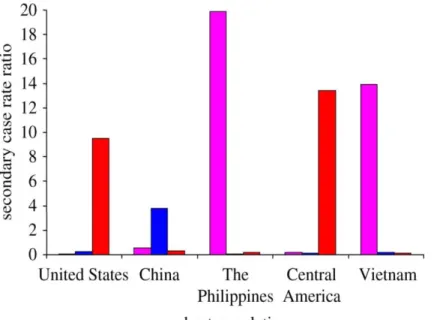
At the epidemiological level, Gagneux et al. (2006) and Reed et al. (2009) demonstrated that the transmission dynamics of TB in highly urbanized settings (i.e. San Francisco [48] and Montreal [81]) were not random. Instead, MTBC lineages frequently found in established ethnic communities (namely
7
Chinese, Filipino and Vietnamese) were the ones that predominated in the corresponding countries of origin (Figure 3) [48,81]. Interestingly, Hirsh et al. (2004) had previously referred that the host’s ethnic origin could predict the MTBC lineage of the infecting strain [46].
At the clinical level, de Jong et al. (2008) observed that lineages 2 and 4 had a higher rate of progression to active TB comparatively to lineage 6 in Gambia [83] and a multivariate analysis performed by Caws et al. (2008) showed that lineage 2 strains were more commonly found in Vietnamese patients with a particular polymorphism on the TLR2-encoding gene [84]. In Ghana (West Africa), Herb et al. (2008) reported a statistically significant association between a polymorphism (G254K) on the Arachidonate 5-Lipoxygenase (ALOX5) gene and increased predisposition to develop TB due to lineage 6 [85]; Intemann et al. (2009) linked a nucleotide variant upstream of the Immunity-related GTPase M (IRGM) coding region with protection from TB caused by lineage 4 (and not by other MTBC lineages) [86]; and Thye et al. (2011) found that the LYQC haplotype of the Mannose-binding lectin 2 (MBL2) gene conferred an exclusive protective effect against lineages 5 and 6 [86]. Interestingly, the MBL2 LYQC haplotype is rarely found outside of sub-Saharan Africa (which encompasses West Africa region), suggesting that it could have had increased its frequency over time owing to its protection to endemic MTBC lineages. [87]. On the other hand, the exonic variant G254K of ALOX5 is highly frequent in Africa relative to other world regions [85], indicating that lineage 6 strains (abundant in West Africa) might have a selective advantage in hosts harbouring the referred polymorphism.
Figure 3. Transmission dynamics ofTB in San Francisco, United States of America (2001-2009). The number
of secondary TB cases is plotted as a function of the number of index cases (secondary case rate ratio) Pink, lineage 1 or Indo-Oceanic lineage; blue, lineage 2 or East Asian lineage and red, lineage 4 or Euro-American lineage. Adapted from [82].
8
Overall, described findings indicated that MTBC lineages tend to be more successful (i.e. more adapted) upon infection of humans that share the same ancestral geographic region (sympatric) than with hosts of foreign ascendency (allopatric) [48,81,82,88].. Furthermore, genetic variants associated with host-pathogen adaptation in TB might have arisen due to natural selection, rather than by chance (i.e. genetic drift) [89]. Consequently, the detection of molecular signatures of adaptation could reveal the basis underlying the success of M. tuberculosis and reveal potential targets for therapy design.
Detection of diversifying selection
Diversifying selection (also referred as positive selection [90]), is the directional action of natural selection to increase the prevalence of beneficial genetic variants over time (Figure 4) [91]. More importantly, diversifying selection is thought to be the main mechanism of adaptation in disease contexts [92]. Classical examples are the high frequency of sickle-cell anaemia (caused by a mutation in Hemoglobin-B gene) in malaria endemic regions [93] and the development of drug-resistance during treatment in HIV [94].
The detection of diversifying selection is not easy if the selective pressure and its targets are not known. To try to mitigate these issues, numerous computational methods were developed to detect selection in genomic sequences by taking advantage of its increasing availability. Briefly, the methods can be divided in two types: the ones based on population genetics data and comparative genomics [91]. The population genetics-based methods apply statistical models in order to distinguish if variation among a representative sample of the population was randomly generated or fixed by selection. Typical evaluated datasets include linkage disequilibrium [96] or allelic frequency [97], for instance. Despite its popularity in human genetics, these methodologies are highly dependent on assumptions about the population demography, such as population size, that are not easily estimated in pathogen populations [91,98]. Furthermore, the resolution provided is rarely to the codon level [98,99]. In contrast, comparative genomics methods rely
Figure 4. Schematic representation of the action of diversifying selection. Adapted from [95]. Reproduced with
9
on measuring information within sequence alignments of protein-coding DNA [91]. The main parameter assessed is the ratio (ω) of the number of nonsynonymous nucleotide substitutions (i.e. that change the encoded aminoacid) per site (dN) over the number of synonymous nucleotide substitutions (i.e. with no aminoacid changing) per site (dS) (ω=dN/dS) [90]. The interpretation of the ω ratio is based on the principle that most mutations are synonymous and evolutionary neutral, i.e. without functional consequences. In that sense, a ω = 1 means that an aminoacid change is neutral and a ω < 1 will reflect negative selection (meaning that aminoacid change was probably deleterious and its fixation rate was reduced). Evidence for diversifying selection is inferred when ω is significantly higher than 1 [90]. Initial applications of the method averaged the ω ratio over all codons in a gene and thus only provided information for the whole-protein [100]. Nevertheless, the action of positive selection is thought to be aimed at only a few number of sites [101–103]. To address this matter, new statistical models were developed to allow estimation of different ω ratios in distinct codons [101,104]. In each model, the variation of ω over all sites can be described by a specific statistical distribution. A likelihood ratio test (LRT) is then used to evaluate what model fits better to the data. To check for positive selection, LRT can compare a model that does not admit ω ≤ 1 (null hypothesis) with one that admits ω > 1 (alternative hypothesis) [103]. Two pairs of models incorporated in Phylogenetic analysis by maximum likelihood (PAML) package were shown to be the most reliable for this purpose (null versus alternative hypotheses, respectively): M1a versus M2a and M7 versus M8 [105]. The LRT is statistically significant (null hypothesis rejected) when twice the log likelihood difference between the two compared models is above the previously described critical values of a chi-squared distribution with two degrees of freedom [105]. The parameters in the statistical distributions of M2a and M8 models are grouped into classes, with one of them allowing ω > 1 [105]. When this condition is met, a post-inference method designed Bayes Empirical Bayes (BEB) is used to calculate the probability of a codon to come from the class with ω > 1 [106]. A more recent method named Fast Unbiased Bayesian AppRoximation (FUBAR) [107] also applies a codon model to infer the synonymous and the nonsynonymous substitution rates (designated as α and β by the authors, respectively). FUBAR differs from PAML due to the adoption of some computational shortcuts and the use of a distinct method to assess site-specific posterior probabilities for being under diversifying selection, indicated as the posterior probability of β > α [107]. Codon models were previously shown to be powerful and robust in the absence of recombination [108]. Indeed, PAML and FUBAR have unveiled positively selected sites in human pathogens, such as in genes encoding for surface glycoproteins of the influenza H5N1 virus [109], as well as a specific siderophore in Pseudomonas aeruginosa [110]. PAML has also revealed sites under diversifying selection in the genome of M.
10
tuberculosis related to drug-resistance [111,112] and to other evolutionary pressures [111]. Particularly, evidences of positive selection were identified in a putative antigenic region, suggesting that human immune system can be responsible for the selection of some variants in the genome of the pathogen [111].
The role of T cells in the immune response against TB
The immunity against TB is tightly dependent on T lymphocytes, namely CD4+ and CD8+ T cells [1,113].
Indeed, the low numbers of CD4+ T cells characteristic of HIV-patients are associated with high
susceptibility to TB [114] and mice that are deficient in CD4+ T cells die rapidly from the disease [115].
CD8+ T cells also contribute to disease resistance in humans [116], rhesus macaques [117] and mice
[115]. Owing to the importance of these two types of T lymphocytes in the control of TB, it is very relevant to detail the characteristics of the T cell-mediated TB protective immune responses. After aerosol inhalation, tubercle bacilli are phagocytized by antigen-presenting cells (APC) such as alveolar macrophages (AM) (Figure 5A) and interstitial dendritic cells [1]. On the surface of APC, peptides generated by proteolysis of M. tuberculosis proteins (i.e. the antigens) are bounded to major histocompatibility complex (MHC) proteins (also known as human leukocyte antigen [HLA] in humans). HLA class I molecules present antigenic peptides to CD8+ T cells and HLA class II molecules to CD4+ T
cells (Figure 5B) [118]. T cell receptor then recognize a specific complex formed by the HLA molecule and the presented peptide (i.e. the T cell epitope) [119,120]. After this step, T cells can stimulate bacterial killing in macrophages by the secretion of inflammatory mediators such as interferon-gamma (IFN-γ) [1] (Figure 5A). Some T cells also differentiate into a memory phenotype that provide more rapid protection
Figure 5. Key players in the immunity against TB. (A) Phagocytosis of M. tuberculosis and secretion of IFN-γ by T cells. Adapted from [122]. (B) Recognition of epitope-MHC II complex by a CD4+ T cell. Adapted from [121].
11
upon new contacts with the same pathogen peptide. This property turns T cells attractive to be explored in the context of vaccine design [113].
Principles and techniques for T cell epitope discovery
The formation of a epitope-HLA complex with the right affinity and stability is a strong requirement to elicit a T cell response [123]. This makes the step of formation of the epitope-HLA complex an important focus of epitope discovery. Nevertheless, identifying the epitope with proper characteristics is a difficult task. First, T cell epitopes can be derived from virtually all proteins expressed in an organism [124]. Second, HLA molecules can accommodate peptides of several lengths in their binding groove, i.e. 8-15 amino acids in the case of HLA class I and 8-20 in HLA class II [125]. Laboratorial approaches to identify T cell epitopes rely on the synthesis of overlapping peptides encompassing antigens of interest. Synthesized peptides are then used to stimulate sensitized T cells ex vivo and the best epitope candidates are inferred through the quantification of correlates of T cell immune response, such as cytokine production [126] or T cell proliferation [127]. The complexity, time and cost of these experimental steps are huge constraints to a more widespread application [125].
In the last two decades, several computational methods were developed to analyse and model the properties of the immune system, giving rise to the field of immunoinformatics [124,128]. T cell epitope prediction, also known as reverse vaccinology [129], is a specific branch of immunoinformatics that offers a large potential to accelerate the process of epitope discovery [123]. The main principle of T cell epitope prediction is to find peptides with high binding affinity to HLA molecules, since these have more probability to be presented to T cells [130,131]. This pretension is complicated by the existence of thousands of HLA class I (HLA-DR, -DQ and -DP) and class II (HLA-DR, -DQ and -DP) alleles in the human population, each one with a distinct binding specificity [132]. Furthermore, HLA alleles have different frequencies across the worldwide population [133]. This means that a certain T cell epitope can elicit a response in some human populations but not in others [134]. Taking this into account, Bui et al. (2006) developed an algorithm that calculates the population coverage of an epitope, i.e. the fraction of individuals in a population in which it will be putatively effective [135].
In addition to the HLA molecule, binding affinity also depends on the amino acid sequence of the antigen epitopes [136,137]. In the context of TB, it was previously shown that a large set of experimentally validated MTBC T cell epitopes are evolutionarily conserved [138]. However, the known repertoire of MTBC epitopes is likely to be incomplete, thus impairing a full view of the evolution across the MTBC
12
immunoproteome [139,140]. In fact, only a small percentage of M. tuberculosis proteins has been shown to include epitopes [140,141].
Among the variety of in silico algorithms that predict T cell epitopes (reviewed in [123] and [142]), the most accurate ones are trained with large sets of epitopes with experimentally determined binding affinity [143]. Particularly, NetMHCpan [144] and NetMHCIIpan [145] were shown to be the best overall predictors in independent benchmarks [146–148]. Both methods only need the amino acid sequences of interest as input to generate predictions for all known HLA molecules. Quantitative prediction of the binding affinity is extrapolated from a training data set through advanced computational algorithms [144,145]. It is also possible to predict the binding affinity to HLA proteins with no known experimental binding data on the basis of other HLA molecules with similar binding pocket residues [143].
The bulk of the immune response is aimed at a small fraction of the possible epitopes [119,120], a feature called immunodominance [149]. In this regard, it was described that a higher kinetic stability of the HLA-peptide complex is a good predictor for immunodominance in both HLA class I [150] and HLA class II [151–153]. In M. tuberculosis, besides antigen 6-kDa early secretory protein antigenic target (ESAT-6) [154], only a few other immunodominant epitopes were validated in TB patients [155]. Another factor that affects immunodominance is the relative abundance of peptides available for binding. Genes with high in vivo expression levels in the context of infection constitute good candidates for the identification of relevant T cell epitopes since their encoded peptides are more likely to be presented than other low abundance peptides with similar HLA-binding affinity and stability [156]. In the M. tuberculosis genome, Talaat and colleagues found a genomic region of 44,849 nucleotides that harbour several genes highly expressed in vivo [157,158]. The region is constituted by 43 contiguous genes and was designated as in vivo-expressed genomic island (iVEGI) [157]. Interestingly, iVEGI is upregulated upon infection in wild-type mice models but not in immune compromised animals or during in vitro growth of M. tuberculosis [157]. This suggests a role for iVEGI in the interaction with host immune cells, highlighting T cell responses as a possible driving force of natural selection in this region.
13
A
IMS
Co-evolution between humans and M. tuberculosis strains have produced numerous examples of clinical and epidemiological heterogeneity (reviewed in [159] and [88]). However, the molecular basis underlying host-pathogen interactions in TB remain poorly understood. Indeed, despite the critical importance of T cells to the control of TB, the impact of the genetic diversity selected during the MTBC evolution on the full repertoire of M. tuberculosis T cell epitopes is poorly study. Still, the existing studies show evidence for genetic variation in known T cell epitopes [138] and for diversifying selection in a predicted antigenic region [111]. The identification and functional characterization of the immune responses elicited by variable epitopes are of critical importance from a vaccine-development point of view since these might correlate with protective responses. Highly in vivo-expressed genes mediate most of the host-pathogen interactions and are thus thought to be major targets of immune-related selective pressure. Therefore, M. tuberculosis iVEGI, a genomic region found to be upregulated in vivo in immunocompetent mice models of infection [157], is a good candidate region to identify these epitopes.
Taking this into account, the aim of this thesis is to analyse the iVEGI for evidence supporting the presence of M. tuberculosis epitopes under T cell driven positive selection. In order to address this goal, a research workflow composed of four main tasks was designed:
(i) Comparative genomics analysis, to investigate the nucleotide diversity of 43 genes highly expressed in in vivo models of infection;
(ii) Detection of diversifying selection, to search for signatures of molecular adaptation among the genetic diversity unveiled at (i);
(iii) Prediction of CD4+ and CD8+ T cell epitopes in protein sequences encoded by genes with sites under
diversifying selection;
(iv) In vitro HLA-peptide binding assays, to provide the experimental validation of the best candidates from preceding tasks.
15
R
ESULTS
High nucleotide diversity and evidence for diversifying selection in the iVEGI
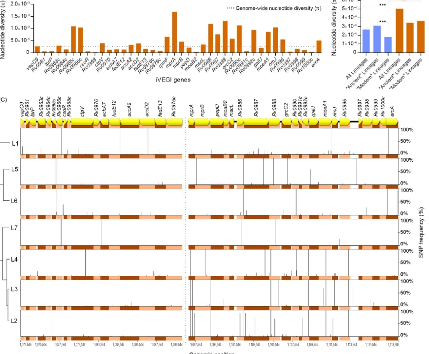
The in vivo expression pattern of iVEGI suggests a role in host-pathogen interactions during infection and the consequent possibility of evolution in response to host pressures. To investigate this, the sequence diversity on the iVEGI was characterized. This region was extracted from the genomes of 270 isolates belonging to the seven MTBC lineages and comparative genomics analysis was performed. The iVEGI was significantly more diverse (π = 5.04 x 10⁻⁴, p < 0.001) than the rest of the genome (π = 2.65 x 10 -4), with the majority of the genes analysed (21 out of 40) displaying levels of nucleotide diversity (π) above the genome-wide mean (Figure 6A). The level of genetic diversity in the iVEGI was very similar (p > 0.05) between ‘ancient’ (lineages 1, 5, 6 and 7; π = 3.42 x 10⁻⁴) and ‘modern’ (lineages 2, 3 and 4; π = 3.62 x 10⁻⁴) MTBC strains. This is in contrast with the whole-genome data, which showed that the ‘ancient’ lineages (π = 3.10 x 10⁻⁴) had a significantly higher nucleotide diversity level (p < 0.001) when compared to the ‘modern’ lineages (π = 1.82 x 10⁻⁴) (Figure 6B). A more detailed analysis revealed a total of 320 SNPs, of which 291 were located in coding regions and 29 in intergenic regions (Figure 6C and Supplementary Table 1). SNPs were detected in all the seven MTBC lineages and with highly variable frequencies. Homoplastic SNPs were also found, 13 located in genes and two in intergenic regions (Supplementary Table 1).
16
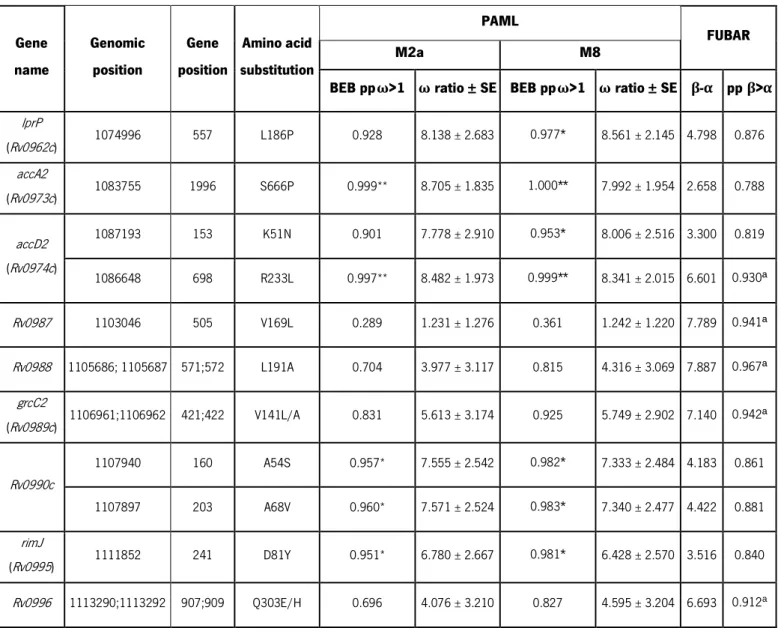
Considering that SNPs under diversifying selection may reflect targets of molecular adaptation driven by host-related pressures, the iVEGI genes were screened for diversifying selection with PAML and FUBAR [105,107]. The site models M2a and M8 included in PAML package were applied and Bayes Empirical Bayes (BEB) analysis revealed significant evidence for diversifying selection (M2a pp > 0.95 or M8 pp > 0.95) in seven codon sites corresponding to amino acid positions 186 of LprP (M2a pp = 0.928 and M8 pp = 0.977), 666 of AccA2 (M2a pp = 0.999 and M8 pp = 1.000), 51 and 233 of AccD2 (M2a pp = 0.901 and M8 pp = 0.953; M2a pp = 0.997 and M8 pp = 0.999, respectively), 54 and 68 of Rv0990c (M2a pp = 0.957 and M8 pp = 0.982; M2a pp = 0.960 and M8 pp = 0.983, respectively) and 81 of RimJ
Figure 6. Comparative genomics analysis of the iVEGI. (A) Nucleotide diversity of individual iVEGI genes. (B) Nucleotide diversity (π) of the genome (in
blue) versus iVEGI (in orange) in strains from the ‘ancient’, ‘modern’ and all the seven MTBC lineages. (C) Schematic representation of the sequence variation found in iVEGI. The relative frequency of the nucleotide substitutions in each of the seven MTBC lineages (right yy axis) is represented on the left yy axis. Genomic positions (xx axis) are according to the reference genome H37Rv (GenBank accession number NC00962.3). Pe_pgrs16, pe_pgrs17 and pe_pgrs18 were excluded from the analysis and are not represented (stroke line). ** p < 0.01; *** p < 0.01.
17
(M2a pp = 0.950 and M8 pp = 0.981) (Table 1). FUBAR identified four additional putative targets of diversifying selection (pp (β>α) > 0.9) in amino acid residues 169 of Rv0987 (pp β>α = 0.941); 191 of Rv0988 (pp β>α = 0.967); 141 of GrcC2 (pp β >α = 0.942) and 303 of Rv0996 (pp β>α = 0.918) (Table 1). An overlap between the predictions of PAML and FUBAR was found for the amino acid 233 of AccD2. To test a possible influence of recombination, nucleotide sequences were analysed with RDP, GENECONV and Bootscan, with none of the methods detecting signals supportive of recombination (data not shown). Overall, evidences for diversifying selection were detected by PAML or FUBAR in 11 codons of nine distinct iVEGI genes (Table 1). These data support high levels of nucleotide diversity and the occurrence of targeted events of diversifying selection in the iVEGI.
BEB, Bayes Empirical Bayes; pp, posterior probability; β, nonsynonymous substitution rate; α, synonymous substitution rate; *p < 0.05; **p < 0.01
a pp above 0.900 considered as highly supportive of diversifying selection by FUBAR.
Table 1. Amino acid substitutions under diversifying selection in iVEGI.
Gene name Genomic position Gene position Amino acid substitution PAML FUBAR M2a M8
BEB ppω>1 ω ratio ± SE BEB ppω>1 ω ratio ± SE β-α pp β>α
lprP (Rv0962c) 1074996 557 L186P 0.928 8.138 ± 2.683 0.977* 8.561 ± 2.145 4.798 0.876 accA2 (Rv0973c) 1083755 1996 S666P 0.999** 8.705 ± 1.835 1.000** 7.992 ± 1.954 2.658 0.788 accD2 (Rv0974c) 1087193 153 K51N 0.901 7.778 ± 2.910 0.953* 8.006 ± 2.516 3.300 0.819 1086648 698 R233L 0.997** 8.482 ± 1.973 0.999** 8.341 ± 2.015 6.601 0.930ª Rv0987 1103046 505 V169L 0.289 1.231 ± 1.276 0.361 1.242 ± 1.220 7.789 0.941ª Rv0988 1105686; 1105687 571;572 L191A 0.704 3.977 ± 3.117 0.815 4.316 ± 3.069 7.887 0.967ª grcC2 (Rv0989c) 1106961;1106962 421;422 V141L/A 0.831 5.613 ± 3.174 0.925 5.749 ± 2.902 7.140 0.942ª Rv0990c 1107940 160 A54S 0.957* 7.555 ± 2.542 0.982* 7.333 ± 2.484 4.183 0.861 1107897 203 A68V 0.960* 7.571 ± 2.524 0.983* 7.340 ± 2.477 4.422 0.881 rimJ (Rv0995) 1111852 241 D81Y 0.951* 6.780 ± 2.667 0.981* 6.428 ± 2.570 3.516 0.840 Rv0996 1113290;1113292 907;909 Q303E/H 0.696 4.076 ± 3.210 0.827 4.595 ± 3.204 6.693 0.912a
18
Specific iVEGI variants under diversifying selection impact T cell epitope
prediction
The sustained high in vivo expression of iVEGI in mouse models of M. tuberculosis infection [157,158] makes its encoded proteins good candidates for antigen presentation. Taking this into account and considering its genetic diversity, the genomic island was next tested for the presence of unknown T cell epitopes that could be the underlying cause of diversifying selection. For this purpose, the binding affinity of the peptides overlapping the iVEGI sites under diversifying selection was predicted to a set of 72 class I and 41 class II HLA molecules (Supplementary Table 2). The analysis was performed with NetMHCpan [144] and NetMHCIIpan [145]. A total of 1530 putative high binding affinity peptides were identified, including 244 for class I HLA and 1286 for class II HLA. To allow class I versus class II comparisons, the results were normalized for the number of peptide lengths and HLA molecules tested and expressed as the percentage of the maximum possible number of high binding affinity peptides (Figure 7 and Supplementary Figure 1). The majority of the sites with significant evidences for being under diversifying selection (eight out of 11) were found to be included in regions encoding peptides predicted to bind with high affinity to at least one HLA of both classes (Figure 7A). The exceptions were the amino acids AccA2 S666, with no predicted high binding affinity peptides, and AccD2 K51 and Rv0990c A68, with high binding affinity peptides detected only to class I HLAs. HLA class II predictions identified in mean five times more immunogenic peptides than HLA class I predictions (Figure 7A). Interestingly, about 90% of the putative CD4+ T cell epitopes were concentrated in the overlapping regions of three sites (Rv0987
19
To evaluate the functional impact of sequence diversity on T cell epitope prediction, the HLA-binding affinity of wild-type peptide sequences was compared to that of the variant peptides under selection (Figure 7B). Variations in class II HLA were highly predominant and responsible for 84% of the total
Figure 7. Immunoinformatics analysis of the sites under diversifying selection in iVEGI. (A) Number of putative
CD4+ and CD8+ T cell epitopes in amino acid sequences including sites under diversifying selection. High binding affinity
peptides were predicted by NetMHCpan and NetMHCIIpan to a representative set of HLA class I (in blue) and class II (in red) alleles (Supplementary Table 2). The number of predicted immunogenic peptides is represented as percentage of the total possibilities for each HLA class (Supplementary Figure 1). (B) Variation in the number of high binding affinity peptides due to diversifying selection in the iVEGI. a More than one amino acid substitution in the same codon; only the substitution resulting
20
differences registered. Strikingly, three substitutions (AccD2 R233L, Rv0987 V169L and Rv0988 L191A) contributed to 72% of all the predicted alterations in class II HLA-binding (Figure 7B). Indeed, variants AccD2 R233L and Rv0987 V169L increased the number of potential CD4+ T epitopes by 2.5% and 2.1%,
respectively. Contrarily, Rv0988 L191A decreased this number by 2.2% when compared to the wild-type sequence. Overall, the data revealed three amino acid substitutions (AccD2 R233L, Rv0987 V169L and Rv0988 L191A) with high probability to be under CD4+ T cell driven diversifying selection.
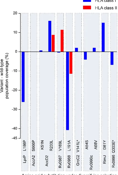
Figure 8. Variations in the worldwide population coverage in sites under diversifying selection. The percentage of the human population with potential to recognize predicted T cell epitopes (worldwide population coverage) was estimated for HLA class I (in blue) and HLA class II (in red). The difference between the population coverage of the variants under diversifying selection relative to the wild-type is on the yy axis. a More than one amino acid substitution in the same codon;
21
Geographic correlations between population coverage and MTBC lineage
frequency
T cell epitopes might be differentially recognized by diverse individuals due to the variable frequencies of HLA alleles across the human population, thus resulting in distinct population coverage. For this reason, the fraction of the human population with potential to recognize the putative epitopes under study (worldwide population coverage) was predicted. Next, the variation resulting from the presence of amino acids under diversifying selection was calculated (Figure 8 and Supplementary Table 3). Despite the low number of high binding affinity peptides to HLA class I (Figure 7), the highest alterations in population coverage imposed by the variant epitopes were found for HLA class I (Figure 8). Of particular note were four amino acid substitutions (LprP L186P, AccD2 R233L, Rv0988 L191A and RimJ D81Y) which showed a high impact on the HLA class I worldwide population coverage (-26%, +16%, -41% and +15%, respectively, as compared to the wild-type sequence) (Figure 8).
The largest variations in class II HLAs worldwide population coverage were attributed to AccD2 R233L and Rv0987 V169L (which were expected to increase coverage by 9% and 11%, respectively) and to Rv0988 L191A (expected to decrease the coverage by 11%). The analysis was expanded by investigating if the regional variations in population coverage were associated with the geographic distribution of the MTBC lineages harbouring the selected variants (Figure 9). For that purpose, the frequency of the MTBC lineages and the alterations in regional population coverage induced by the variants under diversifying selection were compared across 15 world geographic regions (Supplementary Figure 2). No correlations were found for the HLA class I. In contrast, three statistically significant correlations were found between the variations in HLA class II population coverage for AccD2 R233L (rτ = 0.39, p < 0.05), Rv0987 V169L (rτ = 0.47, p < 0.05) and Rv0988 L191A (rτ = -0.41, p < 0.05) and the frequency of lineage 1 and lineage 2 strains (Figure 9 and Supplementary Table 4). AccD2 R233L occurred in all the lineage 1 strains analysed (Figure 6C and Supplementary Table 1) and the variant amino acid induced a calculated increase in regional population coverage ranging from 1% in Central America to nearly 30% in Southeast Asia (Figure 9A). This increase in population coverage significant and positively correlated with the percentage of lineage 1 frequency in the 15 tested regions (graph in Figure 9A). Rv0987 V169L was found in 10% of the lineage 1 strains and 8% of the lineage 2 strains analysed (Figure 6C and Supplementary Table 1) and resulted in a predicted increase of the regional population coverage ranging from 3% in Central America to 29% in Southeast Asia (Figure 9B). The estimated increase in coverage was also significant and positively correlated with the regional prevalence of lineage 1 and 2 strains (graph in Figure 9B). In contrast, Rv0988 L191A, that was found in 69% of the lineage 2 strains (Figure 6C and
22
Supplementary Table 1), was estimated to decrease the regional population coverage from 3% in Central America to 29% in Southeast Asia (Figure 9C). This was significant and negatively correlated with the regional frequency of the lineage 2 strains (graph in Figure 9C). Overall, the results suggest that the amino acid substitutions AccD2 R233L, Rv0987 V169L and Rv0988 L191A were likely relevant to the sympatric adaptation of MTBC strains to certain host populations.
23 Figure 9.Association between differential population coverage of predicted epitopes and the geographic distribution of the MTBC. The frequency (in pie charts) of the MTBC lineages harboring sites under diversifying selection
(in xx axis of each graph) correlated with the variations in population coverage due to differential binding to human HLA class II (green and red color gradients) of peptides harbouring three amino acid substitutions: (A) AccD2 R233L (rτ= 0.39, p <
24
In vitro
validation of the predicted CD4
+T cell epitopes under diversifying
selection
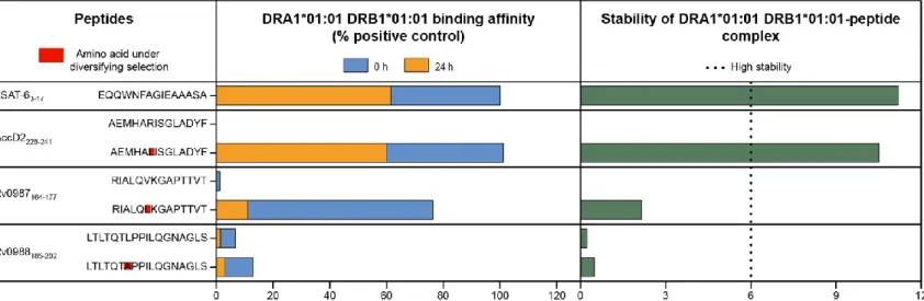
Typical T cell epitopes are expected to bind with high affinity and stability to an HLA molecule [123]. Thus, large alterations in the HLA-binding properties are anticipated in epitopes under T cell driven diversifying selection. Taking this into account, the in vitro HLA-binding affinity and stability were compared for the set of wild-type and variant peptides that i) were predicted to be under diversifying selection; ii) showed large variation in the predicted HLA-binding affinity and iii) impacted the population coverage and correlated with the geographical distribution of the MTBC lineages. This set was formed by six peptides with the amino acid substitutions AccD2 R233L, Rv0987 V169L and Rv0988 L191A. The HLA class II DRA1*01:01 DRB1*01:01 was chosen for the in vitro assays since this was the HLA with the broadest population coverage [160] among the ones with variable predicted binding affinity. The binding affinity and stability of each peptide was compared to a validated DRB1*01:01 CD4+ T cell epitope
of the M. tuberculosis ESAT-6protein [155]. The results confirmed the variant forms of AccD2228-241 and Rv0987164-177, but not of Rv0988185-202, as being epitopes with strong binding affinity to DRB1*01:01. In agreement with the in silico analysis, radical alterations (> 58%) in the binding affinity between wild-type and variant peptides were observed for AccD2228-241 and Rv0987164-177 (Figure 10). Most importantly, the variant AccD2228-241 peptide, in addition to displaying a binding affinity as high as ESAT-63-17, also formed a highly stable complex with DRA1*01:01 DRB1*01:01. This suggests that the variant AccD2228-241, but notthe wild-type peptide, has the potential to function as a previously unidentified immunodominant CD4+
T cell epitope in individuals expressing the HLA DRA1*0101 DRB1*01:01.
Figure 10. In vitro validation of the candidate CD4+ T cell epitopes under diversifying selection. Amino acid substitutions with the highest impact
on HLA class II binding predictions (AccD2 R233L, Rv0987 V169L and Rv0988 L191A) were subjected to in vitro confirmation of the estimated HLA-binding affinity. The HLA class II molecule with the broadest population coverage among the ones with variable predicted binding affinity (DRA1*01:01 DRB1*01:01) was used. The binding affinity of wild-type and variant peptide sequences was measured at 0 h and 24 h after incubation with the DRA1*01:01 DRB1*01:01 protein and compared with a validated M. tuberculosis CD4+ T epitope (ESAT-6
3-17). The stability index of the different DRA1*01:01 DRB1*01:01-peptide
25
D
ISCUSSION
Control of TB, a global disease that affects millions of people every year, highly relies on the development of better preventive strategies as the only vaccine in use (BCG) is of limited efficiency [5,6]. In fact, much effort has been put on the development of new vaccines but so far the results have been at best similar to BCG [161,162]. To achieve better outcomes, a deep understanding of the characteristics of protective immune responses and of how they are triggered and modulated is critical [162,163]. In this context, and considering that the efficiency of host T cell-mediated immunity is determinant for infection control [1,113], it is greatly relevant to obtain a full picture of the factors that might influence T cell responses in TB. Among these factors, the genetic and phenotypic diversity associated with the MTBC has been neglected. Indeed, several studies showed that strains of M. tuberculosis differently interact with innate immune cells [64,65,69,164,165], thus potentially being able to modulate the outcome of the acquired immune response [166]. Another factor with a potential impact on the type and quality of T cells responses is the existence of varying epitopes within the MTBC. It has been previously described that several T cell epitopes are evolutionarily hyperconserved [138], suggesting that the T cell responses induced in the host are similar across the MTBC diversity. However, the sympatric associations between MTBC lineages and human populations [48] and its disruption in HIV-1 infected individuals [114] suggests that at least an unknown set of relevant and variable MTBC CD4+ T cell epitopes may exist. Furthermore,
considering the long co-evolution of MTBC with its human host [51,88], it is likely that the host immune system represents a strong pressure shaping the evolution of the MTBC complex and namely at the level of the epitope-HLA interaction. In fact, different HLA haplotypes have been associated with resistance or susceptibility to TB caused by uncharacterized strains [167–169]) or even associated to TB caused by specific MTBC phylogenetic taxa [170]. In this thesis, a pipeline of molecular evolution, immunoinformatics and in vitro techniques was applied to reveal novel T cell epitopes in MTBC, taking into consideration the genetic variation within MTBC and its phylogeographic adaptation to the human host. Herein presented data constitute the first evidences for T cell driven diversifying selection in MTBC, namely within the genomic island iVEGI.
The iVEGI is a good candidate region within the M. tuberculosis genome for the discovery of MTBC-varying epitopes, due to its in vivo expression profile [157] and high genetic diversity as shown herein. Screening in vivo-expressed regions for the discovery of novel M. tuberculosis antigens has previously allowed the successful identification of a novel antigen encoded by Rv2034 [171]. The iVEGI contains several genes implicated in the survival and pathogenesis [158] of M. tuberculosis and with prolonged high expression
26
levels in immunocompetent animal models of TB infection but not in immunosuppressed animals or during in vitro growth [157]. Data for high genetic diversity in the iVEGI were firstly reported in a study comparing 20 genomes across four MTBC lineages, which highlighted a three genes operon (Rv0986-Rv0988) in this region with high SNP density [89]. Using 270 genomes from all the known seven MTBC lineages, it was revealed that a total of 21 genes in the iVEGI had a level of nucleotide diversity above the whole-genome average. The results also showed that SNPs in the iVEGI were evenly distributed across the MTBC lineages, which is in contrast with the genome-wide data in support for higher genetic conservation among the ‘modern’ (lineages 2, 3 and 4) when compared to ‘ancient’ lineages (lineages 1, 5, 6 and 7). This finding points to differences in the evolutionary process of this particular region. In addition, evidence supporting the action of diversifying selection was found in 11 iVEGI loci. Thus, genetic diversity and molecular evolution analyses strongly support the involvement of this genomic island in the long-term evolution of the MTBC population. Subsequent immunoinformatics data highlighted T cells as one of the driving forces for this evolution. Some variant iVEGI peptides were predicted to have differential binding affinity to class I or class II HLAs, with the more pronounced alterations found in HLA class II. This is congruent with previous studies showing that the contribution of CD4+ T cells for the immunity
against TB is more relevant than the one of CD8+ T cells [115,172]. Distinct host-pathogen interactions
could be the cause of failure in producing a universal vaccine with high worldwide population coverage. Indeed, three distinct TB vaccine candidates were found to have considerably different overall population coverage, thus threatening their efficacy in certain regions of the globe [173]. In this context, much of the existing T cell epitope discovery studies in M. tuberculosis field have been using HLA molecules with high frequency in white individuals, which is likely to have a negative impact in the efficacy of the vaccine in several world regions with high TB burden [141]. The in silico-based approach followed in this thesis addresses this bias by including in the predictions HLA alleles that are predominant in TB endemic regions. The largest variations in HLA-binding affinity were concentrated in three specific codons of three distinct iVEGI-encoded proteins and were estimated to have a large impact in the world population coverage. Interestingly, for these three amino acid substitutions, there were significant correlations between the alterations in HLA class II population coverage and the frequency of the MTBC strains harbouring the selected variants. Therefore, the referred substitutions may represent evolutionary markers in MTBC of sympatric adaptation driven by CD4+ T cells. The binding affinity and the stability of
wild-type and variant forms of the peptides containing these markers was measured in vitro. The in vitro assays showed that the single amino acid substitutions predicted to be under diversifying selection in both AccD2228-241 and Rv0987164-177induced large alterations (> 58%) in the binding affinity between
wild-27
type and variant peptides, which was not observed for Rv0988185-202. However, the most striking finding was that although the wild-type sequence of AccD2228-241 did not bind to DRB1*01:01, the lineage 1-associated variant peptide displayed a level of binding affinity and stability as high as the experimentally confirmed immunodominant epitope ESAT-63-17 [155]. Since the stability of the HLA-peptide complex was associated with immunodominance [150–153], the variant AccD2228-241 peptide may constitute a relevant lineage-specific CD4+ T cell epitope.
29
C
ONCLUSION AND FUTURE PERSPECTIVES
The workflow described in this thesis allowed the identification of predicted T cell epitopes under diversifying selection in iVEGI, their study in terms of envisaged HLA-binding, impact in worldwide population coverage and geographic association between the human population and MTBC and, finally, the functional validation of the most promising candidates. The use of these approaches provided evidence for the existence of HLA-restricted T cell epitopes evolving under diversifying selection across the MTBC in response to immune pressure. From the 11 amino acid substitutions predicted to be under diversifying selection in iVEGI, three (AccD2 R233L, Rv0987 V169L and Rv0988 L191A) were associated with immune system pressure in silico. Of this, strong evidences to be a variable T cell epitope were confirmed in vitro for a peptide encompassing AccD2 R233L. However, T cells cannot be discarded as drivers of Rv0987 V169L and Rv0988 L191A, as performing the in vitro binding assays with other HLA predicted to have variable binding affinity (or even with other peptide) could yield different results. For the eight remaining sites, the responsible selective pressure has yet to be uncovered. Protein signature recognition tools such as InterProScan [174] could be the starting point to understand the cause and importance of these substitutions to M. tuberculosis. These tools perform homology search for conserved motifs, enzymatic active sites and many other features [174], and are thus a vital aid in the functional characterization of a protein.
The variant form of AccD2228-241 was confirmed to have different binding affinity and stabilities comparatively to the wild-type. Nevertheless, further experiments are needed to ensure that the peptide is presented to T cells and is differentially immunogenic in humans infected with lineage 1 strains. To achieve this, wild-type and mutant peptides could be synthesised and used to stimulated blood collected from TB patients that i) express the HLA class II of interest and ii) were infected with lineage 1 strains. Assuming that memory T cells are created upon the recognition of the AccD2228-241-DRB1*01:01 complex, differences in the levels and type of T cell responses are expected upon stimulation. For instance, this could be assessed by the quantification of cytokines known to be associated with TB disease, such as IFN-γ and interleukin-10 (IL-10) [1]. The implications of this study can also be extended to vaccination field, i.e. the BCG vaccine strains harbour the wild-type AccD2228-241 peptide that was found not to bind to DRB1*01:01. It is thus tempting to speculate that boosting the BCG-induced immune response with the variant AccD2228-241 could influence the protection to infections caused by lineage 1 MTBC strains.
![Figure 1. Pathology and death rate of human TB. (A) Gross appearance of TB dissemination in the lungs of a 35-year old male individual infected with HIV [8]](https://thumb-eu.123doks.com/thumbv2/123dok_br/17577015.818476/20.892.105.786.112.370/figure-pathology-death-gross-appearance-dissemination-individual-infected.webp)

![Figure 4. Schematic representation of the action of diversifying selection. Adapted from [95]](https://thumb-eu.123doks.com/thumbv2/123dok_br/17577015.818476/26.892.114.768.590.760/figure-schematic-representation-action-diversifying-selection-adapted.webp)