Radiol Bras. 2014 Jul/Ago;47(4):245–250 245
Intraventricular mass lesions at magnetic resonance imaging:
iconographic essay – part 2
*
Lesões expansivas intraventriculares à ressonância magnética: ensaio iconográfico – parte 2
Castro FD, Reis F, Guerra JGG. Intraventricular mass lesions at magnetic resonance imaging: iconographic essay – part 2. Radiol Bras. 2014 Jul/Ago; 47(4):245–250.
Abstract
R e s u m o
The present essay is illustrated with magnetic resonance images obtained at the authors’ institution over the past 15 years and discusses the main imaging findings of intraventricular tumor-like lesions (colloid cyst, oligodendroglioma, astroblastoma, lipoma, cavernoma) and of inflammatory/infectious lesions (neurocysticercosis and an atypical presentation of neurohistoplasmosis). Such lesions represent a subgroup of intracranial lesions with unique characteristics and some imaging patterns that may facilitate the differential diagnosis.
Keywords: Neoplasms; Cerebral ventricle neoplasms; Central nervous system; Magnetic resonance imaging.
Ilustramos este ensaio iconográfico com imagens de ressonância magnética obtidas em nosso serviço nos últimos 15 anos e discutimos as principais características de imagem de lesões intraventriculares, de etiologia tumoral (cisto coloide, oligodendroglioma, astroblastoma, lipoma, cavernoma) e de etiologia inflamatória/infecciosa (neurocisticercose e uma incomum apresentação da neuro-histoplasmose). Estas lesões representam um subgrupo de lesões intracranianas com características próprias e alguns dos padrões de imagem que podem facilitar o diagnóstico diferencial.
Unitermos: Neoplasias; Neoplasias do ventrículo cerebral; Sistema nervoso central; Ressonância magnética.
* Study developed at Hospital de Clínicas – Universidade Estadual de Campinas (Unicamp), Campinas, SP, Brazil.
1. MD, Resident of Radiology and Imaging Diagnosis at Hospital de Clínicas – Universidade Estadual de Campinas (Unicamp), Campinas, SP, Brazil.
2. PhD, Docent responsible for the Division of Neuroradiology, Professor, Depart-ment of Radiology, Universidade Estadual de Campinas (Unicamp), Campinas, SP, Brazil.
3. Graduate Student of Medicine, School of Medical Sciences, Universidade Estadual de Campinas (Unicamp), Campinas, SP, Brazil.
Mailing Address: Dr. Fabiano Reis. Faculdade de Ciências Médicas – Universidade Estadual de Campinas, Departamento de Radiologia. Rua Tessália Vieira de Camargo, 126, Cidade Universitária Zeferino Vaz. Campinas, SP, Brazil, 13083-887. Caixa Pos-tal: 6111. E-mail: [email protected].
Received March 3, 2013. Accepted after revision September 9, 2013.
Primary neoplasms of this tissue are highly vascularized and are commonly associated with hydrocephalus due to increased production of cerebrospinal fluid. Such lesions occur in a benign form, the choroid plexus papilloma, and less fre-quently in a malignant presentation, the choroid plexus car-cinoma. Tumors such as meningiomas and metastases may also occur in this location.
Masses are more frequently found in the posterior por-tion of the lateral ventricles(2), but their location may vary according to the type of tumor. Choroid plexus papillomas occur mainly in children, with predilection for the lateral ventricles in this age range, while in adults it is usually more frequently found in the fourth ventricle. Ependymomas oc-cur more frequently in the posterior fossa in children, and in adults they are generally supratentorial.
Many times, inflammatory/infectious lesions are ob-served within the ventricular system and among them neuro-cysticercosis is very common in Brazil. Other less frequent conditions, such as histoplasmosis, may also be observed.
In the present essay, the authors have gathered images obtained over the past 15 years at the Radiology Service of Hospital de Clínicas – Universidade Estadual de Campinas, São Paulo, Brazil. The study was duly approved by the Com-mittee for Ethics in Research of the Institution.
RADIOLOGICAL FINDINGS Colloid cyst
It is a benign lesion that usually develops in the antero-superior aspect of the third ventricle, adjacent to the Monro’s foramen(1) and is the most common lesion in this region(3).
Felipe Damásio de Castro1, Fabiano Reis2, José Guilherme Giocondo Guerra3
INTRODUCTION
Intraventricular tumors represent a subgroup of intrac-ranial lesions with typical and unique features, which can be considered apart from the classical subdivisions into intra-and extra-axial tumors(1). Although they are easily visualized, the differential diagnosis among lesions may be difficult without the knowledge of the types of tissues which origi-nate such tumors(2).
The ventricles are surrounded by a layer of ependymal cells and a subependymal plate formed by glial cells. Such layers give origin to ependymomas, subependymomas and subependymal giant cell astrocytomas. Such a lining and the septum pellucidum that is located between the corpus callo-sum and the fornix, separating the lateral ventricles, also give origin to the central neurocytoma, a unique glial neuronal tumor of the ventricular systems(2).
They are rounded-shaped or ovoid lesions, with smooth walls, and may be associated with obstructive hydrocephalus, ei-ther intermittent or not. At computed tomography (CT), the cysts may be either iso- or hyperdense, with no contrast up-take, or with peripheral contrast uptake. At magnetic reso-nance imaging (MRI) (Figure 1) the signal intensity is vari-able, depending upon the contents of the cyst(3), with hyper-or isosignal on T1-weighted sequences, as compared with the cerebrospinal fluid; and with hypo- to hypersignal on T2-weighted sequences, with mixed signal in some cases(1).
Oligodendroglioma
Oligodendrogliomas manifest as well defined, round-shaped or ovoid masses involving the cortex or the subcorti-cal white matter. At intraventricular location, they are adja-cent to the septum pellucidum and present imaging and patho-logical features very similar to those of central neurocytoma, and are differentiated only at immunohistochemical study. At CT, they may be hypodense, isodense and even hyperdense. Calcifications are observed in 20–91% of cases. Cystic or hemorrhagic degeneration may be found. MRI is superior to CT in the evaluation of tumor extent, and the tumor usually is hypointense in relation to the gray matter on T1-weighted sequences and hyperintense on T2-weighted sequences. Heterogeneity of signal intensity is the rule (Figure 2)(1,2).
Astroblastoma
It is a rare tumor, which usually presents as a large pe-ripheral supratentorial tumor. At MRI (Figure 3), they are solid-cystic tumors with a characteristic bubbly feature of the solid component and isosignal on T2-weighted sequences. One observes a mild perilesional hypersignal, disproportion-ate to the tumor size(4).
Lipoma
Intracranial lipoma is a rare congenital malformation that more commonly occurs in the pericallosal region. At images, they are identified as well defined lobulated lesions, with fat density/intensity, located on the medial line. At CT, such tumors present as lesions with fat density (–50 to –100 UH), sometimes with calcifications, mainly the tubulonodu-lar type, with no contrast uptake. At MRI, they present with hypersignal on T1-weighted sequences, with signal intensity drop on T1-weighted sequences with fat suppression, and hyposignal on T2-weighted sequences(5) due to striking chemical shift artifact (Figure 4).
Cavernoma
Also named cavernous malformation or cavernous an-gioma, it is a congenital or acquired vascular abnormality that occurs in 0.5% of the general population. More rarely
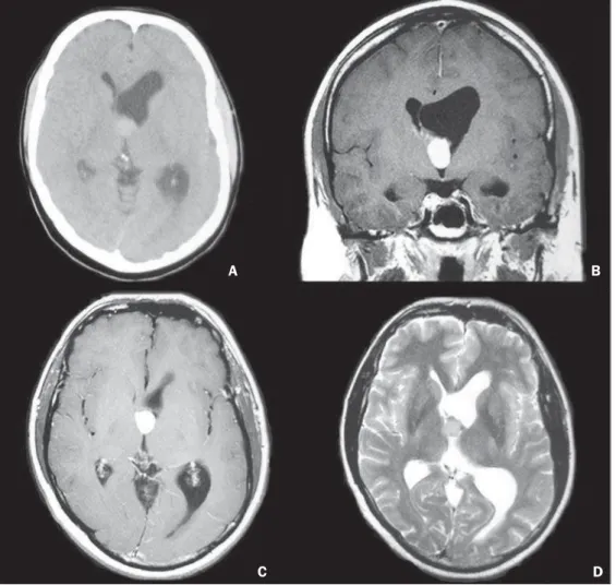
Figure 1. A male, 36-year-old patient. Non contrast-enhanced axial CT image (A) showing spontaneously hyperdense lesion located in the Monro´s foramen and in the anterior portion of the third ventricle, determining left lateral ventricle ectasia. Coronal (B) MRI and contrast-enhanced, axial T1-weighted image (C) demonstrate the presence of a solid lesion with no apparent contrast uptake. On axial T2-weighted images (D) the lesion presents with isosignal. anatomopathological study revealed colloid cyst.
A B
C D
A B
Figure 2. A male, 31-year-old patient. Coronal MRI T1-weighted sequence (A) shows a heterogeneous solid, cystic lesion, with low signal intensity, with some hypersignal foci (hemorrhage) and hyposignal (calcifications) located in the left lateral ventricle. On the contrast-enhanced T1-weighted sequence (B,C) there is intense uptake in the solid portion. On the T2-weighted sequence (D), the lesion presents with hypersignal. There are signs of extension/infiltration of the adjacent encephalic parenchyma. Biopsy revealed oligodendroglioma.
Figure 3. A male, 12-year-old patient. Contrast-enhanced, axial MRI, T1-weighted sequence (A) reveals the presence of a solid lesion, with no sign of necrosis, located in the septum pellucidum with intense contrast uptake (B,C). On the T2-weighted sequence (D), the lesion presents with isosignal and extensive signal change involving the adjacent encephalic parenchyma. In spite of the atypical imaging pattern in relation to that described in the literature, the biopsy revealed astroblastoma.
A B
they may be intraventricularly located. Classically, caverno-mas are found on T2-weighted images as popcorn ball-like lesions with a hyposignal halo due to hemosiderin deposi-tion. Subacute hemorrhage and blood degradation products produce a hypersignal halo on T1-weightes sequences, a
find-ing that helps in the differentiation from hemorrhagic tu-mors and other intracranial hemorrhages (Figure 5). At CT, such malformations are seen as hyperdense, well delimited lesions, with calcifications in 40%-60% of the cases, with no contrast uptake(5).
Figure 4. A male, 70-year-old patient. Coronal MRI, T1-weighted sequence (A) shows a solid lesion with hypersignal on T1-weighted sequence, located on the floor of the third ventricle, with no contrast enhancement (B). On axial (C) and coronal (D) T2-weighted sequences, the lesion presents with hyposignal. The imaging findings are compatible with lipoma.
A B
C D
Figure 5. A male, 60-year-old patient. Axial MRI, T1-weighted sequence (A) demonstrates the presence of a heterogeneous solid lesion, with focal areas of globuliform hypersignal located in the third ventricle. On T2-weighted sequence (B) a hyposignal halo is observed surrounding the lesion that is also demonstrated on the DP and FLAIR sequences (C,D). Anatomopathological analysis was not performed but the lesion is a typical cavernoma.
A B
Neurocysticercosis
Intraventricular neurocysticercosis corresponds to 0.7– 33% of all cases, with a predilection for the fourth ventricle (50%), followed by the lateral ventricles (35%), third ven-tricle (10%) and aqueduct (5%). It generally causes obstruc-tive hydrocephalus because of ventriculitis caused by ependy-mal inflammatory response or adhesions(6).
Neurocysticercosis is seen at CT as a cystic lesion that is initially isodense in relation to the cerebrospinal fluid, and therefore are not clearly visualized. It may be associated with asymmetries in the ventricular system or ventricular dilata-tion. On the other hand, at MRI (Figure 6), the cysts are well defined, due to signal intensity subtly different from that of the cerebrospinal fluid at T1- and T2-weighted images. However, the cysts may appear isodense in relation to the cerebrospinal fluid and, in such cases, 3D CISS sequences are very useful in the characterization of the cysts and scolex demonstration(6). Depending upon their dimensions, neurocysticercosis can exert considerable expansile effect on the compartments where the lesions are located.
Histoplasmosis
Disseminated histoplasmosis refers to multiple organs infection by the Histoplasma capsulatum fungus. The most
common sites of involvement are the skin, respiratory and intestinal tracts. Central nervous system involvement is a rare complication that generally manifests as meningitis, typically occurring in immunosuppressed patients and in extreme age groups(7).
Imaging findings (Figure 7) are nonspecific and the di-agnostic hypothesis of histoplasmosis should be raised for patients with signs of meningitis or cerebritis at MRI stud-ies(7). Also, single or multiple granulomas may be found, usually isointense on T1-weighted and hypointense on T2-weighted sequences, with homogeneous enhancement after intravenous contrast medium injection. Granulomas, as in the present case, may be located inside the ventricular system.
CONCLUSION
Intraventricular mass lesions have a wide variety of pre-sentations at imaging studies, which may be a consequence of the different types of tissues found in the central nervous system, involved in the development of such lesions. There-fore, the study of the skull, particularly by MRI, plays a rel-evant role in the attempt to define differential diagnoses based on their location, signal characteristics on the different se-quences, as well as in the detection of hemorrhage elements and calcifications. In the present study, the authors have aimed
Figure 6. A male, 67-year-old patient. Axial MRI T1-weighted sequence (A) demonstrating the presence of cystic lesions with isosignal in relation to the cerebrospinal fluid, with isointense walls in relation to the brain cortex, with no contrast uptake (B,C), located in the posterior horns and in the third ventricle. The lesions are more clearly identified at axial MRI FLAIR sequence (D). Ventricular derivation is observed. Cerebrospinal fluid analysis was positive for anti-cysticercus antibodies, confirming the diagnosis of neurocysticercosis.
B A
at reviewing the main intraventricular lesions approaching from the most common ones to those more rarely found (par-ticularly those of inflammatory/infectious etiology), which can, however, be included in the differential diagnosis.
REFERENCES
1. Leite CC, Sequeiros IM, Lacerda MTC, et al. Tumores intraventri-culares: achados à ressonância magnética. Rev Imagem. 2001;23:73– 85.
2. Koeller KK, Sandberg GD; Armed Forces Institute of Pathology. From the archives of the AFIP. Cerebral intraventricular neoplasms: radiologic-pathologic correlation. Radiographics. 2002;22:1473– 505.
3. Tien RD. Intraventricular mass lesions of the brain: CT and MR find-ings. AJR Am J Roentgenol. 1991;157:1283–90.
4. Port JD, Brat DJ, Burger PC, et al. Astroblastoma: radiologic-patho-logic correlation and distinction from ependimoma. AJNR Am J Neuroradiol. 2002;23:243–7.
5. Osborn AG. Neuroglial cyst. In: Osborn AG, Salzman KL, Katzman G, et al, editors. Diagnostic imaging brain. 1st ed. Salt Lake City: Amirsys; 2004. p. 7–20.
6. Kimura-Hayama ET, Higuera JA, Corona-Cedillo R, et al. Neuro-cysticercosis: radiologic-pathologic correlation. Radiographics. 2010; 30:1705–19.
7. Zalduondo FM, Provenzale JM, Hulette C, et al. Meningitis, vascu-litis, and cerebritis caused by CNS histoplasmosis: radiologic-patho-logic correlation. AJR Am J Roentgenol. 1996;166:194–6.
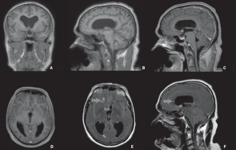
Figure 7. A male, 54-year-old patient. Coronal (A) and sagittal (B) MRI T1-weighted sequence demonstrating the presence of homogeneous, solid nodular lesions with isosignal in relation to the cerebral parenchyma, located in the right Monro’s foramen, with intense contrast enhancement (C,D). On the FLAIR sequences, a lesion with similar characteristics is highlighted in the anterior horn of the right lateral ventricle (arrows on E,F). Necropsy revealed histoplasmosis granulomas.
A B C