ABSTRACT
To evaluate the indings on chest CTs in 16 patients (8 men and 8 women) with laryngotracheobronchial papillomatosis. Methods: This was a retrospective study involving patients ranging from 2 to 72 years of age. The evaluation of the CT scans was independently performed by two observers, and discordant results were resolved by consensus. The inclusion criteria were presence of abnormalities on the CT scans, and the diagnosis was conirmed by anatomopathological examination of the papillomatous lesions. Results: The most common symptoms were hoarseness, cough, dyspnea, and recurrent respiratory infections. The major CT indings were nodular formations in the trachea, solid or cavitated nodules in the lung parenchyma, air trapping, masses, and consolidation. Nodular formations in the trachea were observed in 14 patients (87.5%). Only 2 patients had lesions in lung parenchyma without tracheal involvement. Only 1 patient had no pulmonary dissemination of the disease, showing airway involvement only. Solid and cavitated lung nodules were observed in 14 patients (87.5%) and 13 (81.2%), respectively. Masses were observed in 6 patients (37.5%); air trapping, in 3 (18.7%); consolidation in 3 (18.7%); and pleural effusion, in 1 (6.3%). Pulmonary involvement was bilateral in all cases. Conclusions: The most common tomography indings were nodular formations in the trachea, as well as solid or cavitated nodules and masses in the lung parenchyma. Malignant transformation of the lesions was observed in 5 cases.
Keywords: Papilloma; Tomography, X-ray computed; Lung diseases.
Laryngotracheobronchial papillomatosis:
chest CT indings
Helena Ribeiro Fortes1, Felipe Mussi von Ranke2, Dante Luiz Escuissato3,
Cesar Augusto Araujo Neto4, Gláucia Zanetti1,5, Bruno Hochhegger6,
Klaus Loureiro Irion7, Carolina Althoff Souza8, Edson Marchiori1,2
Correspondence to:
Edson Marchiori. Rua Thomaz Cameron, 438, Valparaiso, CEP 25685-120. Petrópolis, RJ, Brasil. Tel.: 55 24 2249-2777. Fax: 55 21 2629-9017. E-mail: [email protected]
Financial support: None. INTRODUCTION
Laryngotracheobronchial papillomatosis (LTBP) is a disease caused by HPV, characterized by the appearance of papillomas in any part of the aerodigestive tract.(1-5)
Although LTBP affects the larynx more commonly, the central airway can be involved in less than 5% of cases, and distal dissemination to the lung parenchyma occurs in about 1% of cases.(2,4,6,7)The disease has a higher incidence
in children and is the most common benign laryngeal neoplasia in this age group.(4,6-9)It is assumed that the
infection is more commonly acquired during birth, during the passage through the birth canal in mothers infected by the virus. Adults may also present LTBP, in whom the contamination by the virus is usually related to sexual contact.(1,3,4,10,11)Although a presumptive diagnosis can
be made based on the history and clinical-radiological
indings, the inal diagnosis is made by histopathological
analysis of the laryngeal or tracheal lesions, collected by bronchoscopy.(4)The course of the disease is unpredictable,
ranging from spontaneous remission to aggressive disease with pulmonary dissemination and the need for multiple surgical procedures to maintain airway patency.(5)
The objective of the present study was to evaluate the CT scans of 16 patients with LTBP in order to identify the
most common tomography indings. In addition, some
clinical and epidemiological aspects of the disease have been reported.
METHODS
This was a retrospective study involving 16 patients with LTBP and their CT scans of the chest. These scans were randomly collected through personal contacts with radiologists, originating from eight different institutions,
located in ive Brazilian states and in Canada. The diagnosis of LTBP was conirmed by the association of
clinical, radiological, and histopathological data. The CTs of the chest, due to the multiple institutions involved, were performed in different scanners, and, in all cases, using the high resolution technique.
Ten-millimeter HRCT scans were taken using ine axial
sections, 1-2 mm in thickness, from the lung apices to the bases during inspiration, with the patient in the supine
position, a high spatial resolution ilter being used for
image reconstruction. In some cases, iodinated contrast
1. Programa de Pós-Graduação em Radiologia, Universidade Federal do Rio de Janeiro, Rio de Janeiro (RJ) Brasil.
2. Disciplina de Radiologia, Universidade
Federal Fluminense, Niterói (RJ) Brasil.
3. Disciplina de Radiologia, Departamento de Clínica Médica, Universidade Federal do Paraná – UFPR– Curitiba (PR) Brasil.
4. Departamento de Medicina e Apoio
Diagnóstico, Universidade Federal da Bahia – UFBA – Salvador (BA) Brasil.
5. Disciplina de Clinica Médica, Faculdade
de Medicina de Petrópolis, Petrópolis (RJ) Brasil.
6. Disciplina de Diagnóstico por Imagem,
Universidade Federal de Ciências da Saúde de Porto Alegre (RS) Brasil.
7. Liverpool Heart and Chest Hospital NHS
Foundation Trust, Liverpool, England.
8. Department of Diagnostic Imaging, The Ottawa Hospital, University of Ottawa, Ottawa, Ontario, Canada.
Submitted: 21 November 2016. Accepted: 17 March 2017.
medium was injected intravenously. The images were obtained and reconstructed in a matrix of 512 × 512, window openings ranging from 1,000 to 1,500 HU,
and levels between −650 and −750 HU. The images
were digitized and photographed for the evaluation
of the lung ields. Scans were also performed using
a mediastinal window with a width between 350 and 400 HU and a center between 40 and 60 HU for the evaluation of the mediastinum.
The evaluation of the HRCT scans was independently performed by two observers, and discordant results were resolved by consensus. Regarding the pattern
of the indings, air trapping was deined as reduced
attenuation of the lung parenchyma, evidenced mainly by a lower density than usual and absence of reduced
lung volume; consolidation was deined as increased
attenuation of the lung parenchyma that prevented the visualization of vessels and external contours of
the bronchial walls; cavitation was deined as gas-illed spaces, with or without air-luid level, within a nodule, mass, or pulmonary consolidation; mass was deined
as any expansive pulmonary, pleural, mediastinal, or chest wall lesion presenting density of soft, fatty, or bony tissue greater than 3 cm in diameter, regardless of its contours or the heterogeneity of its contents; and
nodule was deined as a focal opacity that is rounded,
or at least partially delineated, smaller than 3 cm in
diameter, and generally presenting soft tissue or calciied tissue density. The criteria for deining these indings are those reported in the Fleischner Society glossary of
terms,(12) and the terminology used is that suggested
in the terminology consensus by the Department of Imaging of the Brazilian Thoracic Association.(13)The
scans were also evaluated for lesions in the central airways, pleural effusion, or any other associated pulmonary or extrapulmonary abnormalities.
RESULTS
Clinical and epidemiological characteristics
Sixteen patients with LTBP were evaluated, 8 (50%) being male and 8 female (50%). Regarding the age
group, our sample ranged from 2 to 72 years (mean = 25.7 years; median = 15.0 years, interquartile range [IQR]: 6-42 years). Regarding clinical symptoms,
hoarseness was reported in 8 patients (50.0%); cough,
in 7 patients (43.7%); and dyspnea, in 6 patients (37.5%). Recurrent pneumonias were also reported in 6 patients (37.5%). Of the 16 patients, 5 (31.2%) developed malignancy (squamous cell carcinoma), all being female, with ages ranging from 7 to 72 years. All of the patients presented weight loss at the time of diagnosis. Four patients underwent tracheostomy during the course of the disease, 2 being male (6 and 11 years of age) and two, female (2 and 5 years of age).
Tomography indings
The major tomography patterns were nodular lesions in the trachea and solid or cavitated nodules in the lung
parenchyma. Other less prevalent indings were mass,
consolidation, air trapping, and pleural effusion (Figures 1, 2 and 3). In relation to the lower airways, tracheal involvement was found in 14 patients, whereas main bronchus involvement was found in only 4 patients. The CT images revealed that this involvement of the lower airways was represented by nodular thickening of the trachea and of the main bronchi and by nodular lesions
of the walls. Solid nodules in the lung parenchyma were found in 14 patients (87.5%), and cavitated nodules were found in 13 patients (81.2%). Masses
were observed in 6 patients (37.5%); air trapping,
in 3 (18.7%); and pleural effusion, in only 1 (6.2%).
Only 1 patient presented no pulmonary dissemination of the disease, showing airway involvement only. The 6 patients who presented masses on the CT images were submitted to pulmonary biopsy, and squamous cell carcinoma was diagnosed in 4 of these patients. One of the patients who presented consolidation was also later diagnosed as having malignant degeneration. Pulmonary involvement was bilateral in all cases.
DISCUSSION
LTBP has a characteristic bimodal distribution, affecting children and young adults.(1,14)The juvenile
form of the disease begins before the age of 20 years and is most often diagnosed before the age of 5.(3,15,16)
The adult form begins after the age of 20, being more common in males in the third or fourth decades of life.(3,4,9,17,18)Orlamd et al.,(19) studying 224 patients,
found that 174 (77.7%) had the juvenile form of the disease, whereas 50 (22.3%) had the adult form.
Among the children in that study, 81% were male. In
adults, males also prevailed, comprising 62% of the cases. In our sample, the age of the patients ranged from 2 to 72 years (mean = 25.7 years; median =
15 years; IQR: 6-42 years), 8 (50%) being male and 8 (50%), female. Nine patients developed LTBP up to
20 years of age, presenting the juvenile form of the disease. Although the other 7 patients were older than 20 years of age, they can not be said to have the adult form of the disease, since the ages corresponded to the time of CT scanning and not necessarily to the time of the onset of the disease.
The estimated incidence of LTBP is approximately 4:100,000 in children and 2:100,000 in adults.(1,3,14,15)
The rates vary according to some factors, such as the age at the onset of the disease, the country studied, and the socioeconomic status of the patients.(1)The
incidence is higher in places with lower socioeconomic level, due to the higher prevalence of HPV infection.
(5)The most common clinical manifestations are
hoarseness, cough, stridor, dyspnea and recurrent infections. Abdulrazak et al.(21) reported that all of
the 31 patients in their study showed hoarseness as a symptom, followed by dyspnea in 9 patients (29%). In our sample, the most common symptom was also
hoarseness, in 8 patients (50.0%); followed by cough,
subsequently diagnosed with malignant lesions by biopsy. Tracheostomy may be necessary when there is a serious risk of airway obstruction by papillomatous lesions. In the literature, it is estimated that the need for tracheostomy varies from 13% to 21% in juvenile papillomatosis cases and from 4% to 6% in adult papillomatosis cases. In our study, there were 4 children who needed tracheostomy throughout the course of the disease.
LTBP has the potential to become malignant, especially as squamous cell carcinoma of the lung.(3,4,16,22)The
estimated malignancy rate of LTBP is approximately 3-7% in adults and less than 1% in children.(17)In our
sample, 5 cases (31.3%) progressed to malignancy, all of which in the form of squamous cell carcinoma of the lung. These 5 patients were female, 4 of whom were adults, ranging from 32 to 72 years of age, and 1 was a child (7 years of age).
Chest CT is the imaging method of choice for the
identiication and characterization of tracheobronchial
polypoid lesions and for the recognition of small nodules during the pulmonary dissemination phase. Chest x-rays may eventually demonstrate solid or cavitated lung nodules; however, tracheal or bronchial lesions are
dificult to be identiied by this method. Focal or diffuse
nodular narrowing, as well as nodular, pedunculated, or sessile polypoid lesions, are generally found in the trachea and the main bronchi, whereas nodules of varying sizes, usually multiple and well circumscribed, are observed in the lung parenchyma. The nodules may be solid or cavitated, with thin or moderately thick
walls (2-3 mm or more). Most nodules are small and
homogeneous when discovered early, but they can develop large air cavities as they grow and become
conluent.(4,7,15,23,24) When there are superimposed
infections or airway obstruction, there may be cavitated
nodules containing air-luid level/debris, consolidations,
atelectasis due to airway obstruction, air trapping, and bronchiectasis.(24-27)
In our sample, 15 patients had lesions in the lung parenchyma, and only 1 of these showed airway lesions only, with no pulmonary dissemination.
Nodular formations in the trachea were observed in 14 patients (87.5%), and parietal nodular formations were also observed in the main bronchi in 4. Solid
nodules in the lung parenchyma were found in 14
patients (87.5%), and cavitated nodules, in 13 (81.2%). These nodules had irregular internal contours
and walls of varying thicknesses. The lesions were
multilobulated and conluent in 8 (50.0%) and 8
patients, respectively. Air trapping was observed in
3 patients (18.7%), and pleural effusion, in only 1
(6.2%). An association with mass was found in 6
patients (37.5%), and consolidation, in 3 (18.7%).
Five patients were subsequently diagnosed with squamous cell carcinoma.
Our study had some limitations. The study was retrospective and observational. The analysis of some cases was transversal, without any evaluation of the evolution of and possible complications due to LTBP. The techniques used for CT scanning varied according to the protocol of each institution involved in the research. However, we believe that this variation had no impact on the results. Despite these limitations, we found no case series in the literature that focused
on the tomography indings of as many LTBP cases
as in our study.
Figure 1. Male patient, 6 years old. Axial CT scans at the level of the upper (in A) and lower lobes (in B), as well as a coronal
CT scan of the lungs (in C), showing nodulation in the trachea (white arrow), sparse areas of air trapping in both lungs (white asterisks), and bilateral parenchymal nodules of varying sizes (some solid or cavitated) disseminated in the lungs.
A
B
In conclusion, the most common tomography
indings were nodular formations in the trachea, solid
or cavitated nodules in the lung parenchyma, masses, consolidations, and air trapping. Cavitated nodules had irregular borders and walls of varying thicknesses.
Most were multilobulated and conluent. Malignant
transformation of the lesions was observed in 5 cases, all of which were female.
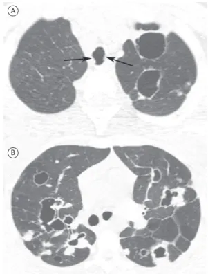
Figure 3. Axial CT scans at the level of the upper lobes (in A) and below the bronchial bifurcation (in B), showing solid and cavitated nodules in both lungs, with thick or thin walls. Nodular formations are also observed in the walls of the trachea (arrows).
A
B
A
B
Figure 2. Male patient, 4 years old. Axial CT scans (lung
window) above (in A) and below (in B) the bronchial bifurcation. In both scans, there are areas of air trapping (white asterisks) and multiple diffuse solid and cavitated nodules in the lungs. In A, there is an irregular narrowing of the tracheal lumen due to polypoid formations (arrow). In B, there is also a mass in the lower lobe of the left lung (black asterisk). The histopathological study of the mass revealed malignant transformation (squamous cell carcinoma).
REFERENCES
1. Carii M, Napolitano D, Morandi M, Dall’Olio D. Recurrent respiratory papillomatosis: current and future perspectives. Ther Clin Risk Manag. 2015;11:731-8. https://doi.org/10.2147/TCRM.S81825 2. Chang CH, Wang HC, Wu MT, Lu JY. Virtual bronchoscopy
for diagnosis of recurrent respiratory papillomatosis. J Formos Med Assoc. 2006;105(6):508-11. https://doi.org/10.1016/S0929-6646(09)60192-3
3. Katsenos S, Becker HD. Recurrent respiratory papillomatosis: a rare chronic disease, dificult to treat, with potential to lung cancer transformation: apropos of two cases and a brief literature review. Case Rep Oncol. 2011;4(1):162-71. https://doi. org/10.1159/000327094
4. Marchiori E, Araujo Neto Cd, Meirelles GS, Irion KL, Zanetti G, Missrie I, et al. Laryngotracheobronchial papillomatosis: indings on computed tomography scans of the chest. J Bras Pneumol. 2008;34(12):1084-9. https://doi.org/10.1590/S1806-37132008001200016
5. Venkatesan NN, Pine HS, Underbrink MP. Recurrent respiratory papillomatosis. Otolaryngol Clin North Am. 2012;45(3):671-94. https://doi.org/10.1016/j.otc.2012.03.006
6. Ağgünlü L, Erbaş G. Recurrent respiratory papillomatosis with lung involvement. Diagn Interv Radiol. 2009;15(2):93-5.
7. Kramer SS, Wehunt WD, Stocker JT, Kashima H. Pulmonary manifestations of juvenile laryngotracheal papillomatosis. AJR
Am J Roentgenol. 1985;144(4):687-94. https://doi.org/10.2214/ ajr.144.4.687
8. Li J, Zhang TY, Tan LT, Wang SY, Chen YY, Tian JY, et al. Expression of human papillomavirus and prognosis of juvenile laryngeal papilloma. Int J Clin Exp Med. 2015;8(9):15521-7.
9. Martina D, Kurniawan A, Pitoyo CW. Pulmonary papillomatosis: a rare case of recurrent respiratory papillomatosis presenting with multiple nodular and cavitary lesions. Acta Med Indones. 2014;46(3):238-43. 10. Fusconi M, Grasso M, Greco A, Gallo A, Campo F, Remacle M,
et al. Recurrent respiratory papillomatosis by HPV: review of the literature and update on the use of cidofovir. Acta Otorhinolaryngol Ital. 2014;34(6):375-81.
11. Omland T, Akre H, Lie KA, Jebsen P, Sandvik L, Brøndbo K. Risk factors for aggressive recurrent respiratory papillomatosis in adults and juveniles. PLoS One. 2014;9(11):e113584. https://doi. org/10.1371/journal.pone.0113584
12. Hansell DM, Bankier AA, MacMahon H, McLoud TC, Müller NL, Remy J. Fleischner Society: glossary of terms for thoracic imaging. Radiology. 2008;246(3):697-722. https://doi.org/10.1148/ radiol.2462070712
in chest CT scans. J Bras Pneumol. 2010;36(1):99-123. https://doi. org/10.1590/S1806-37132010000100016
14. Wiatrak BJ. Overview of recurrent respiratory papillomatosis. Curr Opin Otolaryngol Head Neck Surg. 2003;11(6):433-41. https://doi. org/10.1097/00020840-200312000-00005
15. Reeves WC, Ruparelia SS, Swanson KI, Derkay CS, Marcus A, Unger ER. National registry for juvenile-onset recurrent respiratory papillomatosis. Arch Otolaryngol Head Neck Surg. 2003;129(9):976-82. https://doi.org/10.1001/archotol.129.9.976
16. Franzmann MB, Buchwald C, Larsen P, Balle V. Tracheobronchial involvement of laryngeal papillomatosis at onset. J Laryngol Otol. 1994;108(2):164-5. https://doi.org/10.1017/S0022215100126180 17. Goon P, Sonnex C, Jani P, Stanley M, Goon P, Sonnex C, Jani P,
Stanley M. Recurrent respiratory papillomatosis: an overview of current thinking and treatment. Eur Arch Otorhinolaryngol. 2008;265(2):147-51. https://doi.org/10.1007/s00405-007-0546-z 18. Taliercio S, Cespedes M, Born H, Ruiz R, Roof S, Amin MR, et
al. Adult-onset recurrent respiratory papillomatosis: a review of disease pathogenesis and implications for patient counseling. JAMA Otolaryngol Head Neck Surg. 2015;141(1):78-83. https://doi. org/10.1001/jamaoto.2014.2826
19. Omland T, Lie KA, Akre H, Sandlie LE, Jebsen P, Sandvik L, et al. Recurrent respiratory papillomatosis: HPV genotypes and risk of high-grade laryngeal neoplasia. PLoS One. 2014;9(6):e99114. https:// doi.org/10.1371/journal.pone.0099114
20. Wilcox LJ, Hull BP, Baldassari CM, Derkay CS. Diagnosis and management of recurrent respiratory papillomatosis. Pediatr Infect Dis J. 2014;33(12):1283-4. https://doi.org/10.1097/
INF.0000000000000551
21. Abdulrazak A, Shuaibu IY, Ahmed AO, Hamisu A. Outcome of treatment in patients with recurrent respiratory papillomatosis in Kano: a 10 years retrospective analysis. Niger J Basic Clin Sci. 2016;13(1):36-40. https://doi.org/10.4103/0331-8540.172148 22. Donne AJ, Hampson L, Homer JJ, Hampson IN. The role of HPV type
in recurrent respiratory papillomatosis. Int J Pediatr Otorhinolaryngol. 2010;74(1):7-14. https://doi.org/10.1016/j.ijporl.2009.09.004 23. Marchiori E, Pozes AS, Souza Junior AS, Escuissato DL, Irion KL,
Araujo Neto Cd, et al. Diffuse abnormalities of the trachea: computed tomography indings. J Bras Pneumol. 2008;34(1):47-54. https://doi. org/10.1590/S1806-37132008000100009
24. Xiao Y, Wang J, Han D, Ma L. A Case of the Intrapulmonary Spread of Recurrent Respiratory Papillomatosis With Malignant Transformation. Am J Med Sci. 2015;350(1):55-7. https://doi. org/10.1097/MAJ.0000000000000370
25. Marchiori E, Zanetti G, Mauro Mano C. Tracheobronchial papillomatosis with diffuse cavitary lung lesions. Pediatr Radiol. 2010;40(7):1301-2. https://doi.org/10.1007/s00247-010-1573-2 26. Prince JS, Duhamel DR, Levin DL, Harrell JH, Friedman PJ.
Nonneoplastic lesions of the tracheobronchial wall: radiologic indings with bronchoscopic correlation. Radiographics. 2002;22 Spec No:S215-30. https://doi.org/10.1148/radiographics.22.suppl_1. g02oc02s215