ORIGIN
AL RESEAR
CH
Efects of upper cervical manipulation on the
electromyographic activity of the masticatory
muscles and the opening range of motion of the
mouth in women with temporomandibular disorder:
randomized and blind clinical trial
Efeitos da manipulação cervical alta sobre a atividade eletromiográica dos músculos
mastigatórios e amplitude de movimento de abertura da boca em mulheres com disfunção
temporomandibular: ensaio clínico randomizado e cego
Los efectos de la manipulación cervical en la actividad electromiográica de los músculos
masticatorios y la amplitud del movimiento de apertura de la boca en mujeres con trastorno
temporomandibular: un ensayo clínico aleatorizado y ciego
Gustavo Luiz Bortolazzo1, Paulo Fernandes Pires2, Almir Vieira Dibai-Filho3,
Kelly Cristina dos Santos Berni1, Bruno Mascella Rodrigues4, Delaine Rodrigues-Bigaton5
Mailing address: Delaine Rodrigues Bigaton. Avenida Jaime Pereira, 3701 – CEP: 13403-900 – Piracicaba (SP), Brazil.
E-mail: [email protected] – Phone: (19) 97872013 – Presentation: Oct. 2015 – Accepted for publication: Dec. 2015 – Approved by the Ethics Research Committee of the Universidade Metodista de Piracicaba (UNIMEP), under protocol number 01/09 and registered in the Brazilian registry of clinical trials (RBR-4j6xfx).
Study performed in the Laboratory of Therapeutic Resources – School of Health Sciences of the Universidade Metodista de Piracicaba (UNIMEP) – Piracicaba (SP), Brazil.
1PhD, Graduate Program in Oral and Dental Biology, Universidade de Campinas (UNICAMP) – Piracicaba (SP), Brazil.
2PhD student, Graduate Program in Human Movement Sciences, Universidade Metodista de Piracicaba (UNIMEP) – Piracicaba (SP), Brazil.
3PhD student, Graduate Program in Rehabilitation and Functional Performance, Universidade de São Paulo (USP) – Ribeirão Preto (SP), Brazil.
4Undergraduate in Physiotherapy (IC), Universidade Metodista de Piracicaba (UNIMEP) – Piracicaba (SP), Brazil.
5Professor, PhD by the Graduate Program in Human Movement Sciences, Universidade Metodista de Piracicaba (UNIMEP) – Piracicaba (SP), Brazil.
ABSTRACT | We evaluated the efects of upper cervical manipulation on the surface electromyographic activity (sEMG) of masticatory muscles and range of motion of the opening movement of the mouth in women with temporomandibular disorders (TMD). We evaluated 10 women with myogenic a TMD diagnosis, according to the Research Diagnostic Criteria for Temporomandibular Disorders (RDC/TMD) and divided randomly into an experimental group (EG) n=5, which received upper cervical manipulation, and a placebo group (PG) n=5, which received maneuvers without therapeutic efects. Five interventions were performed in both groups, once a week, with performance of pre-intervention assessments, post-immediate assessments (after 1st intervention) and post-delayed assessments (48 hours after the 5th intervention). The sEMG activity was processed using the root mean square and normalized by the peak value (RMS EMGn). We used for comparison the Student’s
426
t-test and ANOVA two-way repeated measures, adopting as signiicance the amount of 5%, and the Cohen d for treatment efect size. We found a signiicant interaction of group vs time (p<0.05) in the RMS EMGn of the left and right temporal muscles at rest, as well as for all masticatory muscles during maximal isometric contraction during jaw-elevation and jaw-depression. Treatment efect size, high to moderate, was observed in the EG, especially in the post-delayed assessment. We also observed a signiicant increase (p<0.05) and a high treatment efect during mouth opening in the EG. The upper cervical manipulation demonstrated a balance of the RMS EMGn of the masticatory muscles and increase the opening range of motion of the mouth in women with myogenic TMD.
RESUMO | Avaliou-se os efeitos da manipulação cervical alta sobre a atividade eletromiográica de superfície (sEMG) dos músculos mastigatórios e amplitude do movimento de abertura da boca em mulheres com disfunção temporomandibular (DTM). Foram avaliadas 10 mulheres com diagnóstico de DTM miogênica, segundo o Research Diagnostic Criteria for temporomandibular disorders (RDC/TMD), divididas, aleatoriamente, em grupo experimental (GE) n=5, que recebeu manipulação cervical alta e grupo placebo (GP) n=5, que recebeu manobra sem efeito terapêutico. Cinco intervenções foram aplicadas para ambos os grupos uma vez por semana, e avaliações de pré-intervenção, pós-imediato (após a 1ª intervenção) e pós-tardio (48 horas após a 5ª intervenção) foram realizadas. A atividade sEMG foi processada via raiz quadrada da média e normalizada pelo valor de pico (RMS EMGn). Utilizou-se para comparação os testes t de Student e ANOVA two-way (medidas repetidas), adotando-se como signiicância o valor de 5%, e o Cohen’s d para tamanho de efeito de tratamento. Constatou-se a interação signiicativa grupo × tempo (p<0,05) no RMS EMGn dos músculos temporal direito e esquerdo, na condição de repouso, assim como para todos os músculos mastigatórios durante contração isométrica máxima de elevação e depressão da mandíbula. Os tamanhos de efeito de tratamento moderado a alto foram observados no GE, destacando-se na avaliação pós-tardia. Foi observado também um aumento signiicativo (p<0,05) e um alto efeito de tratamento na abertura da boca para o GE. A manipulação cervical alta demonstrou equilibrar o RMS EMGn dos músculos mastigatórios e aumentar a amplitude de movimento de abertura da boca em mulheres com DTM miogênica.
Descritores | Manipulação da Coluna; Eletromiograia; Amplitude de Movimento Articular; Transtornos da Articulação Temporomandibular.
RESUMEN | En este estudio se evaluaron los efectos de la manipulación cervical alta sobre la actividad electromiográica de supericie (SEMG) de los músculos masticatorios y de amplitud del movimiento de apertura de la boca en mujeres con trastorno temporomandibular (TTM). Se evaluaron 10 mujeres con diagnóstico de TTM miogénico, con base en el Research Diagnostic Criteria for temporomandibular disorders (RDC/TMD), las que fueron aleatoriamente divididas en grupo experimental (GE) n=5, que recibió manipulación cervical alta, y grupo placebo (GP) n=5, que recibió maniobra sin efecto terapéutico. Se aplicaron cinco intervenciones para ambos grupos una vez por semana, y se realizaron evaluaciones preintervención, posintervención inmediata (después de la 1a. intervención) y posintervención tardía (48 horas después de la 5a. intervención). La actividad SEMG fue calculada mediante raíz cuadrada de la media y normalizada por el valor de pico (RMS EMGn). Se empleó para comparación los test t de Student y ANOVA two-way (medidas repetidas), y el nivel de signiicancia de 5%, y para el efecto del tratamiento el Cohen’s d. Se encontró la interacción signiicativa grupo × tiempo (p<0,05) en el RMS EMGn de los músculos temporales derecho e izquierdo, en reposo, así como para todos los músculos masticatorios durante la contracción isométrica máxima de elevación y depresión de la mandíbula. Se observaron efectos de tratamiento moderado a alto en el GE, destacando en la etapa posevaluación tardía. También se observó un aumento signiicativo (p<0,05) y un alto resultado del tratamiento en la apertura de la boca en el GE. La manipulación cervical alta demostró equilibrar el RMS EMGn de los músculos masticatorios y aumentar la amplitud del movimiento de la apertura de la boca en mujeres con TTM miogénica.
Palabras clave | Manipulación de la columna; Electromiografía; Amplitud del Movimiento Articular; Trastornos de la Articulación Temporomandibular.
INTRODUCTION
Temporomandibular disorder (TMD) is a term assigned to a subgroup of orofacial pains whose signs and symptoms include limited joint range of motion, pain or discomfort, clicking and noise in the temporomandibular joints (TMJ), which may be accompanied by neck pain, diiculty in chewing and headaches1.
It is known that there are patterns of coordinated movements between the TMJ, the atlanto-occipital joint and the cervical zygomatic-epiphyseal joints, determined by the intrinsic sensorimotor connection
via trigeminocervical complex2. herefore, any change in
one of these structures can trigger changes in the other. Several therapeutic modalities have been used for the treatment of TMD, such as massages3, electrotherapy4
and spinal manipulation5.
Spinal manipulation is notable for establishing joint mobility6-8, promote analgesia9,10 and change
the muscular activity10,11. A recent systematic review
better clinical evidence regarding the efects of the technique12.
Generally speaking, studies have shown that manipulation applied to the upper cervical spine (atlas/axis) shows promising efects in clinical practice, such as increased mouth opening range of motion and the pressure pain threshold in individuals with TMD13, increased joint range of motion of the cervical
spine and reduced pain in people with neck pain and atlantoaxial osteoarthritis7,14,15. However, some authors
warn about the risk of injury to the vertebral artery after application of upper cervical manipulation16,
a risk that, ruled out by Erhardt et al.17 when faced
their recent indings after technical application in asymptomatic individuals.
hus, for proper use of applied manipulation to the upper cervical spine, we suggest previous performance of speciic tests to conirm or not the presence of altered blood low of vertebral artery and instability of the upper cervical spine, when conirmed, they contraindicate the performance of manipulation18,19.
herefore, given the lack of studies that evaluate the efects of upper cervical manipulation on the TMD, especially regarding the electrical activity of the masticatory muscles, taking into account the coordinated movement pattern that exists between the TMJ and the cervical spine and the beneicial results of this technique found in individuals with neck pain and TMD, the aim of this study was to evaluate the efects of upper cervical manipulation on the electrical activity of the masticatory muscles and mouth opening range of motion in patients with myogenic TMD.
herefore, the hypothesis is that the upper cervical manipulation increases the mouth opening range of motion and balances the electrical activity of masticatory muscles (anterior temporal, masseter and supra-hyoid) of individuals with myogenic TMD, to increase its electric activities during maximal isometric contraction and to reduce their electrical activity at rest.
METHODOLOGY
his research is characterized by a randomized and blind clinical trial.
We selected 10 women aged between 20 and 37 years (25.8±6.8 years) diagnosed with myogenic TMD according to the axis I of the Research Diagnostic
Criteria for Temporomandibular Disorders (RDC/ TMD), accompanied by pain and/or fatigue in masticatory muscles during functional activities for a minimum period of one year and a maximum of five years. The volunteers who had no change in vertebral artery flow were included (according to the extension and head rotation test)18 and instability of the upper
cervical spine (according to the Sharp-Purser test)19.
We excluded from the study: women with tooth loss that used full or partial dental prosthesis, trauma history in the face and TMJ, with subluxation or TMJ dislocation, those with a diagnosis of IIIb (osteoarthritis) or IIIc (osteoarthrosis) according to the RDC/TMD and those that were receiving any kind of treatment for their TMD.
he randomization of this study was performed by a stratiied sortition, thus allocating the volunteers into two groups: experimental (EG) n=5, and placebo (PG) n=5. he volunteers were blind regarding the group they belonged to, and the evaluators were also blind regarding the type of intervention.
he study was approved by the Research Ethics Committee of the Universidade Metodista de Piracicaba
(UNIMEP), under protocol number 01/09 and registered in the Brazilian register of clinical trials (RBR-4j6xfx).
Experimental procedure
We evaluated the electrical activity of the right and left masseter (RM and LM), right and left anterior temporal (RT and LT) and supra-hyoid (SH) using surface electromyography (sEMG). he mouth opening range of motion was assessed by a caliper rule. he sEMG signals were collected in pre intervention period, soon after the 1st intervention (post-immediate) and 48
hours after the 5th intervention (post-late). he opening
of the mouth was measured in pre-intervention and post-delayed intervention period.
To measure the range of motion (ROM) of the active mouth opening without pain, we used a caliper ruler (mm). he measures were collected 2 times for each volunteer, we used as the inal result, the mean value of the two measurements.
For the sEMG registry, we used an acquisition module of BIO-EMG signals 1000 (Lynx, São Paulo, SP, Brazil) with the same descriptions and parameters described by Berni et al20.
isometric contraction of the jaw-elevation (teeth clenching) and 3) maximal isometric contraction of the jaw-depression (opening mouth against manual resistance provided by the examiner).
For maximum isometric contraction of jaw-elevation, we used the Parailm® (Chicago) materials, as described
by Berni et al20. For maximum isometric contraction of
jaw-depression, we requested each volunteer to lower their jaw as hard as possible (opening the mouth without bending the head) against the manual resistance provided by the examiner. For each of the situations, three registers of electromyographic signal were taken for 5s, with one-minute intervals between them, and with a randomized sequence of contractions obtained by sortition.
he electromyographic signal was processed in the time domain by means of the root mean square (RMS) and then normalized by RMS peak value (RMS EMGn) via the Matlab® software 2014a
(8.3.0.532).
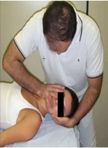
To the EG group, we performed an upper cervical manipulation (occipital, atlas and axis manipulation - OAA), and a right rotation manipulation and another in left rotation. For the manipulative procedure, the volunteers remained in the supine position, and the physical therapist conducted, passively, a slight pull of the head of the volunteers in the upper direction with rotation. After that, a pulse at high speed and short amplitude was accomplished by increasing the rotation parameter7 (Figure 1). he joints were
considered manipulated when noises were produced in one of the three attempts. With lack of cavitation in one of the three attempts, the joints were considered manipulated7.
Figure 1. Positioning for application of the upper cervical manipulation and placebo maneuver
To the PG, a similar maneuver was performed, however without traction and quick boost in rotation8,21.
It is important to note that in the PG, the position without rotation was maintained for 15 seconds on each side. In both groups, there were 5 interventions, one per week, by a physical therapist specialized in Osteopathy with 10 years of experience.
Statistical analysis
he data normality was tested by the Shapiro-Wilk test, which showed normal distribution of data (p>0.05). After inding the normality assumption, we followed up with the comparison of data using the ANOVA two-way test repeated measures to the RMS EMGn variables. he time factor (pre- and post-immediate and post-delayed) was used as within-subject and the group factor (EG and PG) as between-subject. he hypothesis of interest was the group x time interaction. We also used Student’s t-test for intra and inter-group comparison for the mouth opening ROM variable.
he level of signiicance used for the analysis of all statistical tests described was 5%.
he intra-group clinical treatment efect size was assessed using the Cohen d test for all the dependent variables of the research. he “d” values established were: “low treatment efect” (≤0.2), “moderate treatment efect” (≅0.5) and “high treatment efect” (≥0.8)22.
RESULTS
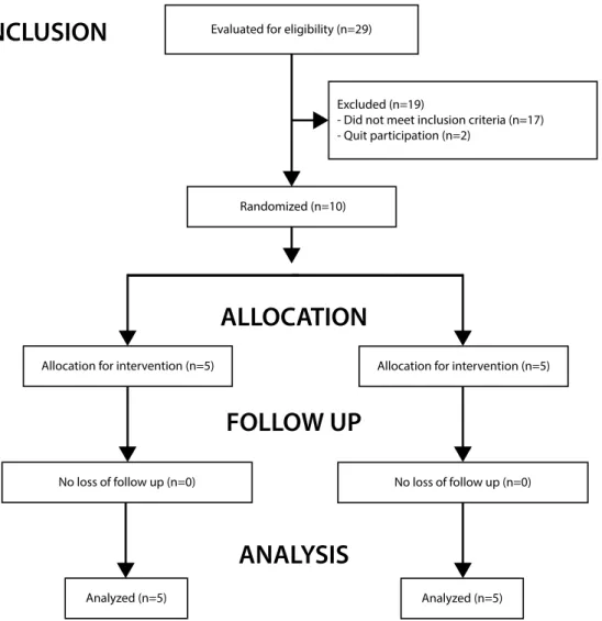
his study was conducted in the laboratory of therapeutic resources of the UNIMEP, between the months of February 2009 and December 2010. Figure 2 shows the lowchart of recruitment, distribution and sample analysis, in which 29 volunteers were previously enrolled, 19 excluded for not meeting the eligibility criteria, i.e., 10 had no diagnosis of myogenic TMD using the RDC/TMD, 3 were already receiving dental treatment during enrollment and 2 dropped out of the study. After volunteer exclusion, the remaining 10 were randomly allocated in the EG and the PG for further analysis.
Evaluated for eligibility (n=29)
Excluded (n=19)
- Did not meet inclusion criteria (n=17) - Quit participation (n=2)
Randomized (n=10)
Allocation for intervention (n=5)
Analyzed (n=5) No loss of follow up (n=0)
Allocation for intervention (n=5)
Analyzed (n=5) No loss of follow up (n=0)
INCLUSION
ALLOCATION
FOLLOW UP
ANALYSIS
Figure 2. Flowchart for sample distribution
Table 1. Intra- and inter-group comparison of the amplitude of mouth opening
PRE
Mean ± SD
POST
Mean ± SD Δ POST x PRE (95%CI)
Experimental Group
27,60±8,56 37,60±11,15 10,00 (3,35;16,65)*, d=1,00
Placebo Group 40,60±11,76 42,40±14,67 1,80 (-4,85;8,45), d=0,13
PRE: pre-intervention assessment; POST: Post-delayed assessment. Δ: diference between the means Student t-test
*Signiicant intra-group diference (p<0.05) Size of the clinical treatment efect - Cohen d
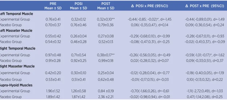
In the ANOVA two-way analysis with repeated measures in mandibular rest (Table 2), we found signiicant group x time interaction only for the LT muscles (F=5.72; p=0.13) and RT muscles (F=7.17; p=0.006). In the Bonferroni correction, there was a signiicant reduction of the RMS EMGn of the LT muscles in post-immediate assessment and RT muscles in the post-delayed assessment for EG; as well as in the moderate to high clinical treatment efect in all
evaluated muscles. In the intergroup analysis, we observed signiicantly lower activity of the LT and RT muscles for the EG in the post-delayed period.
we observed signiicant increase in RMS EMGn in all assessments and muscles of the EG (except for LT in the post-immediate); as well as moderate to high clinical treatment efect on the EG for all muscles. We also found a signiicant increase in the LT muscle activity of the EG in the intergroup post-delayed assessment.
In the ANOVA two-way analysis with repeated measures in maximal isometric contraction of the
jaw-depression (Table 3), we observed a signiicant group x time interaction for the SH muscles (F=34.30, p=0.000). In the Bonferroni correction, we observed a significant increase in RMS EMGn of the SH muscles at all moments of the assessment, and high clinical treatment effect. We also observed significant increase in the muscles activity in the SH of the EG in the intergroup post-delayed assessment.
Table 2. Intra and inter-group comparison of the EMG RMS values of masticatory muscles at rest
PRE
Mean ± SD
POSI Mean ± SD
POST
Mean ± SD Δ POSI x PRE (95%CI) Δ POST x PRE (95%CI)
Left Temporal Muscle
Experimental Group 0.76±0.41 0,32±0,12 0,32±0,10** -0,44(-0,85; -0,02)*, d=-1,45 -0,44(-0,89;0,01), d=-1,49 Placebo Group 0.70±0.37 0,76±0,46 0,79±0,36 0,06(-0,35;0,47), d=0,14 0,09(-0,36;0,54), d=0,24
Left Masseter Muscle
Experimental Group 0.55±0.42 0,26±0,04 0,27±0,08 -0,29(-0,68;0,10), d=-0,99 -0,28(-0,67;0,11), d=-0,93 Placebo Group 0.54±0.32 0,46±0,28 0,52±0,13 -0,08(-0,47;0,31), d=-0,25 -0,02(-0,41;0,37), d=-0,09
Right Temporal Muscle
Experimental Group 0.97±0.48 0,71±0,54 0,38±0,17** -0,26(-0,56;0,05), d=-0,49 -0,59(-1,01;-0,17)*, d=-1,62 Placebo Group 0.91±0.28 0,92±0,25 0,99±0,18 0,02(-0,28;0,32), d=0,07 0,09(-0,33;0,51), d=0,37
Right Masseter Muscle
Experimental Group 0.42±0.20 0,30±0,10 0,25±0,04 -0,12(-0,28;0,04), d=-0,77 -0,18(-0,40;0,05), d=-1,19
Placebo Group 0.53±0.41 0,51±0,41 0,62±0,48 -0,01(-0,17;0,15), d=-0,03 0,10(-0,13;0,32), d=0,22
Supra-Hyoid Muscles
Experimental Group 1.96±1.52 1,26±0,58 0,84 ±0,19 -0,70(-1,66;0,26), d=-0,61 -1,11(-2,72;0,49), d=-1,03 Placebo Group 1.89±1.42 1,87±1,42 2,36 ±2,21 -0,02(-0,98;0,94), d=-0,01 0,47(-1,14;2,08), d=0,25
PRE: pre-intervention assessment; POSI: post-immediate assessment; POST: Post-delayed assessment. Δ: diference between the means ANOVA two-way test repeated measures with Bonferroni correction:
*Signiicant intra-group diference (p<0.05) ** Signiicant inter-group diference (p<0.05) Size of the clinical treatment efect - Cohen d.
Table 3. Intra and inter-group comparison of the EMG RMS values of masticatory muscles during maximal lifting contraction of the jaw and during maximal isometric depression contraction of the jaw
PRE Mean ± SD
POSI Mean ± SD
POST
Mean ± SD Δ POSI x PRE (95%CI) Δ POST x PRE (95%CI)
Left Temporal Muscle - Maximal isometric lifting contraction of the jaw
Experimental Group 0.61±0.25 0.73±0.31 0.84±0.38** 0.13 (-0.00;0.26), d=0.45 0.24 (0.07;0.40)*, d=0.73 Placebo Group 0.69 ±0.35 0.72±0.34 0.65±0.34 0.03 (-0.10;0.16), d=0.09 -0.04 (-0.21;0.13), d=-0.12
Left Masseter Muscle - Maximal isometric lifting contraction of the jaw
Experimental Group 0.49±0.25 0.65±0.26 0.68±0.23 0.16 (0.04;0.28)*, d=0.62 0.20 (0.08;0.31)*, d=0.80
Placebo Group 0.51±0.15 0.52±0.17 0.48±0.18 0.01 (-0.10;0.13), d=0.09 -0.04 (-0.15;0.08), d=-0.21
Right Temporal Muscle - Maximal isometric lifting contraction of the jaw
Experimental Group 0.47±0.16 0.57±0.13 0.59±0.13 0.10 (0.04;0.16)*, d=0.67 0.12 (0.06;0.18)*, d=0.81 Placebo Group 0.52±0.26 0.52±0.23 0.54±0.26 0.01 (-0.05;0.07), d=0.03 0.02 (-0.04;0.08), d=0.08
Right Masseter Muslce - Maximal isometric lifting contraction of the jaw
Experimental Group 0.51±0.35 0.65±0.39 0.73±0.39 0.14 (0.05;0.23)*, d=0.37 0.22 (0.14;0.30)*, d=0.58
Placebo Group 0.46±0.19 0.45±0.17 0.43±0.18 -0.01 (-0.10;0.08), d=-0.03 -0.03 (-0.11;0.05), d=-0.16
Supra-Hyoid Muscles - Maximal isometric depression contraction of the jaw
Experimental Group 0.92±0.09 1.38±0.16 1.73±0.23** 0,46 (0.26; 0.66)*, d=3.52 0.81 (0.62;1.01)*, d=4.62 Placebo Group 1.05±0.20 1.13±0.24 1.08±0.15 0,08 (-0.12; 0.28), d=0.37 0.03 (-0.16;0.23), d=0.18 PRE: pre-intervention assessment; POSI: post-immediate assessment; POST: Post-delayed assessment. Δ: diference between the means
ANOVA two-way test repeated measures with Bonferroni correction: *Signiicant intra-group diference (p<0.05)
DISCUSSION
Individuals with TMD have an increased electrical activity of their masticatory muscles at rest when compared to asymptomatic, and this increase is even more pronounced in the anterior temporal muscle23. Given the above, we observed that the
upper cervical manipulation technique was able to signiicantly reduce the RMS EMGn value of the LT and RT muscles in the EG. We believe that the RT and LT muscles had heightened activity and, therefore, more susceptible to the therapeutic efect of the manipulations. However, despite RMS EMGn not having been signiicantly reduced in the LM and RM muscles, their RMS EMGn values presented themselves much lower in the LG than in the PG after the manipulations.
According to Berni et al.20, the electrical activity
of masticatory muscles at rest is an accurate index for the assessment of individuals with myogenic TMD and without TMD and, therefore, is characterized as an important tool for clinical practice. hus, the results found in this study help to unravel the initial information of the efects of the high cervical manipulation technique on the electrical activity of masticatory muscles at rest in women with myogenic TMD.
In the task of maximal isometric contraction of the jaw-elevation, research shows that the masticatory muscles (LT, RT, RM and LM) have reduced electrical activity when compared to asymptomatic individuals20,23.
Based on this assumption, the study demonstrated that upper cervical manipulation signiicantly increases the RMS EMGn of all masticatory muscles involved in the task, with higher treatment efects in post-delayed assessment to all the muscles in the EG.
In the task of maximal isometric contraction of the jaw-depression, no information was found in the literature that characterizes the diference between TMD and asymptomatic patients. However, Packer et al.5 found a signiicant increase in the activity of SH
muscles in the same task analyzed in TMD patients after a single manipulation of the upper thoracic spine, which is similar to this study that found signiicantly increased RMS EMGn and high clinical treatment efect of the SH muscles (in both moments of assessments) in the EG. Which in fact, suggests the possibility of spinal manipulation techniques having potential and promising efects in the evaluated muscles.
According to Pickar24, spinal manipulation can
modulate the inlux of sensory signals from the paraspinal muscles that are neuroanatomically connected to the manipulated level, and thereby improve the function of these muscles by changing their myoelectric activity. In this study, we believe that a similar modulation process occured, since there was a manipulation efect in all the masticatory muscles, with these muscles connected to the neuroanatomically with the manipulated level via the trigeminocervical complex.
he event described is conirmed by Bicalho et al.25 that found a signiicant increase in the sEMG
potentials of the lumbar paraspinal muscles during the isometric task in spinal extension after vertebral manipulation in the lumbar spine in patients with lumbago, and Camargo et al.26 that observed an increase
in non-normalized electromyographic RMS in the deltoid muscle during the task of isometric contraction in shoulder abduction at 90° after manipulation of the 5th/6th cervical vertebra. On the other hand, Pires et al.27 found no signiicant changes in the non-normalized
electromyographic RMS of the sternocleidomastoid muscles during maximal isometric contraction in shoulder-elevation and head-lexion after high thoracic manipulation, which might be explained by a lack of nerve connection of the evaluated muscles with the segment manipulated.
We also observed that the upper cervical manipulation signiicantly increased the mouth opening range of motion in the EG with high clinical treatment efect. his result parallels with the work of Mansilla-Ferragut7 that evaluated the immediate efects of
manipulation of the upper cervical spine in patients with chronic neck pain and reduced mouth opening ROM, noting increased ROM after the technique application. La Touche et al.28 performed 10 sessions of articulate
mobilization and stabilization exercises in the cervical region in TMD patients, inding a signiicant increase in mouth opening ROM (24 hours and 12 weeks after the intervention).
he limitations found in this study were: 1) conducting assessments only in post-immediate and post-delayed periods (after 48 hours of the last session), since even more satisfactory results could have been observed in long-term assessments due to the chronicity of the TDM evaluated, and the higher clinical efects found in the post-delayed assessment for all tasks and evaluated muscles; 2) assessments of the electrical activity of masticatory muscles only during rest and during maximal isometric contractions, since diferent information could have been determined in more functional joint tasks; 3) small sample size, justiied in part due to the methodological rigor used in this study to evaluate the efects of high cervical manipulation on a sample speciically deined, i.e., with myogenic TMD and 4) lack of assessment of the dominant mastigatoy side of the volunteers, since such information could elucidate diferent information and interpretations of the results.
However, considering the small “n” sample and due care in the interpretation of the results expressed in this study, the same was able to provide novel and positive information about the RMS EMGn values of the masticatory muscles of women with myogenic TMD and, therefore, we suggest that future clinical trials, with a larger sample size, be conducted keeping the methodological rigor used in this study to elucidate the long-term efects of the technique. Finally, regarding the upper cervical manipulation, the study presents relevant information so the clinician physiotherapist may analyze the importance of a potential tool for the treatment of myogenic TMD.
CONCLUSION
We conclude that the hypothesis of this study was conirmed, since manipulation in the upper cervical spine was efective to balance out the RMS EMGn activity of masticatory muscles and increase the mouth opening range of motion in women with myogenic TMD. However, we emphasize the importance of caution in interpreting results due to the small sample size presented in this study.
REFERENCES
1. Bender SD. Temporomandibular disorders, facial pain, and headaches. Headache. 2012;52(1):22-5.
2. Eriksson PO, Haggman-Henrikson B, Zafar H. Jaw-neck dysfunction in whiplash-associated disorders. Arch Oral Biol. 2007;52:404-8.
3. Gomes CA, Politti F, Andrade DV, Sousa DF, Herpich CM, Dibai-Filho AV, et al. Efects of massage therapy and occlusal splint therapy on mandibular range of motion in individuals with temporomandibular disorder: a randomized clinical trial. J Manipulative Physiol Ther. 2014;37(3):164-9.
4. Gomes NCMC, Berni-Schwarzenbeck KCS, Packer AC, Rodrigues-Bigaton D. Efect of cathodal high-voltage electrical stimulation on pain in women with TMD. Rev Bras Fisioter. 2012;16:10-5.
5. Packer AC, Pires PF, Dibai-Filho AV, Rodrigues-Bigaton D. Effect of upper thoracic manipulation on mouth opening and electromyographic activity of masticatory muscles in women with temporomandibular disorder: a randomized clinical trial. J Manip Physiol Ther. 2015;38(4):253-61.
6. Martínez-Segura R, De-la-Llave-Rincón AI, Ortega-Santiago R, Cleland JA, Fernández-de-Las-Peñas C. Immediate changes in widespread pressure pain sensitivity, neck pain, and cervical range of motion after cervical or thoracic thrust manipulation in patients with bilateral chronic mechanical neck pain: a randomized clinical trial. J Orthop Sports Phys Ther. 2012;42(9):806-14.
7. Mansilla-Ferragut P, Fernández-de-Las Peñas C, Alburquerque-Sendín F, Cleland JA, Boscá-Gandía JJ. Immediate efects of atlanto-occipital joint manipulation on active mouth opening and pressure pain sensitivity in women with mechanical neck pain. J Manip Physiol Ther. 2009;32(2):101-6.
8. Vernon H, Humphreys BK. Chronic mechanical neck pain in adults treated by manual therapy: a systematic review of change scores in randomized controlled trials of a single session. J Man Manip Ther. 2008;16(2):42-52.
9. Vicenzino B, Wright A. Efects of a novel manipulative physiotherapy technique on tennis elbow: a single case study. Man Ther. 1995;1(1):30-5.
10. Yu X, Wang X, Zhang J, Wang Y. Changes in pressure pain thresholds and Basal electromyographic activity after instrument-assisted spinal manipulative therapy in asymptomatic participants: a randomized, controlled trial. J Manip Physiol Ther. 2012;35(6):437-45.
11. Herzog W, Scheele D, Conway PJ. Electromyography responses of back and limb muscles associated with spinal manipulative therapy. Spine. 1999;24:146-52.
12. Calixtre LB, Moreira RF, Franchini GH, Alburquerque-Sendín F, Oliveira AB. Manual therapy for the management of pain and limited range of motion in subjects with signs and symptoms of temporomandibular disorder: a systematic review of randomised controlled trials. J Oral Rehabil. 2015;42(11):847-61.
13. Yu H, Hou S, Wu W, He X. Upper cervical manipulation combined with mobilization for the treatment of atlantoaxial osteoarthritis: a report of 10 cases. J Manip Physiol Ther. 2011;34(2):131-7.
manipulation versus nonthrust mobilization in patients with mechanical neck pain: a multicenter randomized clinical trial. J Orthop Sports Phys Ther. 2012;42:5-18.
15. Mansilla Ferragud P, Boscá Gandia JJ. Efecto de la manipulacion de la charnela occipito-atlo-axoidea en la apertura de la boca. Osteopat Cientıica. 2008;3:45-51. 16. Hurwitz EL, Aker PD, Adams AH, Meeker WC, Shekelle
PG. Manipulation and mobilization of the cervical spine: a systematic review of the literature. Spine. 1996;21:1746-59. 17. Erhardt JW, Windsor BA, Kerry R, Hoekstra C, Powell DW,
Porter-Hoke A, Taylor A. The immediate efect of atlanto-axial high velocity thrust techniques on blood low in the vertebral artery: A randomized controlled trial. Man Ther. 2015;20(4):614-22.
18. Cattrysse E, Swinkels RA, Oostendorp RA, Duquet W. Upper cervical instability: are clinical tests reliable? Man Ther. 1997;2(2):91-7.
19. Licht PB, Christensen HW, Høilund-Carlsen PF. Vertebral artery volume low in human beings. J Manip Physiol Ther. 1999;22(6):363-7.
20. Berni KC, Dibai-Filho AV, Pires PF, Rodrigues-Bigaton D. Accuracy of the surface electromyography RMS processing for the diagnosis of myogenous temporomandibular disorder. J Electromyogr Kinesiol. 2015;25(4):596-602.
21. Fernández-de-Las-Peñas C, Alonso-Blanco C, Cleland JA, Rodríguez-Blanco C, Alburquerque-Sendín F. Changes in pressure pain thresholds over C5-C6 zygapophyseal joint after a cervicothoracic junction manipulation in healthy subjects. J Manip Physiol Ther. 2008; 31(5):332-7.
22. Cohen J. Statistical power analysis for the behavioral sciences. 2ª ed. New Jersey: Lawrence Erlbaum; 1988. 23. Hugger S, Schindler HJ, Kordass B, Hugger A. Clinical
relevance of surface EMG of the masticatory muscles. (Part 1): Resting activity, maximal and submaximal voluntary contraction, symmetry of EMG activity. Int J Comput Dent. 2012;15(4):297-314.
24. Pickar JG. Neurophysiological efects of spinal manipulation. Spine J. 2002; 2(5):357-71.
25. Bicalho E, Setti JA, Macagnan J, Cano JL, Manffra EF. Immediate effects of a high-velocity spine manipulation in paraspinal muscles activity of nonspecific chronic low-back pain subjects. Man Ther. 2010;15(5):469-75.
26. Camargo VM, Alburquerque-Sendín F, Bérzin F, Stefanelli VC, Souza DP, Fernández-de-las-Peñas C. Immediate efects on electromyographic activity and pressure pain thresholds after a cervical manipulation in mechanical neck pain: a randomized controlled trial. J Manip Physiol Ther. 2011;34(4):211-20.
27. Pires PF, Packer AC, Dibai-Filho AV, Rodrigues-Bigaton D.Immediate and short-term efects of upper thoracic manipulation on myoelectric activity of sternocleidomastoid muscles in young women with chronic neck pain: A Randomized Blind Clinical Trial. J Manip Physiol Ther. 2015;38(8):555-63.