Open versus endovascular surgery for treatment of popliteal
artery aneurysms: 5 years’ experience at the HCRP-FMRP-USP
Cirurgia aberta e endovascular no tratamento de aneurisma de artéria poplítea:
experiência de cinco anos do HCRP-FMRP-USP
André Felipe Farias Braga1
*
, Rafael Cespedes Catto1, Mauricio Serra Ribeiro1, Carlos Eli Piccinato1, Edwaldo Edner Joviliano1
Abstract
Background: popliteal artery aneurysms (PAAs) account for 70% of peripheral aneurysms. Surgery is indicated for aneurysms that have diameters greater than 2.0 cm or are symptomatic. Repair can be achieved by conventional surgical techniques or using endovascular methods, which are becoming increasingly popular, but for which there is not yet a consensus on indications. Objective: To describe the experience of treating PAAs at the vascular and endovascular surgery department of the Hospital das Clínicas de Ribeirão Preto, ailiated to the Universidade de São Paulo (Brazil). Method: A review was conducted of cases of conventional and endovascular repair of PAAs over the last 5 years, analyzing demographic data, comorbidities, surgical indications, preoperative and early and late postoperative complications, length of hospital stay and patency, during follow-up of up to 1 year. Results: During the period analyzed, ten endovascular surgeries (ES) and 21 open surgeries (OS) were performed. he ES group exhibited a higher frequency of comorbidities. here was a higher frequency of symptomatic patients in the OS group (85%) than in the ES group (40%). he ES group exhibited a lower number of clinical and surgical complications. here were no statistical diferences between the groups in terms of age or length of hospital stay. Primary patency at 1 year was 80% in the ES group and 75% in the OS group. Conclusions: Endovascular treatment for PAAs ofers good results in terms of patency, with acceptable complication rates, in patients with high surgical risk and favorable anatomy. Controlled studies are therefore warranted to validate the endovascular technique and aford it the status of an alternative procedure for use in selected cases.
Keywords: aneurysm; popliteal artery; endovascular; surgery.
Resumo
Contexto: Aneurismas de artéria poplítea (AAPs) correspondem a 70,00% dos aneurismas periféricos. A indicação cirúrgica é para aneurismas com diâmetros maiores que 2,0 cm ou sintomáticos. O tratamento é feito por técnicas cirúrgicas convencionais ou endovasculares. Esta última tem ganho muitos adeptos, mas ainda não há consenso estabelecido sobre sua indicação. Objetivo: Apresentar a experiência da Divisão de Cirurgia Vascular e Endovascular do Hospital das Clínicas de Ribeirão Preto da Universidade de São Paulo no tratamento dos AAPs. Método: Foram revisados casos de reparo convencional e endovascular de AAPs tratados nos últimos cinco anos, avaliando dados demográicos, comorbidades, indicação cirúrgica, complicações pré e pós-operatórias precoces e tardias, tempo de internação e de perviedade em até um ano. Resultados: Foram realizadas no período dez cirurgias endovasculares (CE) e 21 cirurgias abertas (CA). O grupo CE teve maior frequência de comorbidades. Houve maior frequência de pacientes sintomáticos no grupo CA (85,00%) do que no grupo CE (40,00%). O Grupo CE apresentou menor número de complicações clínicas e cirúrgicas. A idade entre os grupos e o tempo de internação de cada grupo não apresentaram diferença estatística. A perviedade primária em um ano no Grupo CE foi de 80,00%, enquanto no Grupo CA foi de 75,00%. Conclusão: O tratamento endovascular para AAPs apresenta bons resultados, em termos de perviedade com taxas de complicações aceitáveis, em pacientes com risco cirúrgico elevado e anatomia favorável, justiicando, assim, a necessidade de mais estudos controlados para modiicar a posição da técnica endovascular como uma terapia alternativa para casos selecionados.
Palavras-chave: aneurisma; artéria poplítea; endovascular; cirurgia.
1 Universidade de São Paulo – USP, Faculty of Medicine of Ribeirão Preto, Clinical Hospital of Ribeirão Preto, Ribeirão Preto, SP, Brazil. Financial support: None.
Conlicts of interest: No conlicts of interest declared concerning the publication of this article. Submitted: May 05, 2015. Accepted: August 03, 2015.
INTRODUCTION
Popliteal artery aneurysms (PAA) are the most common type of peripheral aneurysm and the second most common among all types of aneurysm. In around 50% of cases, involvement is bilateral and there is a strong association with aortic aneurysms.1-3
Popliteal artery aneurysms are most often diagnosed in symptomatic patients, who present with intermittent claudication, critical ischemia of the limb or acute arterial occlusion. Asymptomatic patients are generally diagnosed by screening tests in patients with vascular diseases or those who have been diagnosed with a contralateral aneurysm.1,4,5
The imaging exams generally employed are Doppler ultrasound, primarily for screening, and computed tomography angiography or magnetic resonance angiography, for planning of surgical treatment, irrespective of whether this is accomplished with open surgery (OS) or endovascular surgery (ES). Some cases will also be assessed using arteriography.6,7
Treatment of popliteal aneurysms is indicated in symptomatic patients, when the aneurysm has a diameter greater than 2.0 cm or a diameter of less than 2.0 cm and mural thrombus. Asymptomatic aneurysms smaller than 2.0 cm and diameter without thrombus are monitored periodically using Doppler ultrasound.8,9
Open surgery is most widely employed and the preferred technique is a bypass, using a medial approach, proximal and distal ligature of the aneurysm and an inverted great saphenous vein graft. As endovascular techniques develop, new approaches to management of popliteal aneurysms are being studied, in the hope of achieving lower complication rates.2,10,11
The objective of this study is to describe the last 5 years’ experience of repairing PAAs at the vascular and endovascular surgery department of the Hospital das Clínicas de Ribeirão Preto, affiliated to the Universidade de São Paulo (Brazil). These patients were treated using either endovascular or conventional methods, according to criteria for indication that include anatomy and surgical risk. The study analyzes risk factors, diagnostic methods, indications for procedures, patency, risk of limb loss, postoperative complications and length of hospital stay.
PATIENTS AND METHODS
Data were obtained from medical records held by
the Hospital das Clínicas de Ribeirão Preto (afiliated
to the Universidade de São Paulo) relating to patients treated at the unit between April 1, 2008 and January
31, 2013. Data collection was authorized by the local clinical research committee.
The following data were collected with relation to each patient: sex, age, color, associated diseases, smoking, alcoholism, limb involved, presence of contralateral aneurysm or abdominal aorta aneurysm,
method used to conirm diagnosis, complaints on
presentation, surgical technique employed and postoperative complications, postoperative medication and length of hospital stay.
Patients were deined as having a PAA if there was
focal dilation of the artery greater than 50% of the
expected normal diameter (0.9±0.2 cm), conirmed
by Doppler ultrasonography, digital arteriography, angiotomography or magnetic resonance angiography.12,13
All patients diagnosed with PAA underwent some type of assessment with an imaging method to screen for contralateral and abdominal aneurysms.
For the purposes of analysis, patients were allocated to one of two groups on the basis of clinical complaints. The asymptomatic patients included those with aneurysms larger than 2.0 cm and also those with aneurysms smaller than 2.0 cm with thrombus in their interiors. The symptomatic patients were analyzed
in terms of their complaints, which were classiied
as follows: intermittent claudication; compressive symptoms (venous or neurological, such as edema, pain and/or paresthesias in limbs) and signs and symptoms of ischemia (chronic or acute); or critical ischemic disease, such as cyanosis, pain at rest and trophic lesions, demanding urgent or emergency surgery.
For each case the surgical technique used was
classiied as conventional technique or endovascular
technique. The conventional technique (i.e. open surgery) employed was femoropopliteal bypass with distal and proximal ligature of the aneurysm. All open surgery operations were conducted in surgery centers with spinal anesthesia or general anesthesia. Operations conducted using endovascular techniques were performed in the surgery center or in an angioradiology room with general or local anesthesia, with direct anterograde puncture of the ipsilateral femoral artery or dissection of the ipsilateral femoral artery. Intraoperative arteriography was conducted using iodinated contrast and Viabahn endoprostheses (Gore, Flagstaff, Arizona, USA) were used in all
cases. Aneurysms were deined as repaired if they exhibited no endoleaks or low‑limiting dissections.
All patients treated using the endovascular technique were given a 300 mg dose of Clopidogrel during the immediate postoperative period, were kept on double antiplatelet medication with 75 mg of Clopidogrel for a minimum of 6 months and were prescribed 100 mg/day
choosing the endovascular technique was a high surgical risk for the open technique. The initial condition was a minimum of two patent distal vessels and distal and proximal anchor points with a minimum neck of 1.0 cm. Patients were considered to present a high surgical risk if they were symptomatic or had three or more risk factors associated with cardiovascular disease and functional class III or IV, according to New York Heart Association (NYHA) criteria.
The number of days spent in hospital from the date of the operation until hospital discharge, direct complications of surgery and need for reintervention within 30 days were all analyzed. Additionally, clinical and/or laboratory evidence of clinical complications
during the irst 30 days after the operation, such as
hematoma or surgical wound infections, pneumonia, renal failure requiring dialysis, acute myocardial infarction or clinical heart failure decompensation, were also included in analyses.
All patients were followed-up clinically at 30 days, 90 days, 6 months and 1 year. At these consultations they were assessed by interview, physical examination, ankle-brachial index and ultrasound.
Statistical analyses were performed using GraphPad Prism 6.0, with application of the t test, considering
p < 0.05 to be signiicant.
RESULTS
A total of 28 patients were identiied who had
undergone PAA repair at this service over the previous 5 years. Nine patients underwent ES repairs, one of whom was operated on bilaterally. Eighteen patients underwent conventional treatment, three of them bilaterally, making a total of 21 OS repairs. Patients
with aneurysms that extended to the supericial femoral
artery were excluded from the study.
All of the patients were male. In the ES group, 90% of the sample were over the age of 60 at the time of surgery and in the OS group 77.77% of the patients were less than 75 years old on the day of their surgical procedures. Mean age in the OS group was 70.95 years and mean age in the ES group was 67.6 years. This difference was not statistically
signiicant (p = 0.31).
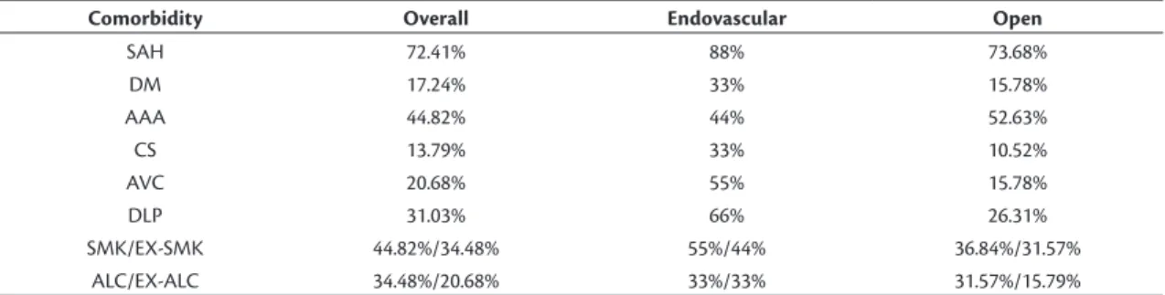
Table 1 contains the distribution of comorbidities. Ultrasound was used as a supplementary diagnostic examination in 80% of the ES group cases and in 61.90% of OS group cases. Preoperative arteriography was employed in 30% of cases in the endovascular group and 71.42% of cases treated with open surgery. Angiotomography was used to plan surgery in 30% of ES group cases and 38.09% of OS group cases (Figures 1a and 1b).
Figure 1. (a) Preoperative tomography; (b) Initial arteriography; (c) Distal bed; (d) Control arteriography. Table 1. Risk factors and concomitant diseases in patients with popliteal aneurysms.
Comorbidity Overall Endovascular Open
SAH 72.41% 88% 73.68%
DM 17.24% 33% 15.78%
AAA 44.82% 44% 52.63%
CS 13.79% 33% 10.52%
AVC 20.68% 55% 15.78%
DLP 31.03% 66% 26.31%
SMK/EX-SMK 44.82%/34.48% 55%/44% 36.84%/31.57%
ALC/EX-ALC 34.48%/20.68% 33%/33% 31.57%/15.79%
Bilateral disorders were detected in 90% of the cases in the ES group and one of these patients underwent ES in the contralateral leg while three other patients were referred for conventional surgery on the contralateral limb. Contralateral disorders were detected in 52.38% of the patients in the OS group.
With regard to surgical indications in the endovascular group, 40% were symptomatic (all with claudication), while 60% were asymptomatic, but had aneurysms with diameters larger than 2.0 cm. Surgical indications in the group of OS patients were acute ischemia in 52.38% of cases and limiting claudication in 33.33% of cases, while 14.29% were asymptomatic but had aneurysms with diameters larger than 2.0 cm (Table 2). The size of symptomatic patients’ aneurysms was not relevant.
In four patients, the endovascular surgical technique employed involved anterograde puncture of the ipsilateral common femoral artery, while in six patients the ipsilateral common femoral artery was dissected for surgical access. Covered stents with lengths ranging from 10.0 cm to 15.0 cm (Viabahn) were used and were not oversized in relation to the healthy native vessels (Figures 1c and 1d). A mean of 1.6 stents were used per patient, observing a 2.0 cm connection area where necessary. Treatment was successful in 90% of cases, with one case requiring an additional stent to correct a proximal endoleak. Early reintervention was necessary in 10% of ES cases because of occlusion of the stent. Just one patient exhibited hematoma as a complication and none of the patients suffered clinical complications.
All of the OS patients were given distal bypasses with inverted great saphenous vein grafts, using a medial access. Three early reinterventions were needed: one fasciotomy, one further bypass and one pseudoaneurysm repair. Two patients in the OS group underwent emergency intraoperative thrombolysis during the procedure, with recanalization of at least one distal recipient vessel. Four patients exhibited infectious complications, including two surgical wound infections, which were resolved with antibiotic therapy, and two patients developed pneumonia, also resolved with antibiotic therapy. One of the patients suffered acute renal failure and required temporary
hemodialysis. None of the ive patients who exhibited
complications after the open technique had been given thrombolysis, but three were operated on as urgent cases while two were asymptomatic patients who underwent elective surgery with just one patent distal vessel, breaking down as 27.27% of the urgent OS and 20% of the elective OS cases (Table 3).
Mean hospital stay was 3.9 days for the ES group and 5.28 days for the OS group, with no statistical
signiicance (p = 0.22) (Table 4 and Figure 2). All of the patients were discharged with prescriptions for
aspirin and statins for an indeinite period and those
in the ES group were also prescribed Clopidogrel for 6 months. In the OS group 66.66% and in the ES group 10% of the patients were kept on Cilostazol during the postoperative period and maintained clinical compensation.
Patients were monitored in outpatients follow-up, including physical examination and ultrasonography
Table 4. Length of hospital stay.
Endovascular Open p-value
Days in
hospital 3.9 5.28 0.22
Table 2. Indications for surgery.
Endovascular Open
Symptomatic
Claudication 40% 33.33%
Acute arterial occlusion 0% 52.38%
Asymptomatic 60% 14.29%
Figure 2. Length of hospital stay.
Table 3. Reinterventions, complications, primary patency and survival of limb by procedure.
Endovascular Open
Early reintervention 1/10 (10%) 3/21 (14.28%)
Complications
Overall 1/10 (10%) 4/21 (19.04%)
Primary patency
1 month 9/10 (90%) 20/21 (95.23%)
6 months 8/10 (80%) 15/20 (75%)
12 months 8/10 (80%) 15/20 (75%)
Survival of limb
30 days 10/10 (100%) 20/21 (95.23%)
depending on patients’ responses to clinical interviews and the results of physical examination and ankle-brachial index assessments. Among the patients treated with endovascular techniques, follow-up revealed 90% primary patency without further intervention at 30 days and 80% after 1 year. Just one case had a failure of patency in less than 1 month and this case was managed by bypass, saving the limb. One of the patients treated with conventional surgery was lost to follow-up after 1 month and was therefore excluded from the statistical analyses of postoperative follow-up longer than 30 days. Just one of the 21 procedures had a failure of patency after 1 month. Among the patients not lost to follow-up, 76.19% of the procedures were still patent after 6 months and remained patent at 1 year (Figure 3). One patient, who had undergone urgent surgery because of acute thrombosis, lost the limb within 1 month (infrapatellar amputation). All of the other patients’ (95.23%) limbs survived beyond 90 days (Table 3). There was no statistical difference in limb salvage rates between the two techniques at
30 or 90 days, with p = 0.30 and 0.47 respectively.
DISCUSSION
Peripheral aneurysms are rare in the general population, and popliteal aneurysms account for 70% of cases of peripheral aneurysms. They are more common among males, at a proportion of up to 30:1, and are also more common in people over the age of 65.4 They are often bilateral, in around 50% of cases,
as can be observed in the majority of series reported. In the cases reviewed here, 68.96% of the patients also had the disease contralaterally and 89.65% were more than 60 years old at diagnosis. All were male. Among these patients, the rate of concomitant
abdominal aorta aneurysms was 44.82%, which is in line with the literature.8,14
Some authors already recommend the endovascular
technique as the irst choice for PAA repair because
of its technical simplicity, percutaneous puncture, shorter hospital stay and lower rate of complications.
However, the irst cases series that compared ES for
popliteal aneurysm revealed that results were inferior to those achieved with conventional surgery, with high rates of complications and lost limbs.15,16 Some
authors attributed the increased risk of complications, which led to thromboses and fractured stents, to the mobility of the knees.17 Initial results have improved
as endovascular techniques have developed and with
the advent of more lexible self‑expanding stents and
those coated with heparin, such as the Viabahn stent used at our service. There are now several literature reviews and cases series reporting good patency and comparable limb salvage rates for conventional surgery and endovascular techniques.3,18-20 Long-term studies
are still needed. At the vascular and endovascular surgery department of the Hospital das Clínicas de Ribeirão Preto, endovascular treatment is only prescribed for cases in which surgery is high risk,
according to the NYHA classiication, and anatomy
is favorable, with at least two patent distal vessels.
We believe that ES is beneicial for these patients
because of the shorter duration of surgery, hospital
stay and recovery, thereby providing the beneits of
minimally invasive surgery to patients at high risk from surgery, with high risk of acute myocardial infarction or congestive heart failure.
Elective repair of popliteal aneurysms has a limb loss rate of less than 5% over 10-year follow-up.21
This compares with an amputation rate secondary to acute thrombosis due to popliteal aneurysms that is higher than 30% in some studies.2,22-25 In our
case series, all of the patients who were operated on urgently (because of acute ischemia) underwent conventional surgery. They accounted for 52.38% of the indications for repair using this technique, with an acceptable amputation rate (4.76% of all OS and 9% of urgent operations). A study by Pulli et al.26
reported that the majority of patients who underwent OS were symptomatic, compared with ES, although some studies report equal success rates for elective and emergency surgery.27 The great majority report
rates of complications and limb loss of 10% to 36% for patients who undergone emergency surgery, which are comparable to the rates for our patients.8,28-30 All of
reaching 69%, and secondary patency of up to 91%, offering a less invasive option for patients at high risk from surgery, even in acute situations.31
Studies report primary patency of 86% to 95% and secondary patency of 96.90% to 100% for aneurysms
repaired with elective endovascular surgery.31-34
Cases repaired with great saphenous vein grafts by conventional surgery have patency of 78.80% to
87.50% in the irst year, with limb salvage rates of
94.30%.20,26 Our patient sample was comparable with
previous studies, with stent patency of 80% during
the irst year and limb survival of 100% over the irst
90 days after ES, while cases treated with OS had
95.23% patency in the irst month and 75% after 1 year,
and a 90-day limb survival rate of 95.23%, including emergency cases. It is important to remember that in the cases treated with the endovascular technique, intraoperative arteriography showed at least two patent
infrapatellar vessels, which indicates good blood low
drainage and contributes to the patency of the stents. In this study, the overall complication rates were different in the two groups – 10% in the endovascular group and 19.04% in the conventional surgery group – which is possibly because the latter included both elective patients and those with critical ischemia. Even differentiating between complications in elective operations and urgent cases, the complications rates were 20% and 27.70% respectively.
In this sample, the lengths of hospital stays were not
signiicantly different between the groups (p > 0.05),
in contrast with published data, which shows that length of hospital stay is shorter among patients treated with ES.9,10,15,19,35-40
Several studies have reported complications including thrombosis, endoleaks, stent migration and stent fractures, which can be as high as 9.60% in the case of type 2 endoleaks, although the great majority are self-limiting and do not lead to expansion of the aneurysm sac.24,39,41 Previous studies have reported
higher rates of reintervention after ES, which was not the case in our series,9 probably because of the small
number of ES and the short follow-up. These rates are the reason why careful monitoring of patients who have undergone popliteal aneurysm repair is necessary, whether treated with endovascular or open surgery.42 In our review, there was just one case of
acute thrombosis of a stent, in which a bypass was performed to save the limb. Retreatment was necessary in 10% of the endovascular cases, compared with the open group, in which 14.28% needed additional intervention within 30 days.
We observed that the clinical groups in our study were highly heterogeneous. The endovascular group
had more comorbidities and were at high risk from surgery, but only cases with favorable anatomy were
selected for this technique, which caused a signiicant
difference between groups in terms of the number of cases. In the group that underwent OS, in addition to the anatomy not being a criterion, patients who underwent emergency surgery with imminent risk of losing the limb to critical ischemia were included in the analysis, causing a clear selection bias and, consequently, skewing the results. The objective of the study was to report the results of the two techniques after application of the preestablished criteria for selecting each method. This is the major limitation of this study. We cannot compare the results of the two groups with each other because the samples in each group were preselected and so we are limited to discussing the absolute results of each. These results are encouraging enough to stimulate prospective studies of favorable cases and the anatomic results of the two techniques are similar.
Randomized studies, albeit with limited numbers of asymptomatic patients with good distal blood drainage, demonstrate that when the techniques were compared over 12 months, assisted primary patency
rates were equal.40 When a longer period of up to
72 months was compared, secondary patencies were also equal.3 However, while offering equal patency,
the endovascular technique had a shorter length of hospital stay and shorter duration of surgery.
A non-randomized multicenter study of 178 patients reported large discrepancies between groups treated with OS and ES, both in terms of clinical presentation and distal drainage. In that study, primary and secondary patencies, time free from reintervention and the rate of limb salvage were all similar.37
While the conventional technique for repair of popliteal aneurysms remains the gold standard,43 this
review of cases treated at the Hospital das Clínicas de Ribeirão Preto reveals data that encourage use of the endovascular technique in view of the low rate of complications and good results over short and medium term follow-up observed among patients with high surgical risk and favorable anatomy. Notwithstanding, we can also conclude that conventional treatment proved effective and had a low relative rate of complications, even including patients with acute ischemia in the analysis.
CONCLUSIONS
Endovascular treatment to repair PAAs exhibited good results in terms of patency, with acceptable complication rates in patients at high risk from surgery and with favorable anatomy. Prospective and controlled studies with longer follow-up times are needed to validate the endovascular technique and afford it the status of an alternative procedure for high risk cases with favorable anatomy.
REFERENCES
1. Trickett JP, Scott RA, Tilney HS. Screening and management of asymptomatic popliteal aneurysms. J Med Screen. 2002;9(2):92-3. http://dx.doi.org/10.1136/jms.9.2.92. PMid:12133930.
2. Marin ML, Veith FJ, Panetta TF, et al. Transfemoral endoluminal stented graft repair of a popliteal artery aneurysm. J Vasc Surg. 1994;19(4):754-7. http://dx.doi.org/10.1016/S0741-5214(94)70052-4. PMid:8164291.
3. Antonello M, Frigatti P, Battocchio P, et al. Endovascular treatment of asymptomatic popliteal aneurysms: 8-year concurrent comparison with open repair. J Cardiovasc Surg. 2007;48(3):267-74. PMid:17505429.
4. Diwan A, Sarkar R, Stanley J, Zelenock GB, Wakefield TW. Incidence of femoral and popliteal artery aneurysms in patients with abdominal aortic aneurysms. J Vasc Surg. 2000;31(5):863-9. http://dx.doi.org/10.1067/mva.2000.105955. PMid:10805875.
5. Claridge M, Hobbs S, Quick C, Adam D, Bradbury A, Wilmink T. Screening for popliteal aneurysms should not be a routine part of a community-based aneurysm screening program. Vasc Health Risk Manag. 2006;2(2):189-91. http://dx.doi.org/10.2147/ vhrm.2006.2.2.189. PMid:17319463.
6. Hall HA, Minc S, Babrowski T. Peripheral Artery Aneurysm. Surg Clin North Am. 2013;93(4):911-23, ix. http://dx.doi.org/10.1016/j. suc.2013.04.008. PMid:23885937.
7. Galizia MS, Ward E, Rodriguez H, Collins J, Carr J. Improved characterization of popliteal aneurysms using gadofosveset-enhanced equilibrium phase magnetic resonance angiography. J Vasc Surg. 2013;57(3):837-41. http://dx.doi.org/10.1016/j.jvs.2012.09.018. PMid:23294506.
8. Lowell RC, Gloviczki P, Hallett JW Jr, et al. Popliteal artery aneurysms: the risk of nonoperative management. Ann Vasc Surg. 1994;8(1):14-23. http://dx.doi.org/10.1007/BF02133401. PMid:8192995.
9. Lovegrove RE, Javid M, Magee TR, Galland RB. Endovascular and open approaches to non-thrombosed popliteal artery aneurysm repair: a meta-analysis. Eur J Vasc Endovasc Surg. 2008;36(1):96-100. http://dx.doi.org/10.1016/j.ejvs.2008.02.002. PMid:18396427.
10. Galiñanes EL, Dombrovskiy VY, Graham AM, Vogel TR. Endovascular versus open repair of popliteal artery aneurysms: Outcomes in the US medicare population. Vasc Endovascular Surg. 2013;47(4):267-73. http://dx.doi.org/10.1177/1538574413475888. PMid:23393086.
11. Medeiros CAF, Gaspar RJ. Correção endovascular do aneurisma de artéria poplítea bilateral. J Vasc Bras. 2006;5(4):303-7. http:// dx.doi.org/10.1590/S1677-54492006000400010.
12. Johnston KW, Rutherford RB, Tilson MD, Shah DM, Hollier L, Stanley JC. Suggested standards for reporting on arterial aneurysms. J Vasc Surg. 1991;13(3):452-8. http://dx.doi.org/10.1067/mva.1991.26737. PMid:1999868.
13. Davis RP, Neiman HL, Yao JST, Bergan JJ. Ultrasound scan in diagnosis of peripheral aneurysms. Arch Surg. 1977;112(1):55-8. http://dx.doi. org/10.1001/archsurg.1977.01370010057010. PMid:831675.
14. Vermilion BD, Kimmins SA, Pace WG, Evans WE. A review of one hundred forty-seven popliteal aneurysms with long term follow-up. Surgery. 1981;90(6):1009-14. PMid:6458912.
15. Stone PA, Armstrong PA, Bandyk DF, et al. The value of duplex surveillance after open and endovascular popliteal aneurysm repair. J Vasc Surg. 2005;41(6):936-41. http://dx.doi.org/10.1016/j. jvs.2005.03.021. PMid:15944589.
16. Curi MA, Geraghty PJ, Merino OA, et al. Mid-term outcomes of endovascular popliteal artery aneurysm repair. J Vasc Surg. 2007;45(3):505-10. http://dx.doi.org/10.1016/j.jvs.2006.09.064. PMid:17275247.
17. Henry M, Amor M, Henry I, et al. Percutaneous endovascular treatment of peripheral aneurysms. J Cardiovasc Surg (Torino). 2000;41(6):871-83. PMid:11232970.
18. Mohan IV, Bray PJ, Harris JP, et al. Endovascular popliteal aneurysm repair: are the results comparable to open surgery? Eur J Vasc Endovasc Surg. 2006;32(2):149-54. http://dx.doi.org/10.1016/j. ejvs.2006.01.009. PMid:16546414.
19. Tielliu IF, Verhoeven EL, Zeebregts CJ, Prins TR, Bos WT, Van den Dungen JJ. Endovascular treatment of popliteal artery aneurysms: is the technique a valid alternative to open surgery? J Cardiovasc Surg (Torino). 2007;48(3):275-9. PMid:17505430.
20. Mohan IV, Stephen MS. Peripheral arterial aneurysms: open or endovascular surgery? prog Cardiovasc Dis. 2013;56(1):36-56. http://dx.doi.org/10.1016/j.pcad.2013.06.001. PMid:23993237.
21. Dawson I, van Bockel JH, Brand R, Terpstra JL. Popliteal artery aneurysms: Long-term follow-up of aneurysmal disease and results of surgical treatment. J Vasc Surg. 1991;13(3):398-407. http://dx.doi.org/10.1067/mva.1991.25131. PMid:1999859.
22. Galland RB. History of the management of popliteal artery aneurysms. Eur J Vasc Endovasc Surg. 2008;35(4):466-72. http:// dx.doi.org/10.1016/j.ejvs.2007.11.011. PMid:18180184.
23. Galland RB, Magee TR. Management of popliteal aneurysm. Br J Surg. 2002;89(11):1382-5. http://dx.doi.org/10.1046/j.1365-2168.2002.02221.x. PMid:12390377.
24. Mahmood A, Salaman R, Sintler M, Smith SG, Simms MH, Vohra RK. Surgery of popliteal artery aneurysms: a 12-year experience. J Vasc Surg. 2003;37(3):586-93. http://dx.doi.org/10.1067/mva.2003.141. PMid:12618697.
25. Ravn H, Wanhainen A, Björck M. Surgical technique and long-term results after popliteal artery aneurysm repair: results from 717 legs. J Vasc Surg. 2007;46(2):236-43. http://dx.doi.org/10.1016/j. jvs.2007.04.018. PMid:17664101.
26. Pulli R, Dorigo W, Castelli P, et al. A multicentric experience with open surgical repair and endovascular exclusion of popliteal artery aneurysms. Eur J Vasc Endovasc Surg. 2013;45(4):357-63. http:// dx.doi.org/10.1016/j.ejvs.2013.01.012. PMid:23391602.
27. Aulivola B, Hamdan AD, Hile CN, et al. Popliteal artery aneurysm: a comparison of outcomes in elective versus emergent repair. J Vasc Surg. 2004;39(6):1171-7. http://dx.doi.org/10.1016/j.jvs.2003.12.023. PMid:15192554.
28. Kauffman P, Puech-Leão P. Tratamento cirúrgico do aneurisma da artéria poplítea: experiência de 32 anos. J Vasc Bras. 2002;1(1):5-14.
30. Shortell CK, DeWeese JA, Ouriel K, Green RM. Popliteal artery aneurysms: a 25-year surgical experience. J Vasc Surg. 1991;14(6):776-9. http://dx.doi.org/10.1067/mva.1991.33214. PMid:1960807.
31. Trinidad-Hernandez M, Ricotta JJ 2nd, Gloviczki P, et al. Results of elective and emergency endovascular repairs of popliteal artery aneurysms. J Vasc Surg. 2013;57(5):1299-305. http://dx.doi. org/10.1016/j.jvs.2012.10.112. PMid:23375609.
32. Thomazinho F, Silvestre JMS, Sardinha WE, Motta F, Perozin IS, Morais D Fo. Endovascular treatment of popliteal artery aneurysm. J Vasc Bras. 2008;7(1):38-43. http://dx.doi.org/10.1590/ S1677-54492008000100007.
33. Etezadi V, Fuller J, Wong S, et al. Endovascular treatment of popliteal artery aneurysms: a single-center experience. J Vasc Interv Radiol. 2010;21(6):817-23. http://dx.doi.org/10.1016/j.jvir.2010.01.041. PMid:20456975.
34. Idelchik GM, Dougherty KG, Hernandez E, Mortazavi A, Strickman NE, Krajcer Z. Endovascular exclusion of popliteal artery aneurysms with stent-grafts: a prospective single-center experience. J Endovasc Ther. 2009;16(2):215-23. http://dx.doi.org/10.1583/08-2412.1. PMid:19456186.
35. Tsilimparis N, Dayama A, Ricotta JJ 2nd. Open and endovascular repair of popliteal artery aneurysms: tabular review of the literature. Ann Vasc Surg. 2013;27(2):259-65. http://dx.doi.org/10.1016/j. avsg.2012.01.007. PMid:22516241.
36. Stone PA, Jagannath P, Thompson SN, et al. Evolving treatment of popliteal artery aneurysms. J Vasc Surg. 2013;57(5):1306-10. http://dx.doi.org/10.1016/j.jvs.2012.10.122. PMid:23375437.
37. Midy D, Berard X, Ferdani M, et al. A retrospective multicenter study of endovascular treatment of popliteal artery aneurysm. J Vasc Surg. 2010;51(4):850-6. http://dx.doi.org/10.1016/j.jvs.2009.10.107. PMid:20138731.
38. Saunders JH, Abisi S, Altaf N, et al. Long-term outcome of endovascular repair of popliteal artery aneurysm presents a credible alternative to open surgery. Cardiovasc Intervent Radiol. 2014;37(4):914-9. http://dx.doi.org/10.1007/s00270-013-0744-6. PMid:24091756.
39. Tielliu IF, Verhoeven EL, Zeebregts CJ, Prins TR, Span MM, van den Dungen JJ. Endovascular treatment of popliteal artery aneurysms: results of a prospective cohort study. J Vasc Surg. 2005;41(4):561-7. http://dx.doi.org/10.1016/j.jvs.2004.12.055. PMid:15874916.
40. Antonello M, Frigatti P, Battocchio P, et al. Open repair verus endovascular treatment for asymptomatic popliteal artery aneurysm: results of a prospective randomized study. J Vasc Surg.
2005;42(2):185-93. http://dx.doi.org/10.1016/j.jvs.2005.04.049. PMid:16102611.
41. Ravn H, Björck M. Popliteal artery aneurysm with acute ischemia in 229 patients: outcome after thrombolytic and surgical therapy. Eur J Vasc. 2007;33(6):690-5. http://dx.doi.org/10.1016/j.ejvs.2006.11.040. PMid:17275362.
42. Wakassa TB, Matsunaga P, Silva ES, et al. Follow-up of the aneurysmal sac after exclusion and bypass of popliteal artery aneurysms. Clinics. 2006;61(2):107-12. http://dx.doi.org/10.1590/ S1807-59322006000200004. PMid:16680326.
43. Hogendoorn W, Schlösser FJ, Moll FL, Muhs BE, Hunink MG, Sumpio BE. Decision analysis model of open repair versus endovascular treatment in patients with asymptomatic popliteal artery aneurysms. J Vasc Surg. 2014;59(3):651-62. http://dx.doi. org/10.1016/j.jvs.2013.09.026. PMid:24246533.
*
Correspondence André Felipe Farias Braga Universidade de São Paulo – USP Campus Universitário, s/n, Monte Alegre CEP 14048-900 - Ribeirão Preto (SP), Brazil Tel.: +55 (11) 96434-5977 E-mail: [email protected]
Author information AFFB and RCC - Resident physicians of Angioradiology and Endovascular Surgery, Hospital das Clínicas de Ribeirão Preto, Faculdade de Medicina de Ribeirão Preto, Universidade de São Paulo (HCRP-FMRP-USP). MSR, CEP and EEJ - Professors, Division of Vascular and Endovascular Surgery, Hospital das Clínicas de Ribeirão Preto, Faculdade de Medicina de Ribeirão Preto, Universidade de São Paulo (HCRP-FMRP-USP).
Author contributions Conception and design: AFFB, RCC, CEP, EEJ Analysis and interpretation: AFFB, RCC Data collection: AFFB, RCC Writing the article: AFFB, RCC Critical revision of the article: EEJ Final approval of the article*: AFFB, RCC, MSR, CEP, EEJ Statistical analysis: AFFB, MSR Overall responsibility: EEJ