135
Anomalous sub aortic brachiocephalic vein: a case report
Radiol Bras. 2008 Mar/Abr;41(2):135–137 Case Report / Relato de Caso
Anomalous sub aortic left brachiocephalic vein: a case
report*
Veia braquiocefálica esquerda subaórtica anômala: relato de caso
Vetri Sudar Jayaprakasam1, Santosh Gurudev1, Klaus Irion2, Ali Nawaz Khan3, Andrew R. Pettit4
The left brachiocephalic vein occasionally follows an aberrant course. It is usually associated with congenital cardiac anomaly. We present a case of anomalous left brachiocephalic vein which followed a sub aortic course, with no cardiac abnormality. Multi detector computed tomography is very useful in accurate diagnosis of this condition and prevents any further investigation in cases of isolated abnormalities.
Keywords: Anomalous left brachiocephalic vein; Mediastinal mass; Computed tomography; Chest.
A veia braquiocefálica esquerda, ocasionalmente, segue curso aberrante. Esta variação freqüentemente está associada com anomalias congênitas do coração. Neste trabalho é apresentado um caso de veia braquioce-fálica esquerda anômala, com trajeto subaórtico, sem anormalidade cardíaca. A tomografia computadorizada com multidetectores é muito útil no correto diagnóstico dessa condição e permite a conclusão diagnóstica, sem quaisquer outras investigações.
Unitermos: Veia braquiocefálica esquerda anômala; Massa mediastinal; Tomografia computadorizada; Tórax.
Abstract
Resumo
* Study developed at The Royal Liverpool University Hospital, Liverpool, United Kingdom.
1. Residents, Radiology Residents (Specialist Registrar in Ra-diology) Mersey Rotation, Liverpool, United Kingdom.
2. PhD, Consultant Radiologist at the Cardiothoracic Centre – Liverpool and The Royal Liverpool University Hospital, Liverpool, United Kingdom.
3. Member of the Royal College of Surgery, Fellow of the Royal College of Radiology, United Kingdom. Charmain Imaging, King Abdulaziz Medical City, Riyadh, Kingdom of Saudi Arabia.
4. Member of the Royal College of Physicians, and of the Royal College of Pathologists, Professor of Haematology, Royal Liverpool University Hospital, Liverpool, United Kingdom.
Mailing address: Dr. Klaus Irion. 912 Manchester Road, Bury. Postcode: BL9 8DW, United Kingdom. E-mail: klaus.irion@ btinternet.com
Received November 12, 2007. Accepted after revision Jan-uary 8, 2008.
left brachiocephalic vein (LBCV) is even rarer. With the advent of non-invasive tech-nologies like multi detector computed tomography (MDCT) and magnetic reso-nance (MRI), there is now an increase in diagnosis of these anomalies. These are usually asymptomatic and the importance of diagnosing this condition is mainly to understand the anatomy during vascular access and mediastinal surgeries. In pa-tients with no other cardiac abnormality, this anomaly may be found incidentally whilst investigation of other causes.
The purpose of this work was to de-scribe this rare case of an anomalous left
Jayaprakasam VS, Gurudev S, Irion K, Khan AN, Pettit AR. Anomalous sub aortic left brachiocephalic vein: a case report. Radiol Bras. 2008;41(2):135–137.
INTRODUCTION
The anatomy of the great vessels in the mediastinum is complex and therefore prone for anomalies. These are usually associated with congenital heart disease. Aberrant right subclavian artery associated with a left aortic arch is the most common vascular anomaly in the mediastinum(1). Persistent left superior vena cava is the commonest venous anomaly with an inci-dence of approximately 0.3% in the general population and 3–5% in congenital heart disease patients(2). An anomaly of the brachiocephalic vein is rare and is usually associated with cardiac anomalies seen in approximately 0.2% to 1% of congenital heart anomalies(3). Isolated anomaly of the
0100-3984 © Colégio Brasileiro de Radiologia e Diagnóstico por Imagem
brachiocephalic vein (ALBCV) in a patient with no other cardiac abnormality, investi-gated with a MDCT with three dimensional reconstructions (3D).
CASE REPORT
A 34 year old male patient with a his-tory of heavy smoking was investigated for lymphocytosis.
An initial chest X-ray (Figure 1) showed mild mediastinal widening which raised the suspicion of an anterior mediastinal mass. A computed tomography (CT) scan of the chest was performed for further evaluation
136
Jayaprakasam VS et al.
Radiol Bras. 2008 Mar/Abr;41(2):135–137 Figure 2. CT images of the anomalous left brachiocephalic vein show-ing sub aortic position. A: Axial view. B: Sagittal view. C: Coronal view. The ascending aorta (AA) is in front of the left brachiocephalic vein (LBCV). The left pulmonary artery (PA) is below the LBCV that is cross-ing from the left to the right to merge with the right brachiocephalic vein (RBCV) to form the superior vena cava (SVC). The descending aorta (DA) lies behind.
A
B C
of the widened mediastinum in this patient with lymphocytosis. The CT revealed an ALBCV (Figure 2). The LBCV followed an obliquely downward course behind the ascending aorta and joined the right brachiocephalic vein forming the superior vena cava (Figure 3). The right brachioce-phalic and the superior vena cava had nor-mal anatomy. No other cardiac anonor-maly was found in this patient. No further inves-tigations or treatment were carried out for the ALBCV.
DISCUSSION
The LBCV is one of the major venous structures within the mediastinum. It is
formed by the union of left subclavian and left internal jugular vein just behind the sternal end of left clavicle. It runs down-ward and to the right side behind the up-per part of manubrium sterni, anterior to the three branches of the arch of aorta and terminates just behind the first right costal cartilage by uniting with the right brachio-cephalic vein, forming the superior vena cava(4).
The normal anatomy and the course of the major veins within the mediastinum are very much dependent on its embryological development. Under the normal circum-stances of fetal development, there are paired anterior cardinal veins that drain the upper body and the extremity. Around
137
Anomalous sub aortic brachiocephalic vein: a case report
Radiol Bras. 2008 Mar/Abr;41(2):135–137 ventral portion. The proximal part of the left cardinal vein disappears completely. The distal portion contributes to the forma-tion of left superior intercostal vein. The proximal part of the right anterior cardinal vein forms the superior vena cava(3).
In contrast, when there are anomalies of the development of arch (e.g., right sided arch) or the pulmonary arteries (atresia or stenosis) this can cause sparing of the in-ferior dorsal portion of the venous plexus and regression of superior ventral portion. This leads to the development of anoma-lous course of the brachiocephalic vein. However the exact mechanism, cause or correlation between these anomalies has not yet been established(3).
There are at least four major patterns of anomalous course of LBCV described by Takada et al.(5). Most of these anomalies are associated with right sided arch. The LBCV can cross the midline above the aortic arch in between the cephalic vessels. It can cross the midline beneath the aortic arch, above the pulmonary artery, as in our patient. Both these can be combined in one patient. The fourth pattern is where the LBCV is beneath the aortic arch, above the
pulmo-nary vein and behind the patent ductus arteriosus.
From the radiology point of view, diag-nosis of an ALBCV is mainly to avoid mis-interpretation of this structure. On an unenhanced CT scan, it can be interpreted as enlarged lymph node(6) or a central pul-monary artery. The descending portion of the ALBCV can be interpreted as persist-ent left superior vena cava. In the presence of the other cardiac anomalies, the diagno-sis can become difficult. However with the availability of MDCT, the vascular channel can be traced from its origin to the termi-nation and correctly identified.
From clinical point of view, knowledge of this anomaly is important when perform-ing central venous catheters or cardiac sur-geries. For anaesthetist this may cause dif-ficulties in introducing left sided central lines unless they know the anatomy. Sur-geons, if not informed already, may not identify the anomalous course which can significantly alter their surgical approach(7). The ALBCV in itself is asymptomatic and is not significant unless associated with other cardiac anomalies. This case report demonstrates the usefulness of MDCT in
demonstrating detailed anatomy of this anomaly and prevents further investigation in asymptomatic cases with no other car-diovascular anomaly.
REFERENCES
1. Donnelly LF, Fleck RJ, Pacharn P, et al. Aberrant subclavian arteries. Cross-sectional imaging find-ings in infants and children referred for evalua-tion of extrinsic airway compression. AJR Am J Roentgenol. 2002;178:1269–74.
2. Bhatti S, Hakeem A, Ahmad U, et al. Persistent left superior vena cava (PLSVC) with anomalous left hepatic vein drainage into the right atrium: role of imaging and clinical relevance. Vasc Med. 2007;12:319–24.
3. Chen SJ, Liu KL, Chen HY, et al. Anomalous brachiocephalic vein: CT, embryology, and clinical implication. AJR Am J Roentgenol. 2005;184:1235–40.
4. Ellis H. The clinical anatomy of the great veins of the neck. Br J Hosp Med (Lond). 2007;68:M5– 6.
5. Takada Y, Narimatsu A, Kohno A, et al. Anoma-lous left brachiocephalic vein: CT findings. J Comput Assist Tomogr. 1992;16:893–6.
6. Chern MS, Ko JS, Tsai A, et al. Aberrant left brachiocephalic vein: CT imaging findings and embryologic correlation. Eur Radiol. 1999;9: 1835–9.
7. Ito M, Kikuchi S, Hachiro Y, et al. Anomalous subaortic position of the brachiocephalic vein associated with tetralogy of Fallot. Ann Thorac Cardiovasc Surg. 2001;7:106–8.
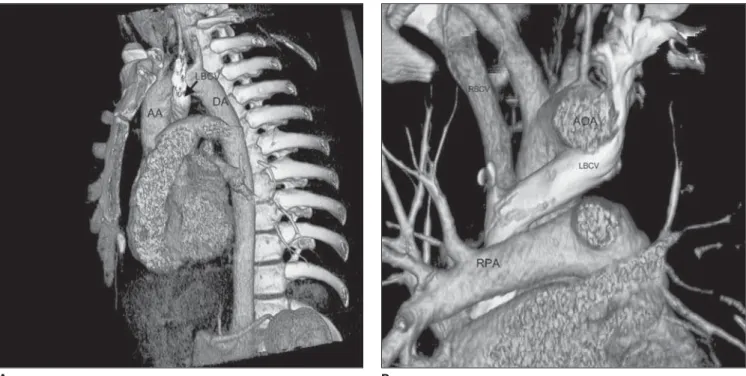
Figure 3. Volume rendering processing of the CT data was used to create the 3D images demonstrating the anomalous course of the LBCV. A: Oblique sagittal view after removing the left chest wall. B: Coronal view after removing the anterior thoracic wall. Ascending aorta (AA), arch of aorta (AOA) descending aorta (DA), left brachiocephalic vein (LBCV), pulmonary artery (PA), right brachiocephalic vein (RBCV), superior vena cava (SVC).