ABSTRACT
Do blood contamination and haemostatic agents
affect microtensile bond strength of dual cured
resin cement to dentin?
1!"#$%&"%'2!()*#%#)+*,-0-34(61, Gokmen ZARARSIZ3!:;#*-"0
1- PhD, Department of Prosthodontics, Faculty of Dentistry, Erciyes University, Kayseri, Turkey.
2- PhD, Department of Restorative Dentistry and Endodontics, Faculty of Dentistry, Erciyes University, Kayseri, Turkey. 3- PhD, DDS, Department of Statistics, Faculty of Medicine, Erciyes University, Kayseri, Turkey.
4- PhD, Professor, Department of Prosthodontics, Faculty of Dentistry, Erciyes University, Kayseri, Turkey.
<**+<< Dr. Kerem Kilic - Department of Prosthodontics, Faculty of Dentistry, - Erciyes University - Kayseri - Turkey - Phone: +90 352 4374937-29082 GSM: +90 3525335253997 - Fax: +90 352 438 06 57 - e-mail: [email protected]
&=>?@;+$!0B=+*C+*;+$!%==C+*;+$!
O
bjective: The purpose of this study was to evaluate the effects of blood contamination and haemostatic agents such as Ankaferd Blood Stopper (ABS) and hydrogen peroxide (H2O2) on the microtensile bond strength between dual cured resin cement-dentin interface. occlusal dentin surfaces with Panavia F under the following conditions: Control Group: no contamination, Group Blood: blood contamination, Group ABS: ABS contamination Group H2O2: H2O2 contamination. The specimens were sectioned to the beams and microtensile specimens were randomly selected from each group, and SEM analyses were performed. !"#$ % &''*$+ differences found between the control and the other groups (p>0.05). Conclusions: Contamination by blood of dentin surface prior to bonding reduced the bond strength between resin cement and the dentin. Ankaferd Blood Stoper and H2O2 could be used safely as blood stopping agents during cementation of all-ceramics to dentin to prevent bond failure due to blood contamination.Key words: Blood contamination. Ankaferd Blood Stopper. Hydrogen peroxide. Microtensile bond strength.
INTRODUCTION
Contemporary restorative dentistry places a certain emphasis on adhesion and resin cements because of all-ceramic restorations becoming increasingly popular34. Long-term survival of all-ceramic restorations depends on the success of a durable bonding between ceramic, adhesive cement and dentin8.
Adhesion can be defined as a flexion force between the molecules at the interfaces of different materials9. The complex organic structure and the dynamic formation and biological activity of dentin prevent a reliable and durable bonding27. Appropriate adhesive agents and luting procedures
a catastrophic decrease in the bond strength at dentin-resin cement interface1,7,18,32,34.
Some treatments have been proposed in an effort to reverse the blood contamination effect during the cementation process: resurfacing with rotary instruments, rinsing with water followed by air drying, rinsing with water and primer re-application or re-etching with phosphoric acid15,17,27. Additionally, clinical and in vivo studies have reported on different hemostatic agents used for the management of hemorrhage in clinical dentistry, such as hydrogen peroxide (H2O2), ferric sulfate, aluminum chloride, trichloracetic acid and Ankaferd Blood Stopper (ABS) (Ankaferd Drug INC, Istanbul, Turkey)14,20-22,31.
ABS is a new, unique folkloric combined medicinal plant extract that has been approved in the management of postsurgery dental bleedings and external hemorrhage, and also could be used to obtain a bloodfree, dry enamel-dentin surfaces in Turkish traditional medicine16,31. ABS can be used as a spray, solution and buffer. ABS is comprised of a standardized mixture of the plants Urtica dioica, Vitis vinifera, Glycyrrhiza glabra, Alpinia RI¿FLQDUXP, and Thymus vulgaris16. Each of these plants has some effect on the endothelium, blood cells, angiogenesis, cellular proliferation, vascular dynamics and cell mediators16,23-24. A recent in vitro study by Goker, et al.16 (2008) showed that ABS exposure resulted in a very rapid formation (less ; $ < This network acts as an anchor for vital physiologic erythrocyte aggregation, covering the classical cascade model of the clotting system without independently acting on coagulation factors and platelets16. ABS was found as effective as Surgicel in achieving hemostasis following partial liver excision in an experimental rat model19.
Few studies have investigated the effects of blood decontamination agents such as ABS and H2O2 on the bond strengths of resin cements to dentin30,31,34. ABS and H
2O2 may affect the sealing and bonding properties of resin cements. In addition, blood contamination may create a thin penetration into the dentinal tubules18,34. In wake of these concerns, the aim of this study was to evaluate the effects of various dentin surface contaminations (no treatment [control], blood, ABS, H2O2) on the μTBS of the resin cement-dentin interface. The null hypothesis was that the μTBS of the resin cement-dentin interface would be similar for all dentin surface conditions.
MATERIAL AND METHODS
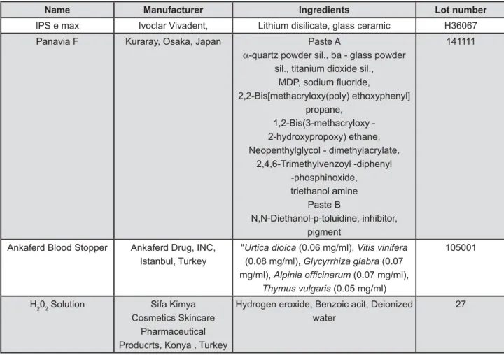
The materials used in this study are presented in Figure 1
Preparation of ceramic blocks
Twelve 5×6×8 mm pressed lithium disilicate glass ceramic rectangular blocks (IPS e.max, Ivoclar Vivadent AG, Schaan, Liechtenstein) were fabricated using the lost wax technique, and ingots were injected into an EP 600 furnace (Ivoclar Vivadent AG) according to the manufacturer’s instructions. After cooling to room temperature, the specimens were divested and air-abraded with 50-= @2O3 particles (Korox, Bego, Bremen, Germany) for 14 s from a distance of approximately 10 mm. "< JJ'+ QR' 600 grit silicon carbide sandpapers to standardize the bonding surfaces. Papers of 220 and 360 grit were used in sequence, each one for 10 s, and a R'' 60 s. The ceramic blocks were ultrasonically cleaned in water for 10 min to ensure contaminant-free surfaces. The bonding surfaces of the ceramic blocks were etched with 37% phosphoric acid (K-Etchant Gel, Kuraray, Tokyo, Japan) for 5 s, rinsed for 30 s with water spray, air-dried and then silanated with % X X Y+ Kuraray, Tokyo, Japan) for 60 s.
Tooth preparation
Twelve caries-free human first mandibular molars were stored in 0.5% chloramin solution at 4°C and used within one month after extraction. @ were removed by cutting off the crown horizontally at the middle, exactly at the top of the pulp chamber, using a low-speed diamond cutting saw (Minitom; Struers, Copenhagen, Denmark). In oder to create a standard smear layer, the exposed dentin surfaces JJ'% QR'% carbide sandpapers in sequence, each for 10 s, and R''% 60 s. The teeth were then rinsed with distilled water to remove any debris.
Experimental design
The 12 prepared teeth were randomly divided into 4 groups according to the following factors \Q $
Control Group: Wet dentin. No contaminant was added.
Group Blood: Fresh capillary human blood (supplied by a single donor) was applied with a microbrush for 20 s, rinsed for 10 s and blotted with absorbent paper.
Group ABS: One drop of ABS solution was applied directly to the dentin surface with a microbrush for 20 s, rinsed for 10 s and blotted with absorbent paper.
Bonding procedures
Panavia F dual-cured resin cement (Kuraray, Tokyo, Japan) was used as adhesive. Equal amounts of ED Primer II Liquids A and B were mixed and applied to the dentin surfaces for 30 s and then the surfaces were thoroughly air dried. Paste A and Paste B of the Panavia F resin cement were mixed and then applied to the ceramic surfaces using a dispenser syringe. A special loading device was used to apply a constant load of 98 N to the ceramic blocks. This load was used to create a uniform resin-luting layer, so as to simulate the
< _ % 10.
Initial light curing was performed for 10 s. Excess cement was carefully removed with an explorer. The resin cement was polymerized from each direction (mesial, distal, buccal, lingual, occlusal) with a LED curing device for 40 s (light intensity: 1,000 mW/ cm2; Elipar FreeLight 2 LED Curing Light, 3M ESPE, MN, USA).
Bond strength test
All specimens were stored in a moisture medium at 37°C for 24 h. Using a low-speed diamond cutting saw (Minitom, Struers, Copenhagen, Denmark) at 3200 rpm (0.085 mm/s) under water cooling, the ceramic-resin cement-tooth sets were cut 1-mm thick slabs, starting at the ceramic side through the
tooth, perpendicular to the bonded interface. The sectioning continued until 1 mm remained to keep { of the teeth were cut horizontally and 1-mm thick slabs were obtained. Subsequently, each slab was pasted to acrylic blocks with sircolant wax, and then slabs were cut perpendicular to the bonded interface to obtain 1.0±0.1 mm2 beam specimens J$ \J*$ from each experimental group were obtained and thermocycled for 6,000 cycles between 5±2°C and 55±2°C, with a dwell time of 20 s and a transfer time of 5 s. The thermocycling process was completed in 4.5 days25. The ends of each beam were attached to a table top material tester (Micro Tensile Tester; Bisco, Schaumburg, IL, USA) using cyanoacrylate (Zapit; DVA, Corona, CA, USA) and subjected to microtensile testing at a crosshead speed of 1 mm/min until the beams fractured. At this point, the load at failure was recorded in N. The debonded beams were carefully removed from the apparatus and the cross-sectional area at the site of failure was measured with a pair of digital calipers (Sylvac Ultra-Cal III; Fowler Co., Inc., Newton, MA, USA) to calculate the bond strengths at failure in MegaPascals.
Name Manufacturer Ingredients Lot number
IPS e max Ivoclar Vivadent, Lithium disilicate, glass ceramic H36067
Panavia F Kuraray, Osaka, Japan Paste A
α-quartz powder sil., ba - glass powder
sil., titanium dioxide sil.,
2,2-Bis[methacryloxy(poly) ethoxyphenyl] propane,
1,2-Bis(3-methacryloxy - 2-hydroxypropoxy) ethane, Neopenthylglycol - dimethylacrylate,
2,4,6-Trimethylvenzoyl -diphenyl -phosphinoxide,
triethanol amine Paste B
N,N-Diethanol-p-toluidine, inhibitor, pigment
141111
Ankaferd Blood Stopper Ankaferd Drug, INC,
Istanbul, Turkey
"Urtica dioica (0.06 mg/ml), Vitis vinifera
(0.08 mg/ml), Glycyrrhiza glabra (0.07 mg/ml), $OSLQLDRI¿FLQDUXP (0.07 mg/ml),
Thymus vulgaris (0.05 mg/ml)
105001
H202 Solution Sifa Kimya
Cosmetics Skincare Pharmaceutical Producrts, Konya , Turkey
Hydrogen eroxide, Benzoic acit, Deionized water
27
Stereomicroscopy and scanning electron microscopy (SEM) examination
Fractured specimens were examined with a stereomicroscope (Olympus SZ-CTV; Olympus, <_+ }$ ~' as adhesive, mixed, or cohesive.
For SEM analysis, two specimens were randomly selected from each group. The debonded beams from each group were sputter-coated (Bal-Tec SCD 050 Sputter Coater; Bal-Tec AG, Balzers,
Liechtenstein) with gold and observed with a scanning electron microscope (LEO 440, Leica-Zeiss, Cambridge, UK).
Statistical analysis
All data sets were subjected to normality tests using the Shapiro-Wilk method, and the Levene’s method to check the assumption of homogeneity of variances. The data were presented as mean and standard deviation. One-way ANOVA test was used to compare the groups and multiple comparisons were performed using Dunnett’s Method. Failure mode distributions were compared using the chi-square test. For all of the analyses, the level of significance was set at p<0.05. Statistical analysis was conducted using SigmaPlot 12.0 software (Systat Software, San Jose, CA, USA). Three samples in Group Blood and one sample in Group ABS that could not be measured because of spontaneous debonding during thermocycling were evaluated as 0 bond strength.
RESULTS
Results of the μTBS test are summarized in Q %_ @@ differences between the groups (p<0.05) and the
post-hoc Dunnett’s Method showed that there were " &''*$+ were found between the control group and the other groups (p>0.05).
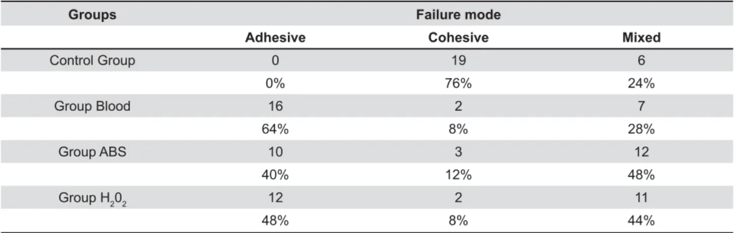
The numbers and percentiles of failure modes in each group are shown in Table 1, There were the groups (p<0.05). The results of failure mode analysis demonstrated that in the control group cohesive type failure mode was predominant than in the other groups (p<0.05). In Group Blood, _ _ than in the ABS and control groups (p<0.05), whereas there were no significant differences between the Blood and H2O2 subgroups in terms of
Groups Failure mode
Adhesive <> Mixed
Control Group 0 19 6
0% 76% 24%
Group Blood 16 2 7
64% 8% 28%
Group ABS 10 3 12
40% 12% 48%
Group H202 12 2 11
48% 8% 44%
Table 1- Failure mode distributions in the groups
Figure 2- Preparation of the microtensile strength test specimens
adhesive type failure mode (p>0.05). In contrast, { _ _ prevailing in ABS Group than in control group (p<0.05).
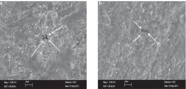
The SEM images of representative debonded beam-shaped specimens from four groups are shown in Figures 4 (a, b) and 5 (a, b). In the $+ observed to be close over the bonding area and smear layer covered the dentin surface, so, the
failure modes of specimens were determined as cohesive failure. Group Blood (Figure 4b), showed in the adhesive failure thatdentinal tubules were obliterated with residues of blood contaminants and _ adhesion. In Groups ABS, H2O2 (Figures 5a and 5b respectively), both residual resin and tubular + specimens were determined as mixed failure. Figure 5- Scanning electron microscopy (SEM) images of the failure modes of debonded beam-shaped specimens (2,500×
!#$&'*; <2O2, smear layer (SL) was partially removed, and both residual resin and tubular
!=>#?
a
b
?; Scanning electron microscopy (SEM) images of the fracture modes of debonded beam-shaped specimens
!QW ! # ? X !;Y !# $ & * # # !Z*\=?X poor adhesion
DISCUSSION
In this study, the effects of blood contamination and various haemostatic agent protocols (ABS and H2O2) on the μTBS of the resin cement-dentin interface were investigated by simulating a clinical try-in procedure. The results obtained in this study did not support the null hypothesis that the μTBS of the resin cement-dentin interfaces are similar for all dentin surface conditions. In the present study, the blood-contaminated group had the lowest μTBS values in all groups.
During cementation of all-ceramic restorations, there is a relatively high risk of blood contamination on the adherent dentin surface, especially for crown margins in the gingival area14. The residual blood remaining on the dentin surface caused an excessive decrease in bond strength at the resin cement-dentin interface, which was shown in previous studies1,18,32,34. In the present study, blood-contaminated group and the control group, which is similar to the previous studies18,34. The can be attributed to its high protein content that, + + obstructing the penetration of the adhesive system into dentin tubules7.
SEM images (Figure 4b) also revealed that dentinal tubules were sealed with residues of blood
_ on
the blood-contaminated specimens compared to the other surface treatments (Figure 5a and 5b), which probably resulted in reduced bond strength due to the lack of interaction of the resin cement with the contaminated surface.
An anionic detergent, H2O2 is one of the most common disinfectant and coagulant materials used in dentistry34.During bonding procedures, H
2O2 might break down to oxygen and water, generating into etched dentin. The oxygen severely inhibits the interfacial polymerization of resin-bonding materials6,34.Although reduction in bond strength of some adhesive systems applied to enamel and dentin may have been caused by the presence of H2O2, as has been shown in some studies6,29,34, some authors have claimed that H2O2 did not affect bond
strength5,29 _+ Q
2O2 did not reduce adhesion when applied for 20 s. However, it is important to point out that it could have been responsible for the complete removal of remaining contaminants by water rinsing from the surface.
ABS induced formation of a protein network with vital erythroid aggregation that covers the entire physiologic hemostatic process. Therefore, ABS could be effectively used both in individuals
with normal hemostatic parameters and in patients _ _
hemostasis16 _
ABS has been previously tested in animals with normal4 and defective hemostasis11. Cipil, et al.11 (2009) concluded that ABS had in vivo hemostatic actions that may provide a therapeutic potential for _ hemostasis in clinical medicine. Experimental studies have established the preclinical and biochemical safety of the oral systemic administration of ABS _ _ product have indicated its sterility and nontoxicity3.
Except from the study by Trakyali and Oztoprak31 (2010), there are no published data about comprehensive observations or intraoral applications concerning the ABS effect on bond strength. According to Trakyali and Oztoprak31 J';'$+ _ observed between shear bond strength values of the ABS contaminated group (9.58±0.95 MPa) and the control group (19.56±1.84 MPa). However, the shear bond strength values of the ABS contaminated group were between 6 and 10 MPa, which is clinically acceptable. The results of _ study. Based on the results of the current study, the μTBS values of the ABS application and control _ result may be connected with removing ABS from the dentin surface by water rinsing and it may have _ dentin surface that provides adhesive penetration into the dentin tubules.
In this study, ABS and H2O2 were applied without blood contamination. No human blood contamination was done before the application of ABS in previous articles either2,31. In the cementation stage of all-ceramics, bleeding mostly occurs around the gingival margin and the clinician applies the haemostatic agent by swab all around the gingival margin as a blood-stopping agent. Therefore, a large part of tooth surface is contaminated just by the haemostatic agent without blood contamination. Additionally, in this study the authors wanted to investigate the effects of haemostatic agents alone on bond strength of resin cement to dentin. For this reason, hemostatic agent subgroups were created without blood contamination. In future studies bond strength can be evaluated by creating subgroups with and without blood contamination on tooth surface before the application of hemostatic agents.
CONCLUSIONS
negatively the μTBS of a dual cured resin cement to dentin. Furthermore, ABS and H2O2 were found + they could be used safely as blood-stopping agents during cementation of all-ceramics to dentin in order to prevent bond failure due to blood contamination.
REFERENCES
;% @ @+ X " _ morphology of one-bottle adhesives to contaminated dentin surfaces. Am J Dent. 1998;11:281-5.
J% @ #+ + @< " Stopper on shear bond strength of bonding systems. Dent Mater J. 2012;31:226-31.
3- Bilgili H, Captug O, Kosar A, Kurt M, Kekilli M, Shorbagi A, et al. Oral systemic adminstration of Ankaferd Blood Stopper has no short-term toxicity in an in vivo rabbit experimental model. Clin Appl Thromb Hemost. 2010;16:533-6.
4- Bilgili H, Kosar A, Kurt M, Onal IK, Goker H, Captug O, et al. _ @< " # model. Med Princ Pract. 2009;18:165-9.
5- Bishara SE, Oonsombat C, Soliman MM, Ajlouni R, Laffoon JF. The effect of tooth bleaching on the shear bond strength of orthodontic brackets. Am J Orthod Dentofacial Orthop 2005;128:755-60.
6- Brauchli L, Eichenberger M, Steineck M, Wichelhaus A. contamination with blood or saliva. Am J Orthod Dentofacial Orthop. 2010;138:435-41.
7- Carvalho Mendonca EC, Vieira SN, Kawaguchi FA, Powers J, @" a self-etching system. Eur J Dent. 2010;4:280-6.
8- Chang SW, Cho BH, Lim RY, Kyung SH, Park DS, Oh TS, et al. Effects of blood contamination on microtensile bond strength to dentin of three self-etch adhesives. Oper Dent. 2010;35:330-6. 9- Chaves CAL, Melo RM, Passos SP, Camargo FP, Bottino MA, Balducci I. Bond strength durability of self-etching adhesives and resin cements to dentin. J Appl Oral Sci. 2009;17:155-60. ;'% X + X #+ Y + + X+ CL, et al. The effect of application sustained seating pressure on adhesive luting procedure. Dent Mater. 2007;23:159-64. 11- Cipil H, Kosar A, Kaya A, Uz B, Haznedaroglu IC, Goker H, et al. In vivo hemostatic effect of the medicinal plant extract Ankaferd Blood Stopper in rats pretreated with warfarin. Clin Appl Thromb Hemost. 2009;15:270-6.
12- Cunha TM, Behrens BA, Nascimento D, Retamoso LB, Lon LF, Tanaka O, et al. Blood contamination effect on shear bond strength of an orthodontic hydrophilic resin. J Appl Oral Sci. 2012;20:89-93. 13- Eiriksonn SO, Pereira PNR, Swift EJ Jr, Heymann HO, Sigurdsson A. Effects of blood contamination on resin-resin bond strength. Dent Mater. 2004;20:184-90.
14- Ercan E, Erdemir AA, Zorba YO, Eldeniz AU, Dalli M, Ince B, et al. Effect of different cavity disinfectants on shear bond strength of composite resin to dentin. J Adhes Dent. 2009;11:343-6. 15- Faltermeier A, Behr M, Rosentritt M, Reicheneder C, Mussig D. An in vitro comparative assessment of different enamel contaminants during bracket bonding. Eur J Orthod. 2007;29:559-63.
16- Goker H, Haznedaroglu IC, Ercetin S, Kirazli S, Akman U, Ozturk Y, et al. Haemostatic actions of the folkloric medicinal plant extract Ankaferd Blood Stopper. J Int Med Hes. 2008;36:163-70.
17- Johnson ME, Burgess JO, Hermesch CB, Buikema DJ. Saliva contamination of dentin bonding agents. Oper Dent. 1994;19:205-10.
18- Kaneshima T, Yatani H, Kasai T, Watanabe EK, Yamashita A. The and an adhesive resin cement. Oper Dent. 2000;25:195-201. 19- Karakaya K, Ucan HB, Tascilar O, Emre AU, Cakmak GK, Irkorucu O, et al. Evaluation of a new hemostatic agent Ankaferd Blood Stopper in experimental liver laceration. J Invest Surg. 2009;22:201-6.
20- Khoroushi M, Tavasoli M. The effect of trichloracetic acid as a hemostatic and etching agent on the morphological characteristics and shear bond strength of resin composite to enamel. Oper Dent. 2010;35:187-93.
21- Kuphasuk W, Harnirattisai C, Senawongse P, Tagami J. Bond strengths of two adhesive systems to dentin contaminated with a haemostatic agent. Oper Dent. 2007;32:399-405.
22- Land MF, Couri CC, Johnston WM. Smear layer instability caused by hemostatic agents. J Prosthet Dent. 1996;76:477-82. JQ% #}+ + # + volatile components in basil (Ocimum basilicum L.) and thyme leaves (Thymus vulgaris L.) and their antioxidant properties. Food Chem. 2005;9:131-7.
24- Matsuda H, Ando S, Kato T, Morikawa T, Yoshikawa M. @ of nitric oxide in lipopolysaccharide-activated macrophages and the structural requirements of diarylheptanoids for the activity. Bioorg Med Chem. 2006;14:138-42.
25- Pisani-Proenca J, Erhardt MC, Valandro LF, Gutierrez-Aceves + "%X + X%# + of ceramic surface conditioning and resin cements on microtensile bond strength to a glass ceramic. J Prosthet Dent. 2006;96:412-7.27.
26- Raffaini, MS, Gomes-Silva JM, Torres-Mantovani CP, Palma-Dibb RG, Borsatto MC. Effect of blood contamination on the shear bond strength at resin/dentin interface in primary teeth. Am J Dent. 2008;21:159-62.
27- Sattabanasuk V, Shimada Y, Tagami J. Effects of saliva contamination on dentin bond strength using all-in-one adhesives. J Adhes Dent. 2006;8:311-8.
28- Sfondrini MF, Gatti S, Scribante A. Effect of blood contamination on shear bond strength of orthodontic brackets and disinclusion buttons. Br J Oral Maxillofac Surg. 2011;49:404-8.
29- Sung EC, Chan SM, Mito R, Caputo AA. Effect of carbamide peroxide bleaching on the shear bond strength of composite to dental bonding agent enhanced enamel. J Prosthet Dent. 1999;82:595-9
Q'% @+ X + #+ "@ blood contamination on bond strength of self-etching adhesive to dental tissue. J Adhes Dent. 2011;13:349-58.
31- Trakyali, G, Oztoprak MO. Plant extract Ankaferd blood stopper effect on bond strength. Angle Orthod. 2010;80:570-4. 32- Xie J, Powers JM, McGuckin RS. In vitro bond strength of two adhesives to enamel and dentin under normal and contaminated conditions. Dent Mater. 1993;9:295-9.
33- Yoo, HM, Pereira PNR. Effect of blood contamination with 1-step self-etching adhesives on microtensile bond strength to dentin. Oper Dent. 2006;31:660-5.