ABSTRACT
TNF-
α
, TNF-β and IL-10 gene polymorphism and
association with oral lichen planus risk in Saudi
patients
Maha Ali AL-MOHAYA1, Fahad AL-HARTHI2, Misbahul ARFIN3, Abdulrahman AL-ASMARI3*
1- Department of Dentistry, Prince Sultan Military Medical City, Riyadh, Saudi Arabia. 2- Department of Dermatology, Prince Sultan Military Medical City, Riyadh, Saudi Arabia. 3- Research Center, Prince Sultan Military Medical City, Riyadh, Saudi Arabia.
Corresponding address: Abudlrahman Al-Asmari - Senior Consultant and Director of Research Center - Prince Sultan Military Medical City - P.O. Box 7897 - Riyadh 11159 - Saudi Arabia - e-mail: [email protected]
Submitted: February 23, 2015 - Modiication: May 7, 2015 - Accepted: May 13, 2015
O
bjectives: Oral lichen planus (OLP) is a chronic inlammatory oral mucosal disease.Cytokines play an important role in the pathogenesis and disease progression of OLP. Various reports have implicated cytokine gene polymorphisms in susceptibility to develop some immune mediated conditions including OLP. The purpose of this study was to investigate the association of tumor necrosis factor (TNF)-α, TNF-β and interleukin (IL)-10 gene polymorphisms with the OLP risk. Material and Methods: Forty two unrelated patients with OLP and 211 healthy volunteers were genotyped for TNF-α (-308 G/A), TNF-β (+252A/G), IL-10 (-1082G/A), IL-10 (-819C/T), and IL-10 (-592C/A) polymorphisms. Results: The frequencies of allele A and genotype GA of TNF-α (-308G/A) were signiicantly higher while allele G and GG genotypes were lower in OLP patients as compared to the
controls (P<0.001). The frequency of GA genotype of TNF-β (+252A/G) was signiicantly
higher in patients than in controls while the AA genotype was completely absent in OLP patients. These results indicated that allele A and genotype GA of TNF-α (-308G/A) as well
as the GA genotype of TNF-β (+252A/G) polymorphisms are associated with OLP risk. The
frequencies of alleles and genotypes of -1082G/A, -819C/T and -592C/A polymorphisms
in IL-10 gene did not differ signiicantly between OLP patients and controls (P>0.05).
However, haplotype ATA extracted from 1082G/A, -819C/T, -592C/A polymorphisms of IL-10 were more prevalent in OLP patients when compared to controls indicating its possible association with OLP susceptibility. Conclusion: It is concluded that TNF-α (-308G/A), TNF-β (+252A/G) and IL-10 (-1082G/A, -819C/T and -592C/A) polymorphisms are associated with the susceptibility of OLP, thus giving additional support for the genetic basis of this disease.
Keywords: Oral lichen planus. Tumor necrosis factors. Interleukin-10. Genetic
polymorphism.
INTRODUCTION
Oral lichen planus (OLP) is a chronic inlammatory oral condition whose etiology is not fully elucidated. It is the prototype of oral lichenoid lesions characterized by T-cell mediated immune responses and abnormal epithelial keratinization cycles6.
OLP manifests as white striations, white papules, white plaques, erythema, erosions, or blisters predominantly affecting the buccal mucosa, tongue and gingival. The OLP lesions may co-exist with
cutaneous and genital lesions, or may be the only disease manifestations. The prevalence of OLP ranges between less than 1 and 3% of the general population17. The prevalence of OLP in Arab
countries including Saudi Arabia has been reported ranging from 0.35 to 1.7%2.
Clinically, OLP may be divided into three subtypes: reticular, erythematous (atrophic), and erosive23
and more than one subtype of OLP may be present in an individual patient. The pathogenesis of OLP is very complex and involves possible antigen presentation by the oral keratinocytes that could be either of an exogenous or an endogenous origin9. This antigenic trigger is accompanied by a
mixed inlammatory response comprising of mainly T-cells, macrophages, and mast cells, as well as the associated cytokines and cytotoxic molecules9,10. It
is a disease associated with middle aged people and is more common among women4.
Cytokines play an important role in the pathogenesis and disease progression of OLP and a strong body of evidence suggests that OLP is a T-cell-mediated disease. The genetic factors that inluence the immune function have been suggested to contribute to the OLP6,12. The gene
polymorphisms of T-helper cell subtype Th1/Th2 cytokines, tumor necrosis factor (TNF)-α, and interleukin-10 (IL-10) have been reported to affect the susceptibility to, and the progression of OLP3,14.
Recently Chauhan, et al.8 (2013) suggested that
proinlammatory cytokines are an important factor in understanding the disease burden of OLP and their comorbid factors. However, immunogenetic studies of OLP have given controversial results and the precise cause of OLP is still unclear.
TNF-α and TNF-β genes are located in tandem on chromosome 6 between the Class I and Class II cluster of the major histocompatibility complex (chromosome 6p21.1–6p21.3). TNF-α-308G/ Apolymorphism (rs1800629) has been reported to be associated with several autoimmune/ inlammatory diseases including OLP3,8. The genetic
variation at position −308 of the TNF-α gene results in two allelic forms in which the presence of guanine (G) deines the common variant and the presence of adenine (A) deines the less common one. The A-allele of TNFα-308G/A polymorphism displays increased gene transcription as compared to the common allele G. It has been shown to produce levels of TNF-α transcription 6–7 fold greater31. TNF-β +252A/G polymorphism (rs909253) has been reported at position +252 within the irst intron of the TNF-β gene, consisting of guanine (TNF-β +252G) on one allele and adenine (TNF-β +252Α) on the alternate allele18. The presence of
G at this position deines the mutated allele known as TNF-β 1 (allele-1) which is a less frequent allele and is associated with higher TNF-α and TNF−β production18.
Interleukin-10 (IL-10) gene maps to the junction of 1q31-q32. It shifts the Th1/Th2 balance by down regulating the Th1 responses and by suppression of pro-inflammatory cytokines, such as TNF-α
and interferon gamma (IFN γ) secretion26. Three
promoter polymorphisms: -1082A/G (rs18000896), -819T/C (rs1800871) and -592A/C (rs1800872) are reportedly involved in the IL-10 transcription rate, thereby directly affecting its production level19. The
-1082G, -819C and -592C alleles (GCC haplotype) have been associated with elevated levels of IL-10 production15 while ACC and ATA haplotypes exhibit
intermediate and low IL-10 gene transcription respectively27. In this study we examined the association of ive single nucleotide polymorphisms
(SNPs) of TNF-α, TNF-β, and IL-10 genes in the etiopathogenesis of OLP in Saudi patients.
MATERIAL AND METHODS
Study group
A total of 253 Saudi subjects visiting Prince Sultan Military Medical City (PSMMC), Riyadh, Saudi Arabia were recruited for this study. Forty two (16 male, 26 female) unrelated patients with oral lichen planus, ages ranging from 27 to 72 years, and 211 (140 male, 71 female) unrelated, healthy patients matched voluntary blood donors with ages ranging from 20 to 65 years from the same population were studied for polymorphisms in TNF-α, TNF-β and IL-10 genes. Patients with any other inlammatory/autoimmune diseases were excluded from the study. This study was approved by the research and ethical committee of PSMMC and written informed consent was obtained from each subject before recruitment.
OLP was diagnosed according to the clinical manifestations and histopathological criteria of the World Health Organization. All patients were diagnosed through a review of the patient’s history, physical examination and histological findings by an oral pathologist. A 4 mm punch biopsy of lesional tissue that extends into the submucosa was performed. Symptoms were bilateral, more or less symmetrical lesions, and a lace-like network of slightly raised gray-white lines (reticular pattern). Erosive, atrophic, bullous, and plaque-type lesions were only accepted as a subplaque-type in the presence of reticular lesions elsewhere in the oral mucosa. Histopathological criterium were the presence of a well-deined band-like zone of cellular iniltration that was conined to the supericial part of the connective tissue, consisting mainly of lymphocytes, signs of “liquefaction degeneration” in the basal cell layer, and the absence of epithelial dysplasia28. No patient was suspected to have
PCR ampliication
Genomic DNA was extracted from the peripheral blood of OLP patients and controls using the QIA ampR DNA mini kit (QIAGEN Hilden, North
Rhine-Westphalia, Germany). TNF-α, TNF-β and IL-10 genes were ampliied using ampliication refractory mutation systems (ARMS)-PCR methodology21 to
detect polymorphisms at positions -308 of TNF-α, +252 in intron1 of TNF-β and at loci -592, -819, and -1082 of IL-10 genes. PCR ampliication was carried out using Ready to Go PCR Beads (GE Healthcare, Little Chalfont, Buckinghamshire, UK). Reactions consisted of 10 denaturation temperature cycles for 15 s at 94°C, annealing for 50 s at 65°C and extension for 40 s at 72°C. Then 25 denaturation cycles of 20 s at 94°C, annealing for 50 s at 59°C and then extension of 50 s at 72°C. Final extension was performed at 72°C for 7 min. A positive control was included in the PCR assay by ampliication of the human growth hormone gene. The ampliied products for the various samples were separated on the 1.5% agarose gel, stained with ethidium bromide and photographed.
Statistical analysis
The frequencies of alleles and genotypes were calculated. Hardy-Weinberg equilibrium was determined using Hardy-Weinberg Equilibrium Calculator for 2 Alleles (http//www.had2know.com/ academics/hardy-weinberg-equilibrium-calculator-2alleles.html). The Bayesian method of the PHASE program (version 2.1) was applied to reconstruct the haplotype (http://stephenslab.uchicago.edu/ software.html). The differences in allele/genotype frequencies between patients and controls were analyzed by Fisher’s exact test using the CalcFisher software13. P values ≤0.05 were considered
signiicant. The strength of the association of the disease with respect to a particular allele/genotype is expressed by odd ratio interpreted as relative risk
(RR) following the Woolf’s method as out lined by Schallreuter, et al.22 (1993). It was calculated only
for those alleles/genotypes which were increased or decreased in OLP patients when compared to the control group. The RR was calculated for all the subjects using the formula given below:
RR=(a×d) (b×c)
a = number of patients with expression of allele or genotype
b = number of patients without expression of allele or genotype
c = number of controls with expression of allele or genotype
d = number of controls without expression of allele or genotype.
Etiologic Fraction (EF) indicates the hypothetical genetic component of the disease. The values 0.0
- 0.99 are of signiicance. EF was calculated for a positive association only where RR>1 using the following formula25.
EF= (RR-1)f , where f =
RR
Preventive Fraction (PF) indicates the hypothetical protective effect of one speciic allele/ genotype for the disease. PF was calculated for negative association only where RR<1 using the following formula25. Values <1.0 indicate the protective effect
of the allele/ genotype against the manifestation of disease.
PF= , where f =
RESULTS
Among OLP patients the male to female ratio was 16:26 (1:1.6). There was no signiicant difference in clinical manifestation or prognosis comparing men to women in our study. Thirty-ive patients (83.333%) had lesions on the buccal mucosa, three (7.14 %) each on the tongue and gingival while one patient (2.39%) had lesions on the palate. The majority of patients (85%) had white lichen. In our patients, reticular type OLP was the commonest (80.95%) followed by erosive (23.81%) and then atrophic types (4.76%).
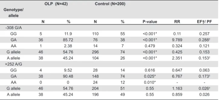
The genotype and allele frequencies of TNF-α (-308G/A) and TNF-β (+252A/G) promoter polymorphisms are presented in Table 1. In both the lichen planus patients and control groups the genotype distributions were in Hardy-Weinberg equilibrium. The frequency of the heterozygous genotype GA was signiicantly higher in OLP patients than in the control (P<0.001) whereas the frequency of homozygous genotypes GG was signiicantly lower in OLP than controls (P<0.001). The frequency of allele-A was signiicantly higher in OLP patients than control subjects (P<0.001). On the other hand, allele-G was signiicantly lower in OLP patients when compared to the control (P<0.001). The difference in frequency of the AA genotype between the two groups was not statistically signiicant.
The frequency of the GA genotype of TNF-β (+252A/G) promoter polymorphism was signiicantly higher in patients group than in the controls. Homozygous AA genotype was completely absent in OLP patients whereas it was present in 12% of the controls. The frequencies of alleles of TNF-β (+252A/G) polymorphism were not signiicantly different between OLP patients and healthy controls (Table 1). Albeit, the frequencies of allele-G were slightly higher in the OLP patients than they were in the control subjects.
(1-RR) f
RR(1-f)+f
The results of SNPs for 10(-1082G/A), IL-10(-592C/A), IL-10(-819C/T), and corresponding alleles and genotypes are summarized in Table 2. The genotype distributions of IL-10 polymorphisms in patient and controls were in Hardy-Weinberg equilibrium. The frequencies of -1082GG and
1082GA genotypes were slightly higher while the frequency of -1082AA genotype was lower in OLP patients when compared to the control subjects, however, the differences were not statistically signiicant.
Similarly the frequencies of alleles and
Genotype/
allele
OLP (N=42) Control (N=186)
N % N % P-value RR EF*/PF
-1082G/A
GG 5 11.9 14 7.53 0.357 1.6 0.098*
GA 33 78.5 140 75.27 0.841 1.2 0.032*
AA 4 9.5 32 12.2 0.251 0.51 0.095
G-allele 43 51.2 168 45.16 0.334 1.3 0.047*
A-allele 41 48.8 204 54.84 0.334 0.78 0.048
-819C/T
CC 12 28.5 88 41.71 0.122 0.559 0.086
CT 25 59.5 102 48.34 0.239 1.5 0.065*
TT 5 11.9 21 9.95 0.78 1.2 0.032*
C-allele 49 62 278 65.88 0.521 0.846 0.026
T-allele 30 38 144 34.12 0.521 1.182 0.172*
-592C/A
CC 12 28.5 88 41.71 0.122 0.559 0.086
CA 25 59.5 102 48.34 0.239 1.5 0.065*
AA 5 11.9 21 9.95 0.78 1.2 0.032*
C-allele 49 62 278 65.88 0.521 0.846 0.026
A-allele 30 38 144 34.12 0.521 1.182 0.172*
N=number of subjects,*statistically signiicant, RR=relative risk, EF=etiologic fraction, PF=preventive fraction Table 2- Genotype and allele frequencies of IL-10 variants in oral lichen planus patients and matched controls
Genotype/
allele
OLP (N=42) Control (N=200)
N % N % P-value RR EF†/ PF
-308 G/A
GG 5 11.9 110 55 <0.001* 0.11 0.257
GA 36 85.72 76 38 <0.001* 9.789 0.288†
AA 1 2.38 14 7 0.479 0.324 0.121
G allele 46 54.76 296 74 <0.001* 0.425 0.153
A allele 38 45.24 104 26 <0.001* 2.351 0.153†
+252 A/G
GG 4 9.52 28 14 0.616 0.647 0.063
GA 38 90.48 148 74 0.025* 6.767 0.173†
AA 0 0 24 12 0.010* -
-G allele 46 54.76 204 51 0.55 1.163 0.026†
A allele 38 45.24 196 49 0.55 0.859 0.026
N=number of subjects,*statistically signiicant, RR=relative risk, EF=etiologic fraction, PF=preventive fraction
genotypes of IL-10 (-819C/T) and IL-10 (-592C/A) polymorphisms did not differ signiicantly between OLP patients and controls (P>0.05). Albeit, the frequencies of genotypes -819 CT, -819 TT, 592 CA and 592 AA were higher in OLP patients while -819 CC and -592 CC genotypes showed a reverse pattern with lower frequency in OLP patients when compared to the controls (Table 2). The frequencies
of haplotypes extracted from 1082G/A, -819C/T, -592C/A polymorphisms of IL-10 are shown in Table 3. The haplotype ATA was signiicantly increased in OLP patients when compared to controls (P=0.008).
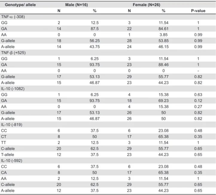
The frequencies of alleles and genotypes of all ive polymorphisms with gender are summarized in Table 4. No statistically signiicant difference was found in the frequencies of alleles or genotypes
Haplotype OLP (N=42) Controls (N=186)
N (Frequency) N (Frequency) P-value
GCC 7 (0.166) 42 (0.225) 0.533 ACC 13 (0.309) 82 (0.440) 0.165 ATA 20 (0.476) 48 (0.258) 0.008* ACA 2 (0.047) 14 (0.075) 0.742
N=number of subjects,*statistically signiicant
Table 3- Haplotype analysis of IL-10 -1082G/A, -819T/C and -592C/A polymorphisms in OLP and control groups
Genotype/ allele Male (N=16) Female (N=26)
N % N % P-value
TNF-α (-308)
GG 2 12.5 3 11.54 1
GA 14 87.5 22 84.61 1
AA 0 0 1 3.85 0.99
G-allele 18 56.25 28 53.85 0.99
A-allele 14 43.75 24 46.15 0.99
TNF-β (+525)
GG 1 6.25 3 11.54 1
GA 15 93.75 23 88.46 1
AA 0 0 0 0
-G-allele 17 53.13 29 55.77 0.82
A-allele 15 46.87 23 44.23 0.82
IL-10 (-1082)
GG 1 6.25 4 15.38 0.63
GA 15 93.75 18 69.23 0.12
AA 0 0 4 15.38 0.27
G-allele 17 53.13 26 50 0.82
A-allele 15 46.87 26 50 0.82
IL-10 (-819)
CC 6 37.5 6 23.08 0.48
CT 8 50 17 65.38 0.35
TT 2 12.5 3 11.54 1
C-allele 20 62.5 29 55.77 0.65
T-allele 12 37.5 23 44.23 0.65
IL-10 (-592)
CC 6 37.5 6 23.08 0.48
CA 8 50 17 65.38 0.35
AA 2 12.5 3 11.54 1
C-allele 20 62.5 29 55.77 0.65
A-allele 12 37.5 23 44.23 0.65
of TNF-α, TNF-β and IL-10 gene polymorphisms between male and female OLP patients.
DISCUSSION
In this study 62% of the OLP patients were women. OLP has previously been reported to be more prevalent in females than males in American, British, Chinese, Iranian, Italian, Japanese and Spanish populations4,5,20. In our patients OLP was
more frequent in the third to fourth decade of life and the median age of the patients group was 42 years. Reticular OLP was the most common (80.95%) with the buccal mucosa as the most affected site (83.33%). These results are compatible with previous studies from various ethnic groups5,20.
There was no signiicant difference in the clinical manifestation or prognosis when comparing men to women however, in our study, these observations should be taken with caution as the study’s sample size is small.
Our results on TNF-α promoter polymorphism in Saudi OLP patients suggested that genotype GA-positive individuals at position -308 of TNF-α are at higher risk for OLP, whereas GG genotypes might be negatively associated with the disease. Data analysis further demonstrated that allele A (TNF-α 2 allele) increases the risk of OLP, whereas allele G (TNF-α 1 allele) reduces OLP risk. Similarly higher frequencies of TNF-α 308 A allele and -308 GA genotype have been reported in Italian7, Indian8, and Chinese
patients with an OLP3,12. Predominance of the AA
genotype was also reported in Thai14 and Brazilian
patients with OLP32. Further, genetic polymorphisms
of cytokine-encoding genes are also known to predispose malignant diseases and have been associated with oral precancerous lesions (OPCLs). TNF-α (-308G/A), IL-6 and TGF-β polymorphisms have been associated with the development of OPCLs in Taiwanese patients11.
Allele A (TNF-α 2 allele) lies on the extended haplotype HLA-A1-B8-DR3-DQ2, which is associated with high TNF-α production. The allele A (TNF-α 2 allele) has been demonstrated to be a much stronger transcriptional activator than the common allele G (TNF-α 1 allele)31. Moreover, TNF-α (-308G/A)
promoter polymorphism has a direct effect on TNF-α gene regulation (increased TNF-α expression) and may be responsible for the association of allele A with the high TNF-α phenotype and more severe diseases31. Further populations bearing a
higher proportion of the allele A (TNF-α 2 allele) are reportedly predisposed to several metabolic, degenerative, inflammatory, and autoimmune diseases1.
The results of this study also show that the GA genotype of TNF-β (+252 A/G) was positively associated with OLP risk, whereas genotype
AA might be protective. This report is the irst to show an association between TNF-β (+252 A/G) polymorphism and OLP. This +252 A/G polymorphism on intron 1 of TNF-β is in linkage disequilibrium with a missense variant (Thr 26 Asn) in exon318. It is also closely linked to TNF-α, which
is in linkage disequilibrium with HLA-A, B8, and DR330 and has been reported to deine a high TNF-α
expressing haplotype in addition to a modifying expression of TNF-β itself18. TNF-β (+252A/G)
polymorphism has also been associated with numerous autoimmune diseases1. High serum and
salivary levels of proinlammatory cytokine and TNF have been detected in OLP patients24. Thalidomide,
a known TNF suppressant has been used to successfully treat OLP showing the implication of high TNF-α expressing genotypes of TNF-α and -β polymorphisms in the pathogenesis of OLP29.
Our results show no signiicant differences in the frequencies of alleles and genotypes of IL-10 (IL-1082G/A), IL-IL-10 (-592C/A), IL-IL-10 (-819C/T) polymorphisms between OLP patients and healthy Saudi controls. However, haplotype ATA from 1082G/A, -819C/T, -592C/A of IL-10 had signiicant association with OLP in Saudi patients. Two other studies dealing with the IL-10 gene polymorphisms in OLP populations could also not ind any difference in the frequencies of IL-10 alleles and genotypes between the patients with OLP and healthy controls3,32. However,the -1082A/-819T/-592A
(ATA) haplotype of the IL-10 polymorphisms, which correlated with a lower serum level of IL-10, has a signiicant association with OLP in a Chinese cohort with Han ethnicity3. Thus, this study together with
the study from China supports that the low producer ATA haplotype of IL-10 polymorphisms is linked to the risk of OLP.
Our results also indicated that the frequencies of alleles and genotypes of all the ive polymorphisms studied are independent of gender (Table 4). However, these results should be taken with caution as the number of males and females is too low for any signiicant conclusion.
Besides TNF-α and IL-10, a number of inflammation and immune regulation-related cytokines, including interferon-gamma (IFN-γ), IL-4, and IL-8, have been suggested to have an important role in the pathogenesis of OLP. Abnormal expression patterns of various inlammation-related cytokines, including IL-10, and TNF-α, in lesions, saliva, serum and peripheral blood mononuclear cells from patients with OLP has also been reported, which may relect the immune dysregulation status of cytokines and emergence as central players in the immunopathogenesis of OLP16. Recently Carrozzo7
basement membrane in OLP lesions.
CONCLUSION
The polymorphisms in TNF-α (-308), TNF-β (+252) and IL-10 (-1082, -819 and -592) gene are associated signiicantly with the risk of OLP susceptibility. However, further studies are required that use a larger sample size to confirm this association.
CONFLICT OF INTERESTS
The authors declare that there is no conlict of interest regarding the publication of this paper.
ACKNOWLEDGEMENTS
The authors thank S. Sadaf Rizvi and Mohammad Al-Asmari for their help with laboratory work.
REFERENCES
1- Al-Harthi F, Zouman A, Arin M, Tariq M, Al-Asmari A. Tumor
necrosis factor-α and -β genetic polymorphisms as a risk factor in Saudi patients with vitiligo. Genet Mol Res. 2013;12:2196-204. 2- Al-Nasser L, El-Metwally A. Oral lichen planus in Arab countries: a review. J Oral Pathol Med. 2014;43:723-7.
3- Bai J, Jiang L, Lin M, Zeng X, Wang Z, Chen Q. Association of polymorphisms in the tumor necrosis factor-alpha and interleukin-10 genes with oral lichen planus: a study in a Chinese cohort with Han ethnicity. J Interferon Cytokine Res. 2009; 29:381-8.
4- Bermejo-Fenoll A, Sánchez-Siles M, López-Jornet P, Camacho-Alonso F, Salazar-Sánchez NJ. A retrospective clinicopathological study of 550 patients with oral lichen planus in south-eastern Spain. Oral Pathol Med. 2010;39:491-6.
5- Carbone M, Arduino PG, Carrozzo M, Gandolfo S, Argiolas MR, Bertolusso G, et al. Course of oral lichen planus: a retrospective study of 808 northern Italy patients. Oral Dis. 2009;15:235-43. 6- Carrozzo M. Understanding the pathobiology of oral lichen planus. Curr Oral Health Rep. 2014;1:173-9.
7- Carrozzo M, Uboldi de Capei M, Dametto E, Fasano ME, Paolo Arduino P, Broccoletti R, et al. Tumor necrosis factor-alpha and interferon-gamma polymorphisms contribute to susceptibility to oral lichen planus. J Invest Dermatol. 2004;122:87-94.
8- Chauhan I, Beena VT, Srinivas L, Sathyan S, Banerjee M. Association of cytokine gene polymorphisms with oral lichen planus in Malayalam-speaking ethnicity from South India (Kerala). J Interferon Cytokine Res. 2013;33:420-7.
9- Farhi D, Dupin N. Pathophysiology, etiologic factors, and clinical management of oral lichen planus, part I: facts and controversies. Clin Dermatol. 2010;28:100-8.
10- Gonzalez-Moles MA, Scully C, Gil-Montoya JA. Oral lichen planus: controversies surrounding malignant transformation. Oral Dis.2008;14:229-43.
11- Hsu HJ, Yang YH, Shieh TY, Chen CH, Kao YH, Yang CF, et al.
Role of cytokine gene (interferon-γ, transforming growth factor-β1,
tumor necrosis factor-α, interleukin-6, and interleukin-10) polymorphisms in the risk of oral precancerous lesions in Taiwanese. Kaohsiung J Med Sci. 2014;30:551-8.
12- Jin X, Wang J, Zhu L, Wang L, Dan X, Zeng X, et al. Association between -308 G/A polymorphism in TNF-α gene and lichen planus: a meta-analysis. J Dermatol Sci. 2012;68:127-34.
13- Khan HA. A visual basic software for computing Fisher’s exact probability. J Stat Softw. 2003;8:1-7.
14- Kimkong I, Hirankarn N, Nakkuntod J, Kitkumthorn N. Tumour necrosis factor-alpha gene polymorphisms and susceptibility to oral lichen planus. Oral Dis. 2011;17:206-9.
15- Koss K, Satsangi J, Fanning GC, Welsh KI, Jewell DP. Cytokine
(TNF alpha, LT alpha and IL-10) polymorphisms in inlammatory
bowel diseases and normal controls: differential effects on production and allele frequencies. Genes Immun. 2000;1:185-90.
16- Lu R, Zhang J, Sun W, Du G, Zhou G. Inlammation-related
cytokines in oral lichen planus: an overview. J Oral Pathol Med. 2015;44:1-14.
17- McCartan BE, Healy CM. The reported prevalence of oral lichen planus: a review and critique. J Oral Pathol Med. 2008;37:447-53. 18- Messer G, Spengler U, Jung MC, Honold G, Blömer K, Pape GR, et al. Polymorphic structure of the tumor necrosis factor
(TNF) locus: an NcoI polymorphism in the irst intron of the
human TNF-beta gene correlates with a variant amino acid in position 26 and a reduced level of TNF-beta production. J Exp Med. 1991;173:209-19.
19- Mormann M, Rieth H, Hua TD, Assohou C, Roupelieva M, Hu SL, et al. Mosaics of gene variations in the interleukin-10 gene promoter affect interleukin-10 production depending on the stimulation used. Genes Immun. 2004;5:246-55.
20- Pakfetrat A, Javadzadeh-Bolouri A, Basir-Shabestari S, Falaki F. Oral lichen planus: a retrospective study of 420 Iranian patients. Med Oral Patol Oral Cir Bucal. 2009;1:E315-8.
21- Perrey C, Turner SJ, Pravica V, Howell WM, Hutchinson IV. ARMS-PCR methodologies to determine IL-10, alpha, TNF-beta and TGF-TNF-beta 1 gene polymorphisms. Transpl Immunol. 1999;7:127-8.
22- Schallreuter KU, Levenig C, Kühnl P, Löliger C, Hohl-Tehari M, Berger J. Histocompatibility antigens in vitiligo: Hamburg study on 102 patients from northern Germany. Dermatology. 1993;187:186-92.
23- Schlosser BJ. Lichen planus and lichenoid reactions of the oral mucosa. Dermatol Ther. 2010;23:251-67.
24- Sklavounou-Andrikopoulou A, Chrysomali E, Iakovou M, Garinis GA, Karameris A. Elevated serum levels of the apoptosis related molecules TNF-alpha, Fas/Apo-1 and Bcl-2 in oral lichen planus. J Oral Pathol Med. 2004;33:386-90.
25- Svejgaard A, Platz P, Ryder LP. HLA and disease 1982 - a survey. Immunol Rev. 1983;70:193-218.
26- Thio CL. Host genetic factors and antiviral immune responses to hepatitis C virus. Clin Liver Dis. 2008;12:713-26.
27- Turner DM, Williams DM, Sankaran D, Lazarus M, Sinnott PJ, Hutchinson IV. An investigation of polymorphism in the interleukin-10 gene promoter. Eur J Immunogenet. 1997;24:1-8. 28- Van der Meij EH, Mast H, van der Waal I. The possible premalignant character of oral lichen planus and oral lichenoid
lesions: a prospective ive-year follow-up study of 192 patients.
Oral Oncol. 2007;43:742-8.
29- Wang Z,Yao H, Cui B, Ning G,Tang GY. Genetic linkage analysis of oral lichen planus in a Chinese family. Genet Mol Res. 2011;10:1427-33.
30- Wilson AG, de Vries N, Pociot F, di Giovine FS, van der Putte LBA, Duff GW. An allelic polymorphism within the human tumor necrosis factor alpha promoter region is strongly associated with HLA A1, B8, and DR3 alleles. J Exp Med. 1993;177:557-60. 31- Wilson AG, Symons JA, McDowell TL, McDevitt HO, Duff GW. Effects of a polymorphism in the human tumor necrosis factor alpha promoter on transcriptional activation. Proc Natl Acad Sci USA. 1997;94:3195-9.