416
Rev. bras. hematol. hemoter. 2007;29(4):416-424 Carta ao Editor
Transferrin saturation as a possible
marker of hematological recovery after
hematopoietic stem cell transplantation
Saturação da transferrina como possível
marcador de recuperação hematológica após
transplante de células-tronco hematopoiéticas
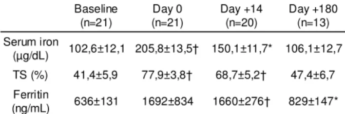
Carta ao Editor / Letter to Editor on day +14 post-BMT. By day +180, serum iron and TS had returned to baseline levels but serum ferritin values remained elevated. Table 1 shows the evolution of iron status parameters over time.
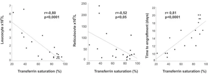
On day +14, TS levels showed a negative correlation
Senhor Editor:
Changes in iron status have been reported after high-dose chemotherapy and hematopoietic stem cell transplantation (HD-HCT) with possible implications towards increased morbidity and mortality after the procedure.1,2
However, the mechanisms implicated in the disorders of the iron metabolism in this setting are poorly understood and a possible association with low hematopoietic activity due to myeloablative treatment is suspected.3
To study iron status changes and its relationship with hematological recovery after HCT, transferrin saturation (TS) along with serum iron and ferritin were evaluated in 21 patients undergoing HD-HCT (allogeneic / autologous: 16 / 05; male / female: 12 / 09; median age: 40 years, range 17-80; ECOG score ≤ 2). HCT indications were: acute lymphoblastic leukemia (n=6), acute myeloid leukemia (n=3), chronic myeloid leukemia (n=5), non-Hodgkin's lymphoma (n=3), Hodgkin's lymphoma (n=1), multiple myeloma (n=1), myelodisplastic syndrome (n=1) and myelofibrosis with myeloid metaplasia (n=1). Twelve patients (57%) were transplanted with their disease in complete remission and thirteen (62%) had been previously transfused with a mean of 13 red blood cell units (range: 1 - 50 units). All iron status parameters were determined before HD-HCT and prospectively on day 0, day +14 and day +180. Hematological recovery was evaluated by absolute leucocyte and reticulocyte counts on day +14 as was time to engraftment (defined by the number of days from day 0 until absolute neutrophil count >500/µL). Paired t-tests were used for pair-wise comparisons and correlation analysis was achieved by the Pearson correlation coefficient. Analysis of variance was used to study the association between categorical and continuous variables.
After high-dose therapy there was a marked increase in all iron status parameters on day 0 which remained elevated
Table 1. Changes in iron status parameters before and after HD-HCT. Values are shown as means ± standard errors of the mean
Baseline (n=21)
Day 0 (n=21)
Day +14 (n=20)
Day +180 (n=13)
Serum iron
(µg/dL) 102,6±12,1 205,8±13,5† 150,1±11,7* 106,1±12,7
TS (%) 41,4±5,9 77,9±3,8† 68,7±5,2† 47,4±6,7
Ferritin
(ng/mL) 636±131 1692±834 1660±276† 829±147* HD-HCT = high-dose chemotherapy with hematopoietic stem cell transplantation; TS=transferrin saturation; *p<0,05 versus baseline †p<0,01 versus baseline
with leucocyte and reticulocyte counts and a strong positive correlation with time to engraftment (Figure 1).
Univariate analysis was performed to ascertain the contribution of disease status before HD-HCT, previous red blood cell transfusions and type of HCT on TS levels on day +14. Only the type of HCT (allogeneic vs autologous) significantly influenced TS levels on day +14 (p<0.05). Patients receiving autologous HCT engrafted faster (mean: 10 days, range 9-12) than those receiving allogeneic HCT (mean: 16 days, range 12-20) (p=0.001).
In this study, substantial increases in all iron status parameters were observed on day 0 and sustained for at least two weeks post-transplant, with a tendency to regain baseline levels after six months. Pronounced changes in iron status levels, specifically for transferrin saturation, were also found early after the start of conditioning therapy in patients undergoing both allogeneic and autologous HCT.1,4,5 This
increasing body of evidence on alterations of iron metabolism in HCT and the possibility of iron overload in this setting are of great concern.
The mechanisms that may lead to temporary changes in plasma iron parameters after HCT have not been elucidated yet. Proposed explanations are focused on the conditioning effects and include reduced iron uptake due to suppression of erythropoiesis, release of intracellular iron from lysis of bone marrow and neoplastic cells, and hepatic toxicity leading to further release of iron.3,6
In our study, TS levels were not influenced by disease status or the number of red blood cell units transfused before HD-HCT and only four of our patients presented hepatic toxicity (Grade I, n=3; Grade II, n=1). Therefore, the strong correlation between TS levels on day +14 and the hematological recovery parameters indicates that suppression of hematopoietic activity is, at least in part, responsible for the main changes observed in iron status after transplant, possibly due to under-utilization of iron by bone marrow erythroid precursors. In accordance with this hypothesis, it was recently demonstrated that the time
Flávio Augusto Naoum1,2 Paulo Cesar Naoum1
417
Carta ao Editor Rev. bras. hematol. hemoter. 2007;29(4):416-424
patients maintained TS levels above 80% after HD-HCT was dependent on the number of days before the start of reticulocyte recovery.7
The participation of time to hematological recovery after HD-HCT in the iron metabolism might explain the significant impact that the type of transplant exerts on TS levels on day +14, as engraftment is faster in patients receiving autologous HCT in relation to those undergoing allogeneic transplantation.
In conclusion, our results confirm that plasma iron parameters increase significantly early after HD-HCT and suggest that TS levels on day +14 are probably dependent on bone marrow suppression caused by the conditioning therapy. Although it would be tempting to propose the utilization of TS as a marker of engraftment after HCT, further and larger studies are needed to validate this assumption.
Resumo
Alterações no perfil de ferro já foram descritas em pacientes sub-metidos à quimioterapia de altas doses com transplante de células precursoras hematopoiéticas (TCPH), e uma possível relação en-tre o metabolismo do ferro e a reconstituição hematopoiética pós-transplante, embora proposta, ainda carece de confirmação. Com o objetivo de avaliar as alterações do perfil de ferro e a sua corre-lação com a recuperação hematológica pós-TCPH, foram determi-nados a saturação da transferrina (ST), ferro e ferritina séricos em 21 pacientes submetidos a TCPH, antes do transplante e prospec-tivamente no dia 0, no dia +14 e no dia +180. Após a quimioterapia de altas doses, todos os parâmetros analisados se elevaram acen-tuadamente no dia 0 e permaneceram ainda alterados no dia +14; após 180 dias, observou-se uma tendência de retorno para valores próximos aos obtidos antes do transplante. No dia +14, os valores de ST apresentaram forte correlação inversa com a contagem ab-soluta de leucócitos (p<0,0001) e de reticulócitos (p<0,05), e uma correlação direta com o tempo de enxertia (p<0,0001). Nossos resultados demonstram que o perfil de ferro se altera de forma aguda e significativa após a quimioterapia de altas doses com TCPH e que tais alterações estão aparentemente correlacionadas com a
atividade hematopoiética após o transplante. Rev. bras. hematol. hemoter. 2007;29(4):416-417.
Palavras-chave: Transplante de células precursoras hematopoiéticas; distúrbios do metabolismo do ferro; transferrina, eritropoiese.
References
1. Gordon LI, Brown SG, Tallman S et al. Sequential changes in serum iron and ferritin in patients undergoing high-dose chemotherapy and radiation with autologous bone marrow transplantation: possible implications for treatment related toxicity. Free Radic Biol Med 1995;18:383-9. 2. Altès A, Remacha AF, Sureda A et al. Iron overload might increase
transplant-related mortality in haematopoietic stem cell transplantation. Bone Marrow Transplant 2002;29:987-9.
3. Evens AM, Mehta J, Gordon LI. Rust and corrosion in hematopoietic stem cell transplantation: the problem of iron and oxidative stress. Bone Marrow Transplant 2004;34:561-71.
4. Sahlstedt L, Ebeling F, von Bonsdorf L et al. Non-transferrin-bound iron during allogeneic stem cell transplantation. Br J Haematol 2001;113:836-8.
5. Bradley SJ, Gosriwitana I, Srichairatanakool S et al. Non-transferrin-bound iron induced by myeloablative chemotherapy. Br J Haematol 1997;99:337-43.
6. Dürken M, Nielsen P, Knobel S et al. Nontransferrin-bound iron in serum of patients receiving bone marrow transplants. Free Radic Biol Med 1997;22:1159-63.
7. Altès A, Remacha AF, Sarda P et al. The relationship between transferrin saturation and erythropoiesis during stem cell transplantation. Haematologica 2006;91:992-3.
Figure 1. Correlation between transferrin saturation levels on day+14 and hematological recovery parameters (absolute leucocyte and reticulocyte counts on day+14 and time to engraftment). Saturation returned to baseline levels but serum ferritin values remained elevated
Avaliação: Editor e dois revisores externos Conflito de interesse: não declarado
Recebido: 24/10/06
Aceito após modificações: 26/04/07
Correspondence: Flávio Augusto Naoum,
Academia de Ciência e Tecnologia Rua Bonfá Natale, nº 1860
15020-1 – São José do Rio Preto-SP – Brasil