252 Rev Bras Med Esporte – Vol. 19, No 4 – Jul/Aug, 2013
EFFECTS OF HIGH-IMPACT EXERCISE TRAINING ON
BONE MECHANICAL PROPRIETIES – AN EXPERIMENTAL
STUDY IN FEMALE WISTAR RATS
ORIGINAL ARTICLE
LOCOMOTOR APPARATUS IN EXERCISE AND SPORTS
Fernando Fonseca de Almeida e Val1
Rodrigo Okubo1
Maurício José Falcai1
Fábio Senishi Asano1
Antônio Carlos Shimano1
1. Medicine School of Ribeirão Preto, University of São Paulo – Ribeirão Preto, SP, Brazil.
Mailing address:
Laboratório de Bioengenharia do Departamento de Biomecânica, Medicina e Reabilitação do Aparelho Locomotor da Faculdade de Medicina de Ribeirão Preto – Universidade de São Paulo.
Av. Bandeirantes, 3.900, Monte Alegre - 14049-900 – Ribeirão Preto, SP, Brasil.
ABSTRACT
Introduction and objectives: Physical activity has well-established benefits on bone mechanical proprieties and is a non-pharmacological treatment strategy against bone weakening processes. Training regimens originate loading stress to bone and promote adaptive responses which enhance bone health. Jump exercises generate high-impact loading on bone structure with positive adaptations. The goal of this study was to investigate the effect of high-impact training regimens with different number of weekly sessions for different time periods in bone mechanical proprieties of female Wistar rats. Methods: fifty-four female Wistar rats, mean age 10 weeks, were randomly allocated in six groups (n=9 each): CGI (four-week sedentary group, control), TGI3 (trained three times per week, four weeks), TGI5 (trained five times per week, four weeks), CGII (eight-week sedentary group, control), TGII3 (trained three times per week, eight weeks) and TGII5 (trained five times per week, eight weeks). The high-impact protocol consisted on the completion of ten 40cm-vertical jumps per session. Results: Bone mechanical proprieties in the groups that underwent higher weekly frequency regimens for longer periods had greater gains in maximum strength and stiffness parameters when compared with the animals that trained less. Conclusions: the present results indicate that performance of high-impact training protocol has beneficial effects on bone mechanical proprieties, even with a low weekly frequency, suggesting hence, that for bone gain, daily work volume is not necessary. However, for greater result, daily exercise does have better outcome.
Keywords: physical activity, mechanical testing, bone tissue.
INTRODUCTION
Throughout life the human musculoskeletal complex (MSC) is subject to multifarious mechanical demands. Physical activities, fractures and immobilization processes are stimuli that cause qualitative and quantitative adaptation responses. Since the MSC is crucial for human movement, protection and anchorage of body structures, Shimano et al., in 20021, stated that studies involving this complex are necessary to better understand its complexity. The diversity of processes to which the MSC is submitted varies with physical and mechanical composition of these stimuli. Type of loading to which MSC is submitted (compression, straining, twisting), frequency of application, level and magnitude, exposure period and stimuli imposition/removal velocity, among others, are components of these stimuli. Hence, MSC adaptations are consequence of the kind of mechanical imposition.
These stimuli may come from a multitude of sources and result in different structural adaptations. For instance, those derived from physical activity result in positive dimensional alterations in width, diameter, perimeter, transversal section, bone volume and weight. Concerning bone tissue, Eastell, in 20032, observed that cortical and trabecular regions of bones of trained individuals presented elevated thickness and resistance to external demands. In contrast, absence of stimuli to MSC derived from paralysis, immobilizations and absence of weight bearing, produced negative alterations to bone. These alterations include loss of biomechanical, structural and mineral proprieties with bone mass loss as consequence3,5.
Natural processes, as aging, or even pathologic processes may cause weakening of bone tissue. Osteoporosis and its consequen-ces, generate loss in MSC integrity. Once these processes have im-portant implications in health, it becomes a public health problem, producing elevated cost to governments. Therefore, efficient treat-ment strategies are necessary to be developed to counter this fact6. Physical activity has beneficial effects on bone. Maintenance of bone mineral density (BMD), or even its increase, are observed effects due to training regimens. Bone overload derived from phy-sical activity in training regimens becomes and attempt to stop degenerative processes of bone tissue7.
In experimental models, performance of physical activity demonstrated increase in bone health during training periods. Although maintenance of results occurred, it happened only for a limited time after cessation of training stimuli8.
Swimming (minimum impact generation) and running (me-dium impact generation) have beneficial effects on biomechanical, mineral and structural proprieties of bones. Animals submitted to exercise protocols with greater weight sustaining, such as running, evidence greater BMD in specific carrying sites when compared to those submitted to a lesser weight sustaining protocol9. Greater benefits are seen in bone tissue submitted to high-impact exerci-se modalities. It is believed that due to higher mechanical stress, with elevated tissue deformation and dynamism and also having short-duration and being a high-intensity stimulus, high-impact modalities deserve special attention10. This modality presents two high generating tension phases, an initial acceleration component
253
Rev Bras Med Esporte – Vol. 19, No 4 – Jul/Aug, 2013
triggered by vigorous muscle concentric contraction, and a final deceleration phase, where impact absorption takes place. Both generate elevated energy loads to bone tissue11.
However, it’s not well established in the literature, which would be a minimum high-impact training regimen for significant bone gain. Therefore, the main objective of this study was to investigate the effect of different high-impact training regimens on bone me-chanical proprieties and verify which regimen would be the most efficient on promoting positive osteogenic effects.
METHODS
Animals
Fifty-four female Wistar rats (Rattus norvegicus albinus), mean age 10 weeks were kept in collective cages at controlled tempera-ture (25±2oC) and received water and food ad libitum. All animals were donated by the Central Animal Facility of the Ribeirão Preto Campus - University of São Paulo (USP-RP). They were allocated in the animal facility of the Bioengineering Laboratory – Medicine School of Ribeirão Preto (FMRP/USP-RP). This project was approved by the Animal Experimentation Ethics Committee (CETEA/FMRP/ USP-RP).
Experimental groups
All animals were randomly allocated in six experimental groups, divided as follows: control groups (CGI and CGII), animals that trai-ned for four weeks (TGI3 and TGI5) and animals that traitrai-ned for eight weeks (TGII3 and TGII5). Total training period, four or eight weeks, characterized the training duration. The control groups remained sedentary for a period of four weeks (CGI) or eight weeks (CGII); the exercise groups that trained during four weeks were divided either in groups that trained three (TGI3) or fives times per week (TGI5); animals that trained during eight weeks were divided similarly, in groups with three (TGII3) or five weekly training sessions (TGII5). All groups were composed by nine animals (n=9), with a total of 54 animals in the experiment.
High-impact training technique
The high-impact training technique consisted of each animal successfully jumping from the bottom of a 40cm deep wooden box to its top edge. The front limbs were used to grip to the top of the box after the jump. In order to initiate each jump, the animals were electrically stimulated. After having completed the each jump, the animals were manually positioned at the bottom of the box for the next jump.
Experimental protocol
The training protocol consisted in the completion of ten jumps per session. The weekly training frequency characterized the trai-ning volume of each group. Three seconds was the time between
jumps, and a period of no longer than one minute was necessary for the completion of one session. The electrical stimulus was only necessary for the first training sessions. After this short period, the animals no longer needed the electrical stimulus to execute the jump. At training protocol completion, each animal was euthanized with an excessive chloral hydrate dose applied to the ventral region. Left femurs were dissected, identified and stored in saline. Dissec-tion only began after ten minutes post excessive chloral hydrate application and confirmation of animal death.
Sample preparation and mechanical assays
In order to analyze mechanical proprieties of femoral neck, we performed flex-compression assays through axial compression of the femoral head of left femurs. Femur distal extremity was fixa-ted in a methyl-methacrylate sphere to better accommodate the body-test in the assay machine. A plumbline was used to assure verticality. The body-test set was then attached to a bench vise on the base of the assay machine. Flex-compression assays were performed in a universal assay machine (EMIC®-DL 10000) at the Bioengineering Laboratory of the Medicine School of Ribeirão Preto. Capacity of the load cell was 50kgf, application velocity of 10mm/ min with a pre-load of 5N and 30 seconds of accommodation time. A load/deformity chart was plotted. Maximal strength and relative stiffness were calculated though TESC software, version 1.4.
Statistical analysis
Data were analyzed with SIGMASTAT software version 3.5 and presented as means ± SD. Data from mechanical assays were con-sidered normal and one-way ANOVA was used, followed by a post--hoc analysis (Tukey multiple comparison test). Student’s T-test was applied for intergroup comparisons. Statistical significance was considered if p<0.05.
RESULTS
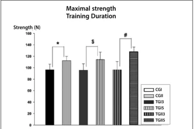
The four-week training regimen caused adaptations to femur maximal strength parameter in both groups (table 1). Regarding this parameter, and taking into account the training volume (weekly training frequency) there was statistical significance between the eight-week training groups CGII, TGII3 and TGII5 (p<0,05) (figure 1A). Statistical significance was also found between the control groups CGI and CGII (p=0,008), groups that trained with a three--weekly sessions frequency for four and eight weeks (TGI3 and TGII3 respectively, p=0,003) and groups that underwent training with a five-weekly session frequency for four and eight weeks (TGI5 and TGII5 respectively, p<0,001) (figure 1B).
Adaptations due to different training regimens were also found for the stiffness parameter (table1). There was no statistical differen-ce between groups that trained for four weeks; though comparison between trained and sedentary groups that trained for eight weeks showed significant bone stiffness adaptations (p<0,05) (figure 1C).
Table 1. Values (MD ± SD) of the mechanical properties of maximal strength and relative stiffness of the femur bones of the experimental groups.
CGI (n = 9) TGI3 (n = 9) TGI5 (n = 9) CGII (n = 9) TGII3 (n = 9) TGII5 (n = 9)
Maximal strength (N) 98,07 ± 8,9 96,94 ± 11,09 98,62 ± 12,98 110,81 ± 6,12 111,26 ± 9,84 127,60 ± 8,42
254 Rev Bras Med Esporte – Vol. 19, No 4 – Jul/Aug, 2013
There was also statistical significance between groups with diffe-rent training duration, but with equal number of weekly sessions (p<0,05) (figure 1D).
Figure 1C. Measurements of the mechanical assays performed. Training volume and relative stiffness. ANOVA revealed the effect of different training frequencies on stiffness (* for p < 0.001 and # for p = 0.005 and $ for p = 0.003). CGI, control/1 month; CGII control/2 months; TGI3, 3x/week/1 month; TGI5, 5x/week/1 month; TGII3, 3x/week/2 months; TGII5, 5x/week/2 months.
Figure 1A. Measurements of the mechanical assays performed. Training volume and maximal strength. ANOVA revealed time effect performance on maximal strength parameter (# for p = 0.002 and * for p = 0.003) without training. CGI, control/1 month; CGII control/2 months; CTI3, 3x/week/1 month; TGI5, 5x/week/1 month; TGII3, 3x/week/2 months; TGII5, 5x/week/2 months.
Figure 1B. Measurements of the mechanical assays performed. Training duration and and maximal strength. ANOVA revealed the effect of different training protocols on the maximal strength parameter (* for p = 0.008, $ for p = 0.003 and # for p < 0.001). CGI, control/1 month; CGII control/2 months; TGI3, 3x/week/1 month; TGI5, 5x/week/1 month; TGII3, 3x/week/2 months; TGII5, 5x/week/2 months.
Figure 1D. Measurements of the mechanical assays performed. Training duration of and relative stiffness. ANOVA revealed the effect on the stiffness of different training protocols (* for p < 0.007 and # for p < 0.001). CGI, control/1 month; CGII control/2 months; TGI3, 3x/week/1 month; TGI5, 5x/week/1 month; TGII3, 3x/week/2 months; TGII5, 5x/week/2 months.
DISCUSSION
The data evidence that the high-impact protocol described in this study positively affected the bone tissue, even for shorter training periods. However, the groups that underwent longer and more frequent training protocols presented more expressive gains in the analyzed biomechanical parameters.
The training volume (three or five weekly sessions) did not signifi-cantly affect the group that underwent a four-week training regimen. Nevertheless, an eight-week training regimen presented higher effec-tiveness in promoting osteogenic alterations. This was evidenced by comparisons between groups that trained with three weekly sessions, five weekly sessions and also sedentary/control groups.
Regarding the duration of the training protocol (four or eight weeks), and taking into account the strength parameter, we observed
Volume de treinamento - força máximaMaximal strength
Training Volume
Strength (N)
Stiffness Training Duration
Stiffness (N/mm)
CGI CGII TGI3 TGI5 TGII3 TGII5 Stiffness
Training Volume
Stiffness (N/mm)
CGI CGII TGI3 TGI5 TGII3 TGII5
Maximal strength Training Duration
Strength (N)
CGI CGII TGI3 TGI5 TGII3 TGII5 CGI CGII TGI3 TGI5 TGII3 TGII5
CGI CGII TGI3 TGI5 TGII3 TGII5
CGI CGII TGI3 TGI5 TGII3 TGII5
statistical difference between groups that underwent different weekly sessions. Nevertheless, statistical difference was also observed for this parameter between the control groups CGI and CGII. This may be explained by a possible interference of the age factor itself, although it was not possible to quantify its influence on osteogenic process. Duration of training regimen similarly affected the bone stiffness para-meter. No statistical differences were found between control groups, suggesting no interference of age in this manner. Consequently, we suggest that alterations in this parameter are originated from high--impact exercise mechanical stress.
255
Rev Bras Med Esporte – Vol. 19, No 4 – Jul/Aug, 2013
Robling et al15, showed that an eight-hour interval was sufficient to restore tissue cell mechanical sensitivity when compared to shorter periods. This demonstrates the importance of recovery periods to maximize osteogenic effects of mechanical overloads, such as those created by exercise.
The present study was conducted with similar interval betwe-en jumps for all experimbetwe-ental groups. Interval differbetwe-ences betwebetwe-en groups consisted of those originated from the number of weekly sessions. For those groups that underwent three weekly sessions, the intervals consisted of 48 hours; for those that underwent five weekly sessions, interval consisted of 24 hours. All training sessions were undertaken at the same time of day. Training periods of four and eight weeks, and also time between sessions might have di-minished the influence of these factors on bone tissue sensitivity. Other factor that affects bone remodeling is the type mechanical load that is given to the tissue. Due to the adopted training pro-tocol, only concentric muscle contraction was the source of me-chanical stress in the hind limbs. No eccentric muscle contraction stimuli were given to the hind limbs of the animals. This is mainly due to the fact that the animals used their front limbs to pull the-mselves to the top of the box after completion of the jump, and also because they were gently repositioned back to the bottom of the box for the next jump.
Studies that evaluated correlations between load magnitude and bone growth, as well as muscle influence over bone tissue, sho-wed that eccentric muscle activity is more effective in promoting BMD gains than concentric muscle contractions. This means that exercise which concentrate more muscle activity in the eccentric phase of the movement are more ostegenically effective than those concentrating muscle activity in concentric phases. Moreover, they also demonstrate that the load magnitude is the main mechanism by which muscle activity influences bone mass adaptations16,17.
REFERENCES
1. Shimano MM, Shimano AC, Volpon JB. Histerese de fêmures de ratas submetidos a ensaios de flexão em diferentes faixas etárias. Rev Bras Eng Biomed 2002;18:89-97.
2. Eastell R. Management of osteoporosis due to ovarian failure. Med Pediatr Oncol 2003;41:222-7. 3. Bain SD, Rubin CT. Metabolic modulation of disuse osteopenia: endocrine-dependent site specificity
on bone remodeling. J Bone Miner Res 1990;5:1069-75.
4. Smith EL, Gilligan C. Mechanical forces and bone. Bone Miner Res 1989;6:139-73.
5. Wronski TJ, Lowry PL, Walsh CC, Ignaszewskin JA. Skeletal alterations in ovariectomized rat. Calcif Tissue Int 1985;37:32-8.
6. Fuchs RK, Snow CM. Gains in hip bone mass from high-impact training are maintained: A randomized controlled trial in children. J Pediatr 2002;141:357-62.
7. Ocarino NM, Serakides R. Effect of the physical activity on normal bone and on the osteoporosis prevention and treatment. Rev Bras Med Esporte 2006;12:149-52.
8. Honda A, Sogo N, Nagasawa S, Kato T, Umemura Y. Bone benefits gained by jump-training are preserver after detraining in Young and adult rats. J Appl Physiol 2008;105:849-53.
9. Huang TH, Lin SC, Chang FL, Hsieh SS, Liu SH, Yang RS. Effects of different exercise modes on
mine-ralization, structure, and biomechanical properties of growing bone. J Appl Physiol 2003;95:300-7. 10. Turner CH. Three rules for bone adaptation to mechanical stimuli. Bone 1998;23:399-407. 11. Nagasawa S, Umemura Y. Bone hypertrophy in rats: effects of jump number and height. Adv Exerc
Sports Physiol 2002;8:87-92.
12. Umemura Y, Nagasawa S, Honda A, Singh R. High-impact exercise frequency per week or day for osteogenic response in rats. J Bone Miner Metab 2008;26:456-60.
13. Burger EH, Klein-Nulend J. Responses of bone to biomechanical forces in vitro. Adv Dent Res 1999;13:93-8. 14. Umemura Y, Sogo N, Honda A. Effects of intervals between jumps or bouts on osteogenic response
to loading. J Appl Physiol 2002;93:1345-8.
15. Robling AG, Burr DB, Turner CH. Recovery periods restore mechanosensitivity to dynamically loaded bone. J Exp Biol 2001;204:3389-99.
16. Hawkins SA, Schroeder ET, Wiswell RA, Jaque SV, Marcell TJ, Costa, K. Eccentric muscle acion increases site-specific osteogenic response. Med Sci Sports Exerc 1999;31:1287-92.
17. Heinonen A, Oja P, Kannus P, Sievanen H, Haapasalo H, Manttari A. Bone mineral density in females athletes representing sports with different loading characteristics of the skeleton. Bone 1995;17:197-203.
In the present study, the adopted protocol almost exclusively obligated the animals to use concentric muscle contractions. This type of muscle contraction demonstrated to be effective in enhan-cing both maximum strength and relative stiffness parameters of the analyzed femoral necks. Despite being scientifically proved that eccentric muscle contraction generates higher tension, - and the-refore, present higher osteogenic gains – the adopted protocol on this study showed that its possible to obtain significant bone gains exclusively through the application of only one type of muscle contraction.
CONCLUSIONS
Through analysis of both maximum strength and relative stiff-ness parameters, obtained through mechanical assays of femoral necks derived from animals that underwent high-impact training protocols, we showed that daily training frequencies for longer pe-riods are beneficial to bone health. We also show that lesser weekly sessions associated to a shorter period is capable of inducing alte-rations in these proprieties. This demonstrates that frequent daily exercises are not necessary to evoke alterations in bone tissue and bone health, although higher frequencies and longer periods have additional benefits.
AKNOWLEDGEMENTS
The authors would like to thank the Bioengineering Laboratory of the Medicine School of Ribeirão Preto, University of São Paulo (FMRP/USP-RP) for the infrastructure and the agencies FAPESP and CNPq, for granting financial aid and scholarships.