The history of the Diego blood group
Pedro C. Junqueira
1Lilian Castilho
2Diego blood group initially, because it appeared to be rare, was considered as a family or private factor. With further investigation, it was possible to trace this blood group from an individual family in Venezuela to the Indians across the continent of America and eventually to the Mongolian race in Asia. This review article follows the developments over the years and the history of the Diego blood group.
Rev.bras.hematol.hemoter.,2002,24(1):15-23
Keywords: Diego, blood group, history
1 - Honorary president of the Brazilian Society of Hematology and Hemotherapy, member of the Brazilian Academy of Medicine
2 - PhD, Researcher and Consultant in Immunohematology, Hemocentro, Unicamp
Correspondência para: Pedro C. Junqueira
R. Prudêncio de Moraes, 985 - apto 104 - Rio de Janeiro - RJ - CEP: 22420-041 - fone: (21) 2522-9951
Artigo Especial
Diego as a Private Factor
In 1953, Miguel Larysse and his co-workers Túlio Arends and R. Dominguez Sisco (from the Maternidad Concepcíon, Caracas) at the Centro de Investigaciones del Banco de Sangue de Caracas studied a serum from a full term male infant who appeared at birth to be clinically and hematologically normal but at that time the billirrubin determination was not carried out. Jaundice was evident after 12 hours, it became increasingly severe and the infant expired at three days. The direct antiglobulin test carried out on the newborns red blood cells was positive. Blood specimens of the positive mother and her Rh-group compatible infant were sent to Philip Levine at his consultation service from Ortho Research Foundation (Raritan, New Jersey) and both samples arrived in excellent conditions in June. Although the infants red cells were strongly coated, no antibody was demonstrable in the maternal serum when it was tested with an extensive panel of selected cells, which, did not include the fathers red blood cells. As ABO and
Rh incompatibility was excluded, the occurrence of a low-incidence blood factor with its corresponding antibody was suspected.
On October 26th, 1953, the father of the dead infant visited Levine in New York. At this time his red blood cells were tested against the maternal serum and a strong agglutination reaction was found. Levine and the father agreed with the name of the blood factor as Diego (Dia).
Levine also demonstrated that Dia was not
identical with two other previously recognized low-incidence blood factors associated with cases of hemolytic disease of the newborn named as Mia and Bea.
The Dia antibody was described as one
Diego as an Indian Factor
In 1955, the mother of the first documented Dia antibody carrier consulted Layrisse about a new pregnancy, which permitted further and more extensive tests in Venezuela and at Raritan, New Jersey. Layrisse and collaborators studying four generations of the original Diego family, noticed that the third and fourth generations seemed to be Caucasoid but their skins were a little dark-brown. However, the members of the second generation and the great grandmother in the first generation had a dark-brown skin and they seemed to be of Mongolian origin. Taking into account this observation, they started to observe the physical characteristics of a population from different countries with 286 individuals who had been tested with an anti-Dia serum by the indirect antiglobulin test.
The frequencies of positive results found in these populations were:
Population Frequencies of
tested Dia found
Caracas 2.26%
Barcelona 3.28%
Carib Indian 35.54%
Arawaka Indian 5.26%
Curiepe mixed Negro 7.33%
From these findings Layrisse et al., 1955 (2), concluded that it was evident that the Diego Factor was not restricted to a single family, and could be placed among the relatively high incidence blood group systems in Venezuela and probably in South America, with apparently genetic, anthropologic and clinical significances. They also commented that since the Diego terminology was meaningless and this factor had probably some anthropological implications it should be changed to a more expressive one like Indian Factor. About this comment we must remember that Chown and Lewis (Nature, 1953) said that what appears to be a rare, private Table 1 Typing of red cell antigens in selected members of the Diego Family
ABO Rh MN Ss Kk Fyª Jkª/Jkb Leª P Diª
I-1 O Cde/cde N nt nt nt nt nt nt +
I-2 A dCe/dce N nt nt nt nt nt nt +
II-1 O dcE/dce N nt nt nt nt nt nt 0
II-2 O dcE/dce MN SS Kk 0 JkªJkb 0 0 +
II-3 O Dce/dce M Ss Kk + JkªJkb 0 + 0
II-4 O Dce/dce N nt nt nt Nt nt nt 0
III-1 O Dce/dcE M Ss Kk 0 JkªJkb 0 0 0
III-3 O dcE/dce MN Ss Kk + Jkª ? 0 0 0
III-5 O dcE/dce M SS Kk + JkbJkb 0 + +
III-6 O DCe/DCe MN ss Kk + Jkªjkb 0 + 0
III-7 O dce/dce M Ss Kk + JkªJkb 0 0 0
III-9 O Dce/dce nt nt nt nt Nt nt nt +
III-10 O ddE/dce M Ss Kk 0 JkªJkb 0 + +
III-11 O dce/dce M Ss Kk + JkbJkb 0 + +
IV-7 O DCe/dcE MN Ss Kk + JkªJkb 0 0 +
IV-8 O DCe/dcE MN Ss Kk 0 JkªJkb + + +
IV-9 O DCe/dce nt nt nt nt nt nt nt +
IV-10 O DCe/dce nt nt nt nt nt nt nt 0
or family antigen in one population might be fairly frequent in another one, and so Layrisse was right.
In 1955, Dr Jean Dausset, a French immunologist, later awarded the Nobel Prize (1980), after working with Layrisse in Caracas, came to Rio de Janeiro to visit me because I was studying the Brazilians Indians. At the time, Dr Dausset brought me a sample of the Diego serum and this serum turned out to be very important to me. In that year we had the opportunity to test two Brazilian Indian groups with the anti-Diego serum using the indirect antiglobulin test: the Carajas living in Santa Izabel, Bananal Island, State of Mato Grosso and the Kaingaques living in a reservation near Palmas, State of Paraná. Thirteen (36%) of the red blood samples from the Carajas tested and 22 (46%) of the Kaingaques samples tested gave positive reactions with the anti-Diego serum. We had also the opportunity to test 200 red blood samples from true Black donors from the Rio de Janeiro Municipal Blood Bank that showed negative results with the anti-Diego serum. We sent our results to Layrisse, in Caracas and to Levine, in Raritan. We had our paper published in Nature, volume 17 on page 41 (3). Levine et al. published their paper in this same volume of the Nature Journal on page 40 (4).
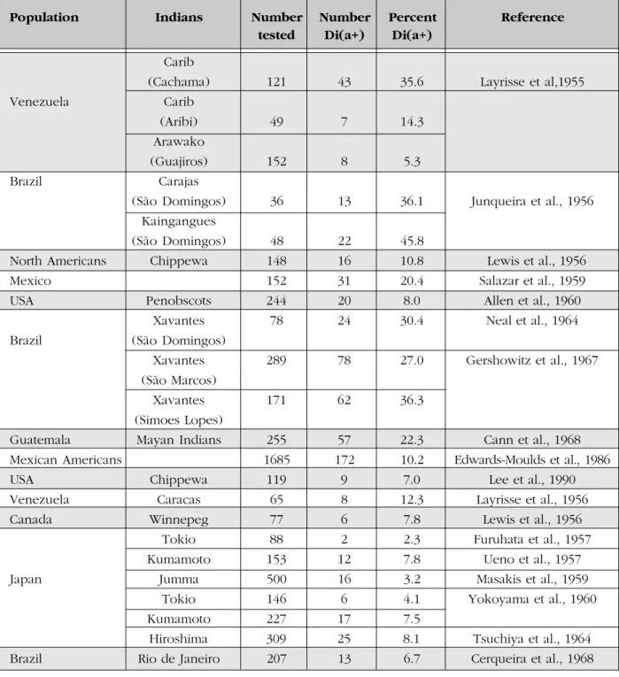
Table 2 shows the incidence of the Dia
antigen in different Indian populations.
Diego as a Mongolian Factor
In 1956, in a paper published in the Nature Journal (5), Layrisse and Arends stated: Since the Indians of the American continent are considered to be anthropological related to the Mongolian people of the old world, we decided to investigate the incidence of the Diego Factor in other available representative Asians living in Venezuela. They tested 100 unrelated males from Canton (China), living in Venezuela, and detected 5 Diego positive individuals (5% of the Chinese tested). They also tested sixty-five unrelated Japanese and found 8 Diego positive
subjects (12.5% of the Japanese tested) (6). These findings indicated that the Diego factor was not restricted to South America and suggested that this antigen was a Mongolian rather than an Indian factor.
In the same year, Lewis et al., (7) showed that the Diego antigen was found to be present in 16 of 148 unrelated Chippewa Indians from North Minnesota and in 6 of 77 unrelated Japanese from Winnipeg. This finding suggested that Diego might be an Asiatic characteristic.
In 1957, Levine and Robinson (8) said that studies carried out by Layrisse and his colleagues on the Diego blood factor in other populations including the Brazilian Indians carried out by Junqueira et al. (3), apparently suspected that the Diego factor could be Mongolian in its origin. Further, they concluded that the term Indian for the Diego blood factor was not appropriate. Levine and Robinson, 1957 (8) also demonstrated that the Dia antigen was
genetically independent from other 15 low incidence factors and from four high incidence factors previously recognized. Furthermore, Layrisse, Sanger and Race, 1959 (9) using evidence from the literature and from nine new families studied, showed that the Dia antigen
had no links with most of the established blood group systems.
Many papers showing the distribution of the Dia antigen considered that it was essentially
a Mongolian characteristic, absent in Whites , Blacks, Australian aborigines and other populations (10-39). Table 3 shows the incidence of the Dia antigen in the Chinese and
Korean populations.
The book named The distribution of Human Blood Groups and other polymorphisms (Mourant et al., 1976) (40) is considered the best one to show the early worldwide race distribution of Dia..
In 1967, thirteen years after the detection of the anti-Dia, Thompson, Childers and Hatcher
identified the anti-Dib (41). As the phenotype
Di(ab) has not been reported yet, we may assume that only two alleles (Dia and Dib) control
the Diego blood group system.
Population Indians Number Number Percent Reference
tested Di(a+) Di(a+)
Carib
(Cachama) 121 43 35.6 Layrisse et al,1955
Venezuela Carib
(Aribi) 49 7 14.3
Arawako
(Guajiros) 152 8 5.3
Brazil Carajas
(São Domingos) 36 13 36.1 Junqueira et al., 1956
Kaingangues
(São Domingos) 48 22 45.8
North Americans Chippewa 148 16 10.8 Lewis et al., 1956
Mexico 152 31 20.4 Salazar et al., 1959
USA Penobscots 244 20 8.0 Allen et al., 1960
Xavantes 78 24 30.4 Neal et al., 1964
Brazil (São Domingos)
Xavantes 289 78 27.0 Gershowitz et al., 1967
(São Marcos)
Xavantes 171 62 36.3
(Simoes Lopes)
Guatemala Mayan Indians 255 57 22.3 Cann et al., 1968
Mexican Americans 1685 172 10.2 Edwards-Moulds et al., 1986
USA Chippewa 119 9 7.0 Lee et al., 1990
Venezuela Caracas 65 8 12.3 Layrisse et al., 1956
Canada Winnepeg 77 6 7.8 Lewis et al., 1956
Tokio 88 2 2.3 Furuhata et al., 1957
Kumamoto 153 12 7.8 Ueno et al., 1957
Japan Jumma 500 16 3.2 Masakis et al., 1959
Tokio 146 6 4.1 Yokoyama et al., 1960
Kumamoto 227 17 7.5
Hiroshima 309 25 8.1 Tsuchiya et al., 1964
Brazil Rio de Janeiro 207 13 6.7 Cerqueira et al., 1968
Table 2 Incidence of the Diª antigen in different indian populations
responsible for the hemolytic disease of the newborn and for hemolytic transfusion reactions (42). Due to the composition of our population, and supported by studies showing that 3.6% of the multi-transfused patients in Brazil have anti-Dia (43) the Brazilian red cell panels used for
antibody screening ought to include a Di(a+). In 1975, Race and Sanger (42) said in the last edition (6th) of their book: The Venezuelan
discovery of Dia will make an outstanding
The Diego blood group system
The Diego blood group system is a rapidly expanding system and today it consists of two pairs of antithetical antigens (Dia and Dib, and
Wra and Wrb) and 17 low incidence antigens
(44) (Table 4).
Spring et al., in 1992 (45) recognized an association between band 3 (anion exchange 1 - AE1), the most abundant integral protein of the red blood cell (RBC) membrane, and the Diego blood group system. They found by SDS-PAGE that Di(a+) red cells always have band 3 variant Memphis, although not all band 3
Memphis red cells are Di(a+). This observation led to the investigation of band 3 from red cells of known Diego blood group in order to ascertain whether the expression of the Dia
antigen is linked to band 3 Memphis, and to define the molecular basis of this variant.
In 1993, the Diego blood group locus was assigned to chromosome 17 by Zelinski et al., (46) and in 1994, Bruce et al. (47) showed by H
2DIDS (4,4 diisothiocyanato-2,2
dihydrostibene disulfonate) binding studies on samples of known Diego phenotypes that the expression of the Dia antigen is associated with
an increased susceptibility of band 3 to labeling
Population Chinese Number Number Percent Reference
tested Di(a+) Di(a+)
Venezuela Canton 100 5 5.0 Layrisse et al., 1956
Taiwan Fukein and Canton 1000 32 3.2 Lin-Chu et al., 1988
Korea Seoul 227 17 6.1 Won et al., 1960
3 Incidence of the Diego antigen in the chinese and korean populations
Table 4 Antigen assigned to Diego blood group system by the ISBT Nomenclature (Daniels et al., 2001)
System Number Symbol Amino Acid Substitution
010DI 0100001 DI 1 Diª Leu 854
010DI 0100002 DI 2 Dib Pro 854
010DI 0100003 DI 3 Wrª Lys 658
010 DI 010004 DI 4 Wrb Glu658
010DI 0100005 DI 5 Wdª Val 557 Met
010DI 0100006 DI 6 Rbª Pro 548 Leu
010DI 0100007 DI 7 WARR Thr 552 lle
010DI 0100008 DI 8 ELO Arg 432 Trp
010DI 0100009 DI 9 Wu Gly 565 Ala
010DI 0100010 DI 10 Bpª Asm 569 Lys
010DI 0100011 DI 11 Moª Arg 656 His
010DI 0100012 DI 12 Hgª Arg 656 Cys
010DI 0100013 DI 13 Ugª Tyr 555 His
010DI 0100014 DI 14 Swª Arg 646 Gln
010DI 0100015 DI 15 BOW Pro 561 Ser
010DI 0100016 DI 16 NFLD Glu 429 Asp
Pro 561 Ala
010DI 0100017 DI 17 Jnª Pro 566 Ser
010DI 0100018 DI 18 KREP Pro 566 Ala
010DI 0100019 DI 19 Tra Lys551Asn
010 DI 0100020 DI 20 Fra Glu480Lys
by H
2DIDS. This provided evidence for a link
between the expression of the Dia antigen and
the presence of band 3 variant Memphis (45). DNA sequence analysis (47) showed that the Dia/Dib polymorphism results from a point
mutation at nucleotide 2561 (C>T) resulting in a single amino acid substitution in position 854, with a proline corresponding to the Dib antigen
and leucine to the Dia antigen. Molecular analysis of band 3 from individuals with red cells expressing the Dia antigen showed the
simultaneous occurrence of the mutations 2561T (854Leu) and 166G (56Glu) responsible for the Band 3 variant Memphis (47).
Carries of band 3-Memphis are asymptomatic and show no morphologic abnormalities of their erythrocytes (48). Studies to determine the frequencies of band 3-Memphis in some populations have been performed, proving that band 3-Memphis is not a rare polymorphism and that the gene frequency of band 3-Memphis varies among different populations, with a high frequency among Indians and the Japanese (48-51).
Bruce et al. (1995) also reported that the Wra /Wrb
polymorphism results from a glutamic acid in position 658 of band 3 corresponding to Wrb and a lysine in the same position,
corresponding to Wra (52).
Evidence suggests that the Wrb antigen is
also associated with the glycophorin A (GPA) because Wrb antigenrequires both band 3 and
GPA for its expression in the red cell membrane (52-54).
The recognition that band 3 carries antigens of the Diego blood group system and the elucidation of the Dia/Dib
and Wra/Wrb
polymorphisms have led several investigators to elucidate the molecular basis of the other low incidence Diego antigens and to create a more accurate structural model of band 3 (55). New studies with band 3 and Diego are being developing and new findings are emerging. We have found in our population by molecular studies a high frequency of 166G mutations (Memphis) and the possibility that the Dia antigen can not be associated with the
band 3 variant Memphis (56).
In the 1980s and 1990s, serological, biochemical and molecular studies have given a new face to Immuno-hematology. Most of the papers published are cooperative works and are improving the understanding of the Diego system. It is important that these studies should continue in different populations to expand the knowledge of the structure and function of band 3.
A história do sistema de grupos sangüíneos Diego
Pedro C. Junqueira, Lilian Castilho
Resumo
O sistema de grupos sangüíneos Diego, devido à sua raridade, era considerado um fator privado familiar. Investigações posteriores, de estudos familiares na Venezuela e em índios do continente americano e mongóis na Ásia, evidenciaram a sua existência.
Neste relato apresentamos o desenvolvimento do conhecimento e da sua história.
Rev.bras.hematol.hemoter.,2002,24(1):15-23
Palavras-chave: Diego, grupos sangüíneos, história
References
1. Levine P, Koch EA, McGee RT. Rare human isoagglutinins and their identification. Am. J. Clin. Path. 1954; 24: 292-304.
2. Layrisse M, Arends T, Domingues Sisico R. Nuevo grupo sanguíneo encontrado en descendents de índios. Acta Med.
Venezuelana 1955; 3: 132-138.
3. Junqueira PC, Wishart PJ, Ottensosser F, Pasqualin R, Lorenso FP, Kalmus H. The Diego blood factor in Brazilian Indians.
Nature. 1956; 177:41.
5. Layrisse M, Arends T. The Diego blood factor in Chinese and Japanese. Nature. 1956; 177: 1083-1084.
6. Arends T and Layrisse M. Investigación del factor Diego en Japaneses y Chinos. Acta Cientif Venez. 1956; 7:11-12.
7. Lewis M, Ayukawa H, Chown B, Levine P. The blood group antigen Diego in North American Indians and in Japanese. Nature
1956; 177:41.
8. Levine P, Robinson EAV. Some observations of the new human blood factor Diª
Blood. 1957; 12: 448-453.
9. Layrisse M, Sangger R, Race RR. The inheritance of the antigen Diego: evidence for its independence of other blood group systems. Am. J. Hum. Genet. 1959; 11: 17-25.
10. Layrisse M, Arends T. The Diego blood factor in Negroid populations. Nature. 1957; 178: 478-479.
11. Loureiro Fernandes J, Junqueira PC, Kalmus H, Ottensooser F, Pasqualin R, Wishart P. P.T.C. threshold, color vision and blood factors of Brazilian Indians Kaingaques.
An. Human Genetics. 1957; 22: 16-21.
12. Junqueira P C, Kalmus H, Wishart P. P.T.C. threshold, color vision and blood factors of Brazilian Indians Carajas. An. Human Genetics 1957; 22: 22-25
13. Lewis M, Kaita H, Chown B. The blood groups of the Japanese population. An. J.
Human Genetics 1957; 9: 274-283.
14. Furuhata T, Yokoyama M, Kuniyuki M. First investigation of the Diego factor in Japanese in Japan, in Tokyo. Proc. Jap. Acad. 1957; 33: 228.
15. Ueno N, Murahato M. Introduction to the Diego blood group and the frequency of the Diego Antigen among Japanese in Kumamoto area. Blood and Transfusion
1957; 4: 146 (in Japanese).
16. Iseki S, Masaki S, Furukawa K, Mohn JF, Lambert RM and Rosamilia HG. Diego and Miltenberger blood factor in Japanese. J. Med. Sci 1958; 2: 120-126.
17. Layrisse M. Anthropological considerations of the Diego (Di) antigen possible application in the studies of Mongolian and hybrid
population. Am. J. Phys. Anthropol. 1958; 16: 173-86.
18. Gershowitz H. The Diego factor among Asiatic Indians, Apaches and West African Negroes; blood types of Asiatic Indians and Apaches.
Am. J. Phys. Anthropol. 1959; 17: 195-200. 19. Masakis S, Furukawa K. The frequency of the Diego factor in 500 Japanese living in the Jumma area. Jap. J. Legal Med. 1959; 13(3): 359.
20. Salazar Mallen M, Arias T. Inheritance of Diego blood group in Mexican Indians.
Science 1959; 130: 164-5.
21. Yokoyama M, Murakata M, Ueno N. The Diego factor in Japanese. Nature 1960: 591. 22. Won CD, Shin HS, Kem SW, Swanson J, Matson GA. Distribution of heredity blood factors among Koreans residing in Seoul, Korea. Am. J. Phys. Anthropol. 1960; 18: 115-124.
23. Layrisse M, Wilbert J. El antigeno del sistema sanguineo Diego. La Fundacion Creole y
la Fundacion Eugenio Mendonza; Editora
Sucre, Caracas, 1960.
24. Allen FH, Corcoran PA. Blood groups of the Penobscot Indians. Am. J. Phys. Anthropol. 1960; 18: 109-114.
25. Neal JV, Salzano F M, Junqueira P C, Keiter F and Maybury Lewis D. Studies on the Xavante Indians of the Brazilian Mato Grosso. Human Genetics 1964; 16: 52-140. 26. Tsuchiya T, Kurata M, Fukuma A, Onishi T, Yokoyama M, Kobayashi H and Tomita K. Anthropological observation of blood groups in Hiroshima. Nature 1964; 204-87.
27. Murakami S. The rare blood types and variants in Japanese. Acta Crim. Jap. 1967; 33: 138-145.
28. Gershowitz H, Junqueira PC, Salzano FM, Neel JV. Further studies on the Xavante Indians III blood groups and ABH-Leª secretor types in the Simoes Lopes and São Marcos Xavantes. Human Genetics 1967; 19: 502-513.
29. Cerqueira AJB, Junqueira PC, Tsum T. Groupos sangüíneos dos sistemas ABO, Rh e Diego em Japaneses. A Folha Médic 1968; 57: 105-109.
AJ. The Diego blood group: anti-Diª and Di(a+) blood group antigen found in Caucasians. Med. J. Aust. 1968; 1: 406-7. 31. Cann HM, VanWest B, Barnett CR. Genetics
of Diego blood groups in Guatemalan Indians: use of anti-serum to Diego a and Diego b antigens. Science 1968; 162: 1391-2. 32. Iseki S, Masaki S, Furukawa K, Lambert RM and Mohn JF. The blood group a n t i g e n D i ª i n J a p a n e s e a n d i t s independence of the ABH secretor system.
Vox Sang. 1970; 19: 483-7.
33. Nakajima H, Hayakawa Z, Ito H. A new example of anti-Diª found in Japanese woman.
Vox Sang. 1971; 20: 271-3.
34. Simmons RT. The apparent absence of the Diego (Diª) and the Wright (Wrª) blood group antigens in Australian aborigines and in Guineans. Vox Sang. 1979; 19: 533-6. 35. Edwards, Moulds JM, Alperin JB. Studies of
the Diego blood group among Mexican Indians. Transfusion 1986; 26: 234-6. 36. Leon S, Mak KH, Chua E. Blood group
distribution among Chinese donors in Hong Kong. Presented at the 19th Congress of International Society of Blood Transfusion, in Sidney Australia. May 1986. 37. Lin Chu M, Broadberry RE, Chang FJ. The distribution of blood group antigens and alloantibodies among Chinese in Taiwan.
Transfusion 1988; 28: 350-2.
38. Yung CH, Chow MP, Hu Hy, Mon LL, Lyou Jy, Lee TD. Blood group phenotypes in Taiwan. Transfusion 1989; 29: 233-5. 39. Lee TD, Zhad TM, Chow MP and Lee G.
HLA, GM, KM and Diego blood group typing of Chippewa Indians. Transfusion 1990; 30: 728-732.
40. Mourant AE, Kopec AC, Domanicewska, Sobezak K, eds. Distribution of the human blood groups and other polymorphisms.
London: Oxford University Press 1976:
608-14.
41. Thompson PR, Childers DM, Hatcher DE. Anti-Dib first and second examples. Vox Sang 1967; 13:314-318.
42. Race RR, Sanger R. Blood Groups in Man, 6th edn. Oxford: Blackwell Scientific
Publications, 1975.
43. Zago-Novaretti MC, Soares MOC, Dorlhiac-Llacer PE, Chamone DAF. Anti-Diego in multitransfused patients. Rev. Paulista Med. 1992;110 (S):IH52.
44. Daniels GL, Anstee DJ, Cartron JP, Dahr W, Fletcher A, Garratty G, Henry S, Jorgensen J, Judd WJ, Kornstad L, Levene C, Lin M, Lomas-Francis C, Lubenko A, Moulds JJ, Moulds JM, Overbeeke M, Reid Me, Rouger P, Scott M, Sistonen P, Smart E, Tani Y, Wendel S, Zelinski T. International Society of Blood Transfusion working party on terminology for red cell surface antigens.
Vox Sang 2001;80:193-196.
45. Spring FA, Bruce LJ, Anstee DJ, Tanner MJA. A red cell band 3 variant with altered stilbene disulphonate binding is associated with the Diego (Dia) blood group antigen. Biochem J 1992; 288:713-716.
46. Zelinski T, Coghlan G, White L, Philips S. Provisional assignment of the Diego blood group locus to chromosome 17. Transfusion
1993, 33 (Suppl.1)47S: Abstract.
47. Bruce LJ, Anstee DJ, Spring FA, Tanner MJA. Band 3 Memphis variant II. Altered stilbene disulphonate binding and the Diego (Dia) blood group antigen are associated with the human erythrocyte band 3 mutation
Pro854→Leu. J. Biol. Chem. 1994a;
269:16155-16158.
48. Ranney HM, Rosemberg GH, Morrison M, Mueller TJ. Frequencies of band 3 variants of human red cell membranes in some different populations.Br. J. Haematol. 1990; 75:262-267.
49. Ideguchi H, Okubo K, Ishikawa A, Futata Y, Hamasaki N. Band 3-Memphis is associated with a lower transport rate of phosphoenolpyrovate. Br. J. Haematol. 1992; 82:122-125.
50. Mueller TJ, Morrison M. Detection of a variant of protein 3, the major transmembrane protein of human erythrocyte.J. Biol. Chem.. 1977; 252: 6573-6576.
band 3-protein: their prevalence in human and non human primates. Hum. Genet. 1990; 86:126-130.
52. Bruce LJ, Ring SM, Anstee DJ, Reid ME, Wilkinson S, Tanner MJA. Changes in the blood group Wright antigens are associated with a mutation at amino acid 658 in human erythrocyte band 3: A site of interaction between band 3 and glycophorin A under certain conditions. Blood 1995; 85:541-547.
53. Ring SM, Tippet P, Swallow DM. Comparative immunochemical analysis of Wra and Wrb red cell antigens. Vox Sang.
1994; 67:226-230.
54. Telen MJ, Chasis JA. Relationship of the human erythrocyte Wrb antigen to an
interaction between glycophorin A and band 3. Blood 1990; 76:842-848.
55. Jarolim P, Rubin HL, Zakova D, Storry J, Reid ME. Characterization of seven low incidence blood group antigens carried by erythrocyte band 3 protein. Blood 1998; 92:4836-4843.
56. Balleotti Jr W, Rios M, Fabron A, Costa FF, Bianco C. Castilho L. Frequency of the DIA/ Memphis II allele among Amazonian Indians, Brazilians of Japanese descent and Brazilians blood donors.Transfusion 2000; 39(S).