649 Address to: Dr. Vinicius Rodrigues Taranto Nunes. Rua São Tomé do Príncipe 130,
Jardim Atlânico, 31550-210 Belo Horizonte, MG, Brasil.
Fax: 55 31 3492-1054
e-mail: [email protected]
Received in 02/10/2011
Accepted in 14/12/2011
INTRODUCTION
CASE REPORT
Bilateral paracoccidioidomycoic iliopsoas abscess associated
with ileo-colonic lesion
Helena Duani
[1], Vinícius Rodrigues Taranto Nunes
[2], Anísio Borges Assumpção
[2],
Isadora Soia Borges Saraiva
[1], Rodrigo Macedo Rosa
[2], Augusto Mota Neiva
[2]and Ênio Roberto Pietra Pedroso
[1][1]. Setor de Infectologia, Hospital das Clinicas, Universidade Federal de Minas Gerais, Belo Horizonte, MG. [2]. Insituto Alfa de Gastroenterologia, Hospital das Clinicas, Universidade Federal de Minas Gerais, Belo Horizonte, MG.
Paracoccidioidomycosis (PCM) is a systemic mycosis endemic in Lain America, whose agent is the dimorphic fungus Paracoccidioides brasiliensis, which manifests itself in a chronic presentaion in over 90% of the cases. Its prevalence is greater among males from 30 to 60 years old, and lungs are the most frequent compromised organs. Its acute/subacute form with generalized lesions involves muliple organs and is rare and severe1,2.
We herein present a case of disseminated PCM, with involvement of lymphatic and gastrointestinal systems, and a subsequent development of bilateral iliopsoas muscle abscess (IPA), which consitutes a rare complicaion of the disease.
A 24-year-old rural man presening fever, asthenia, weight loss (20kg in the period), dull abdominal pain, diarrhea and hematochezia for six months. After four months of the initial symptoms, he developed disseminated subcutaneous nodules, which began draining a purulent secreion when he was admited to our hospital.
On physical examinaion, we found bilateral cervical, inguinal, and axillary lymphadenomegaly and a painful abdomen at supericial palpaion, especially in the right iliac fossa.
The laboratory tests revealed a hemoglobin level of 8.9g/dL; total leukocyte count of 6,000/mm3 (eosinophil count of 60/mm3);
C-reacive protein of 188mg/dL; viral serology negaive for HIV and hepaiis B and C; nonreacive Venereal Disease Research Laboratory (VDRL) test; negaive sputum alcohol acid-resistant bacilli (AARB) test in three samples (Auramine and Ziehl Neelsen); negaive sputum
FIGURE 1 - Colonoscopy revealing the cecum and the ileocecal valve, a large ulcer, and an ulcer with ibrin (arrows).
Revista da Sociedade Brasileira de Medicina Tropical 45(5):649-651, Sep-Oct, 2012
Case Report
ABSTRACT
This case report shows the clinical development of a paient with systemic paracoccidioidomycosis presening with lymphaic-intesinal manifestaion. The paient iniially had a substanial clinical improvement but had a recrudescence ater six months of sulfamethoxazole-trimethoprim oral treatment, with the emergence of feverish syndrome, lumbar pain, and intermitent claudicaion, characterizing a bilateral iliopsoas muscle abscess, necessitaing clinicosurgical therapeuics.
Keywords: Paracoccidioidomycosis. Psoas abscess. Paracoccidioides brasiliensis.
fungal smear; normal renal and hepaic funcion panels; and negaive microbiological stool tests. An abdominal and pelvic computed tomography (CT) demonstrated mesenteric lymphadenomegaly, with a hypodense center. Colonoscopy evidenced muliple deep ulcers with changeable sizes and forms along the whole colon, intercalated by normal mucosa, and a deformed ileocecal valve, which favored the diagnosis of infectious ulcerative pancolitis
(Figure 1). Anatomopathological examinaion of the colonic mucosa revealed some areas of erosion covered in ibrino-leukocyic exudate, inlammatory iniltrate in corion, with some mulinucleated giant cells, and lots of round shaped yeast, in various sizes, compaible with ileum-colonic PCM (Figure 2). A biopsy of a cervical lymph node also was performed, and in the anatomopathological examinaion, we found the Paracoccidioides brasiliensis, with non-caseating granulomas containing mulinucleated giant cells and epithelioid hisiocytes, presening a steering wheel shape, compaible with PCM granulomatous lymphadeniis.
During hospitalization, the patient was treated with venous sulfamethoxazole-trimethoprim (SMX-TMP) and amphotericin B (amphoB) deoxycholate, receiving a cumulaive dose of 1,900mg ater
650
Duani H et al - Bilateral paracoccidioidomycoic iliopsoas abscess
DISCUSSION 38 days. A complete regression of lymphadenopathy occurred, the
paient gained weight, and the diarrhea ceased. Ater 40 days from admission, he was discharged, with a prescripion of a daily use of SMX-TMP 800 + 160mg.
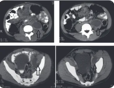
The paient returned 40 days later, presening recrudescence of the iniial symptoms, including pain in the let hip, limited extension of the homolateral hip, limping, and fever. Furthermore, cultures were obtained, and an anatomopathological examinaion of an inguinal lymph node biopsied revealed Paracoccidioides brasiliensis. He was rehospitalized and treated with amphoB plus oral itraconazole and sulphadiazine. Abdominal and pelvic CT showed mesenteric lymphadenomegaly, enlargement of the let iliac and psoas muscles, with inner voluminous liquefacion, which extended from the level of the third lumbar vertebra (L3) to the pelvis. Moreover, a smaller collecion was observed in the right psoas muscle at L3 level, and let pyelocaliceal dilaion was present (Figure 3).
An ultrasound (US)-guided lumbar percutaneous puncture of the collecion was performed at L3 level, and a Cystoix® 12 Fr drain
was placed inside it. There was immediate drainage of 100ml of thick purulent secreion, which revealed the presence of Paracoccidioides brasiliensis. Bacterial and fungalcultures of this specimen was negaive.
The paient kept the iniial symptomatology ater eight days of drainage, being therefore submitted to a new CT scan, that demonstrated reduction of the left psoas abscess, but another collecion was found involving the homolateral iliac muscle. He was then submited to an US-guided inguinal puncture of this abscess, using 14G Jelco, with immediate drainage of 50ml of purulent secreion. The lumbar drain was let in place for 18 days.
Control CT scan 20 days ater drainage showed minimal residual bilateral iliopsoas collecion, and the pyelocaliceal dilaion was no longer observed. Ater having received a cumulaive dose of 2.9g of amphoB and 60 days of oral itraconazole and sulfadiazine, he was discharged.
Ambulatory follow-up six months ater treatment showed no signs of relapse, maintaining oral itraconazole and SMX-TMP usage. FIGURE 2 - Anatomopathological exam: fragments of colonic and ileal mucosa demonstraing areas of erosion covered in ibrino-leukocyic exudate, inlammatory iniltrate in corion, with some mulinucleated giant cells and many round-shaped yeast (compaible with Paracoccidioides brasiliensis) in various sizes (arrows), compaible with ileum-colonic paracoccidioidomycosis.
FIGURE 3 - Abdominal and pelvic computed tomography scan: mesenteric lymphadenomegaly, enlargement of the let iliac and psoas muscles, with inner voluminous liquefacion, which extended from the level of the third lumbar vertebra (L3) to the pelvis (arrows).
Paracoccidioidomycosis can manifest itself in two disinct clinical forms: acute/subacute or chronic. The acute/subacute form represents 3 to 5% of PCM cases, being more common in the irst three decades of life, especially in children3. It evolves rapidly, with signiicant general
state impairment, fever, hepatosplenomegaly, lymphadenomegaly, together with digesive, osteoaricular, and cutaneous manifestaion. Mesenteric lymph node hypertrophy can lead to bowel obstrucion or choledochal compression. Moreover, it can impair oral drugs and nutrient absorpion, aggravaing the nutriional state of the paient.
This report evidences PCM evolvement in an immunocompetent young adult presenting a subacute severe disseminated form, with colitis and systemic lymphadenomegaly, especially in the retroperitoneum, which istulized not only to the skin but also to the lumbar musculature. The maintenance of the Paracoccidioides brasiliensis foci was strictly related to the relapse of a severe form of PCM, requiring prolonged clinicosurgical therapeuics to control the infecion.
The therapeuic choice depends on the severity of the PCM, and the opions are sulfas, azolics, and amphoB. The great eicacy of the sulfa and azolic drugs jusiies the restricted use of amphoB, which is more toxic and is exclusively parenteral. AmphoB is reserved for more severe and systemic forms, which compromise structures beside the lungs, mucosa, and skin, and when there is imminent risk of death. Only a few studies have been held in order to deine its opimal treatment. Only two randomized trials have been published so far, neither of which have collected suicient data to assess response or cure rates4,5. The duraion of treatment is sill a mater of debate,
since no randomized trials have compared different treatment duraions, and are based mainly on data from open studies and specialist opinions6. In this report, the patient had initially been treated
with venous SMX-TMP and amphoB because of the severity of the
651
Rev Soc Bras Med Trop 45(5):649-651, Sep-Oct, 2012
good compliance. However, at this stage of treatment, the disease relapsed, manifesing as cold retroperitoneal abscesses. Therapeuics at that moment was based on anifungal associaion because of the disease severity and life threatness.
The iliopsoas muscle abscess (IPA) is a retroperitoneal collecion in the space deined by the iliac and psoas muscles, usually determined by a bacterial infection. It is considered a rare medical entity; nonetheless, with the advent of CT scan for evaluaion of paients with sepsis with undetermined focus, the diagnosis of IPA has become more frequent. Its emergence is related to two mechanisms: primary, with hematogenic spread, and secondary, when related to adjacent structures infecion.
The diagnosis, evoluion and treatment of IPA are well described for bacterial infecion. Iliopsoas abscess related to PCM is even more rare and there is litle informaion about this PCM enity7. In the
paient reported, the most likely mechanism of IPA formaion was the istulizaion of retroperitoneal lymph nodes into the iliopsoas muscle compartment, simultaneous to cutaneous istulizaion of the supericial lymph nodes.
The classical triad symptoms, characterized by fever, lumbar pain, and claudicaion, are present in only 30% of the cases8. Other
symptoms include inguinal bulge, malaise, anorexia, and weight loss. In 60% of the cases, the US conirms the diagnosis, with the CT scan considered the gold standard for diagnosis.
Historically, the iniial IPA treatment was based on extraperitoneal surgical drainage, associated with adequate animicrobial therapy. The irst IPA radiologic (US, CT) guided percutaneous drainage occurred in 19849, and since then, the best therapeuic opion has been on debate.
So far, the treatment choice is the guided percutaneous drainage, as it is the least invasive and associated with lower morbidity and mortality10; however, many local and systemic factors of each paient
may inluence the therapeuic choice11.
Ater the irst drainage, there was only parial improvement in the paient reported, requiring the repeiion of the CT scan, which showed that the let IPA was composed of two disinct compartments, requiring a new US-guided percutaneous puncture. Therapeuic failure ater percutaneous drainage is rare, as described by Castademir et al, who revealed that this method was eicacious in 21 of 22 paients; nonetheless, when it happens, surgical treatment is indicated12. The
right IPA was of litle volume, and we opted to treat it with anibioics only, a plausible treatment for small abscesses.
The main complicaions of IPA are deep venous thrombosis, bowel obstrucion, ureteral obstrucion, and sepsis. In this case, pyelocaliceal dilaion was due to extrinsic compression and was fully reverted ater drainage, with no renal sequela.
Paracoccidioidomycosis reveals itself as a variable clinical enity that ought to be remembered in the clinical diagnosis of a localized or systemic disease, with possible involvement of the lungs, skin, mucosa, central nervous system, retroperitoneal musculature or lymph nodes, intesine, adrenal, kidneys, or prostate and likely associaion with tuberculosis, neoplasms, and sepsis. In this report, we presented
a paient with muliple organ compromise because of disseminated PCM, apparently with anifungal resistance, necessitaing broad-spectrum anibioics and radiologic-guided percutaneous drainage of cold retroperitoneal abscesses, who evolved with slow recovery despite all therapeuics. Although the irst descripion of PCM goes back over 100 years, its opimal treatment is not yet well-deined. The lack of informaion about diagnosis and treatment may contribute to the development of unusual forms of presentaion that are capable of producing signiicant damage and death.
REFERENCES
1. Coutinho ZF, Silva D, Lazera M, Petri V, Oliveira RM, Sabroza PC, et al. Paracoccidioidomycosis mortality in Brazil (1980-1995). Cad Saude Publica 2002; 18:1441-1454.
2. Shikanai-Yasuda MA, Queiroz-Telles F, Mendes RP, Colombo AL, Morei ML. Guidelines in Paracoccidioidomycosis. Rev Soc Bras Med Trop 2006; 39:297-310. 3. Pereira RM, Bucaretchi F, Barison EM, Barison Ede M, Hessel G, Tresoldi AT.
Paracoccidioidomycosis in children: clinical presentaion, follow-up and outcome. Rev Inst Med Trop Sao Paulo 2004; 46:127-131.
4. Shikanai-Yasuda MA, Benard G, Higaki Y, Del Negro GM, Hoo S, Vaccari EH, et al. Randomized trial with itraconazole, ketoconazole and sulfadiazine in paracoccidioidomycosis. Med Mycol 2002; 40:411-417.
5. Queiroz-Telles F, Goldani LZ, Schlamm HT, Goodrich JM, Espinel-Ingrof A, Shikanai-Yasuda MA. An open-label comparaive pilot study of oral voriconazole and itraconazole for long-term treatment of paracoccidioidomycosis. Clin Infect Dis 2007; 45:1462-1469.
6. Shikanai-Yasuda MA. Pharmacological management of paracoccidioidomycosis. Expert Opin Pharmacother 2005; 6:385-397.
7. Neves MT, Livani B, Belangero WD, Tresoldi AT, Pereira RM. Psoas Abscesses Caused by Paracoccidioides brasiliensis in na Adolescent. Mycopathologia2009; 167:89-93. 8. Mallick IH, Thoufeeq MH, Rajendran TP. Ileopsoas Abscess. Postgrad Med J 2004;
80:459-462.
9. Mueller PR, Ferrucci Jr JT, Witenberg J, Simeone JF, Butch RJ. Iliopsoas Abscess: Treatment by CT-guided Percutaneous Catheter Drainage. AJR Am J Roentgenol 1984; 142:359-362.
10. Gupta S, Suri S, Gulai M, Singh P. Ilio-psoas abscesses: Percutaneous Drainage Under Image Guidance. Clin Radiol 1997; 52:704-707.
11. Yacoub WN, Sohn HJ, Chan S, Petrosyan M, Vermaire HM, Kelso RL, et al. Psoas Abscess Rarely Requires Surgical Intervenion. Am J Surg2008; 196:223-227. 12. Cantasdemir M, Kara B, Cebi D, Selcuk ND, Numan F. Computed Tomography-guided
Percutaneous Catheter Drainage of Primary and Secondary Iliopsoas Abscesses. Clin Radiol 2003; 58:811-815.
ABSTRACT IN PORTUgUESE
Abscesso paracoccidioidomicóico bilateral do iliopsoas associado ao acomeimento íleo-colônico
Este relato de caso descreve a evolução clínica de paciente com paracoccidioi-domicose sistêmica com manifestação linfáica-intesinal. O paciente evoluiu inicialmente com melhora clínica acentuada e recrudescência após seis meses de uso de SMX-TMP pela via oral, com o surgimento de síndrome febril, dor lombar, e claudicação intermitente, caracterizando um abscesso bilateral do músculo íleopsoas, com necessidade de terapêuica clínico-cirúrgica.