INTRODUCTION
Address to: Dr. Antonio Lucio Teixeira. Deptº Clínica Médica/FM/UFMG. Av. Alfredo Balena 190, Santa Eigênia, 30130-100 Belo Horizonte, MG, Brasil.
Phone: 55 31 3409-2651 e-mail: [email protected] Received in 12/03/2012 Accepted in 22/05/2012
METHODS
Paracoccidioidomycosis case series with and without
central nervous system involvement
Vinicius Sousa Pietra Pedroso
[1], Ana Claudia Lyon
[1], Stanley Almeida Araújo
[1],
Juliana Márcia Ribeiro Veloso
[1], Enio Roberto Pietra Pedroso
[1],[2]and Antônio Lucio Teixeira
[1],[2][1]. Programa de Pós-Graduação em Infectologia e Medicina Tropical, Faculdade de Medicina, Universidade Federal de Minas Gerais, Belo Horizonte, MG. [2]. Departamento de Clínica Médica, Faculdade de Medicina, Universidade Federal de Minas Gerais, Belo Horizonte, MG.
ABSTRACT
Introducion: Paracoccidioidomycosis (PCM) is the most important systemic mycosis in South America. Central nervous system involvement is potenially fatal and can occur in 12.5% of cases. This paper aims to contribute to the literature describing eight cases of neuroparacoccidioidomycosis (NPMC) and compare their characterisics with paients without neurological involvement, to idenify unique characterisics of NPCM. Methods: A cohort of 213 PCM cases was evaluated at the Infecious Diseases Clinic of the University Hospital, Federal University of Minas Gerais, Brazil, from October 1976 to August 2008. Epidemiological, clinical, laboratory, therapeuic and follow-up data were registered. Results: Eight paients presented NPCM. The observed NPCM prevalence was 3.8%. One paient presented the subacute form of PCM and the other seven presented the chronic form of the disease. The parenchymatous form of NPCM occurred in all paients. 60% of the paients who proceeded from the north/ northeast region of Minas Gerais State developed NPCM. The neurological involvement of a mother and her son was observed. NPCM paients exhibited demographical and clinical proiles similar to what is described in the literature. When NPCM cases were compared to PCM paients, there were diferences in relaion to origin and posiive PCM family history. Conclusions: The results corroborate the clinical view that the neurological indings are extremely important in the evaluaion of PCM paients. Despite the limitaions of this study, the diferences in relaion to paient’s origins and family history point to the need of further studies to determine the suscepibility factors involved in the neurological compromise.
Keywords: Paracoccidioidomycosis. Neuroparacoccidioidomycosis. Epidemiology. Case series.
Paracoccidioidomycosis (PCM), a systemic infecion caused by the fungus Paracoccidioides brasiliensis (Pb), is the most important deep mycosis in South America1. It can be considered a public health
problem due to the elevated social and economic costs borne not only by the acive disease, but also by the frequent associated sequelae2.
It affects patients of all ages, but is more common among individuals with ages ranging from 30 to 50 years. There are no diferences in incidence between genders unil puberty; however, in adult life, it becomes more frequent among men, in an 11:1 raio3.
The infecion occurs by the inhalaion of propagules of the fungus, resuling in a primary pulmonary complex, usually asymptomaic, which tends to resolve spontaneously, leaving residual lesions that may contain viable fungi for years. Nevertheless, in some cases, the primary infecion develops, leading to a symptomaic pulmonary disease or spreading to other sites by hematological and/or lymphaic routes, afecing virtually any organ3,4. In general, the most afected sites are
the lungs, the skin, mucous membranes, lymph nodes and adrenal glands. In the last decades, with the development of new diagnosic methods, especially neuroimaging, it was veriied that central nervous system (CNS) involvement is much more common than it was thought to be, taking place in approximately 12.5% of the cases5,6 and reaching,
in one study, 36% of them2.
Several authors have associated the neurological form of PCM with prolonged clinical course, diffuse involvement and immunosuppression. However, a few works in the literature make comparisons between paients presening NPCM and paients without neurological involvement. Most of these studies make only diagnosic approaches, comparing the detecion capacity of some anigen in the sera of paients with and without neuroparacoccidioidomycosis (NPCM) or evaluating the detection of these markers in the cerebrospinal luid7-10. Therefore, there is a lack of studies that deine
singular clinical features that could disinguish the development of NPCM.
Thus, the present study reports and analyzes the epidemiological, clinical and therapeuic characterisics of eight NPCM cases assisted at the Infecious Diseases Clinic of the University Hospital of the Federal University of Minas Gerais, Belo Horizonte, Brazil, comparing them to the cases of PCM without neurological involvement assisted at the service.
From October 1976 to August 2008, 213 PCM cases were assisted at the ambulatory. Epidemiological, clinical, laboratory, therapeuic and follow-up data were registered in a database generated by the sotware SPSS 12.0. The analyses were made with this sotware using the Chi-square, the Fisher exact tests or Student’s t-test. Cases had the diagnosis confirmed by complementary methods (biopsy, immunological assay, culture or direct microscopy of clinical specimens) or by necropsy.
Nine of the 213 cases presented neurological symptoms associated with the disease. However, one of these cases was excluded because the paient missed the follow-up and it was not possible to conirm
RESULTS
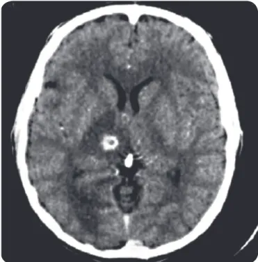
the presence of CNS lesions. The diagnosis of NPCM was made by the presence of typical neuroimaging indings (ring-like contrast enhancement lesions - Figure 1) concomitantly with fungus isolaion from other sites (lungs or skin). In three cases, diagnosis was further conirmed with neurosurgical drainage of brain lesions and fungus isolaion.
FIGURE 1 - Computed tomographic scan showing the typical ring-like appearance of a lesion located in the right thalamus (case 2).
Source: Infecious Diseases Clinic of the University Hospital of the Federal University of Minas Gerais.
The prevalence of NPCM among the observed populaion was 3.8% (95% conidence interval: 1.7-7.0). Seven paients were male, with mean age of 42 years, ranging from 24 to 61 years. One paient presented the subacute form of PCM and the other seven presented the chronic form of the disease, but all of them exhibited the parenchymatous form of NPCM.
Four paients were rural workers, two were bricklayers, one was a homemaker and there was no occupaional informaion in one case.
Six patients had previous histories of non-neurological PCM and the other two paients had their PCM diagnosis made with the neurological manifestaions. The disease was never limited to the CNS and the skin was the most frequently afected site (62.5%), followed by the lungs and lymph nodes (37.5%), and by mucous membranes (25%). Laryngeal and adrenal involvements were also observed. All the cases are summarized in Table 1.
The clinical picture was characterized especially by signs of intracranial hypertension (62.5% of the cases), and headache was
the most common complaint. Motor deicits were observed in 44.4% of the cases; seizures, sensiive and cogniive deicits were exhibited by 33.3% of them.
NPCM’s mean ime to diagnosis, deined as the period between the onset of neurological symptoms unil the search for medical atenion, was 3.8 months, ranging from one week to one year.
All paients underwent computadorized tomography scanning of the skull. Seventy-five percent of the lesions were located at the supratentorial compartment, while 50% were infratentorial. In the supratentorial compartment, the frontal lobes were the most afected sites, with 37.5% of the lesions, followed by the temporal and parietal lobes (25% each) and by the occipital lobes, basal nuclei e thalamus (12.5% each). In the infratentorial compartment, lesions were restricted to the cerebellum.
All paients were treated with more than one drug and the most used combinaion was amphotericin B with sulpha, especially the associaion sulphamethoxazole-trimethoprim (n= 6). The azolic agents were commonly used too, especially Ketoconazole (n= 4). The mean treatment ime was 4.5 years and there were no deaths. Surgical procedures (craniotomy with abscess drainage) were performed in three cases.
Approximately 63% of the paients developed sequelae in the follow-up: 60% of them remained with sensiive deicits, 40% of them, with motor deicits or seizures and 20% with gait disturbances.
Ater the characterizaion of the cases, data from NPCM paients were compared to non-neurological PCM paients.
Demographic proiles from both groups of paients were compared. No staisical diference was observed in gender distribuion or age. When considering origin, it was observed that there were 42 PCM paients who came from the same Minas Gerais state’s macro regions from which NPCM paients came. This distribuion was staisically diferent in the north/northeast region (60% of the paients who proceeded from this region (n=5) developed NPCM (n=3), p<0.001, Fisher exact test). There was also a diference in relaion to the cases which came from other Brazilian States (Bahia, Espírito Santo and Pernambuco (24 PCM cases and 3 NPCM cases), p=0.042, Fisher exact test). The two groups had similar occupaional distribuions.
Clinical aspects were also compared. The mean ime course of the disease was 11.4 months among NPCM paients and 13.83 months among PCM paients, without staisical diference. There were no diferences related to the diagnosis form either.
Many clinical signals and symptoms exhibited by the paients at the irst consultaion were registered in the research protocol form. A posiive associaion was observed between the neurological form of the disease and the presence of headache (p=0.001), sweaing (p=0.004), seizures (p=0.006), dizziness (p=0.028) and focal deicit (p=0.011).
The frequency of non-neurological affected sites was also compared between the two groups. There was no diference among all the various forms of the disease and the development of NPCM, including the mulifocal or disseminated form.
No diferences were observed when considering the paients past medical history, intravenous drug use and previous or current tobacco use or alcoholism. However, there was a posiive relaion between PCM family history and the development of NPCM (p=0.014).
Finally, no diferences related to the relapse of the disease or to the period passed unil the relapse could be observed.
Ethical consideraions
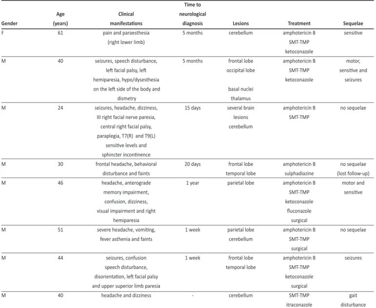
TABLE 1 - Demographic and clinical data exhibited by eight paients with neuroparacoccidioidomycosis assisted at the Infecious Clinic of the University Hospital, Federal University of Minas Gerais, Belo Horizonte, Brazil.
Time to
Age Clinical neurological
Gender (years) manifestaions diagnosis Lesions Treatment Sequelae
F 61 pain and paraesthesia 5 months cerebellum amphotericin B sensiive
(right lower limb) SMT-TMP
ketoconazole
M 40 seizures, speech disturbance, 5 months frontal lobe amphotericin B motor,
let facial palsy, let occipital lobe SMT-TMP sensiive and
hemiparesia, hypo/dysesthesia ketoconazole seizures
on the let side of the body and basal nuclei
dismetry thalamus
M 24 seizures, headache, dizziness, 15 days several brain amphotericin B no sequelae
III right facial nerve paresia, lesions SMT-TMP
central right facial palsy, cerebellum
paraplegia, T7(R) and T9(L) sensiive levels and sphincter inconinence
M 30 frontal headache, behavioral 20 days frontal lobe amphotericin B no sequelae
disturbance and faints temporal lobe sulphadiazine (lost follow-up)
M 46 headache, anterograde 1 year parietal lobe amphotericin B motor and
memory impairment, SMT-TMP sensiive
confusion, dizziness, ketoconazole
visual impairment and right luconazole
hemiparesia surgical
M 51 severe headache, vomiing, 1 week parietal lobe amphotericin B no sequelae
fever asthenia and faints cerebellum SMT-TMP
surgical
M 44 seizures, confusion 1 week frontal lobe amphotericin B seizures
speech disturbance, temporal lobe SMT-TMP
disorientaion, let facial palsy ketoconazole
and upper superior limb paresia surgical
M 40 headache and dizziness - cerebellum SMT-TMP gait
itraconazole disturbance
M: male. F: female; CT: computadorized tomography; SMT-TMP: sulphamethoxazole-trimethoprim.
DISCUSSION
This paper draws together informaion on a cohort of 213 PCM paients admited from 1976 to 2008 in the Infecious Diseases Clinic of the University Hospital of the Federal University of Minas Gerais, which is a referral center for the treatment of PCM in a notably endemic area for this disease in Brazil. Despite the methodological limitaions imposed by the retrospecive analysis of data, the great amount of informaion in this database allows valuable consideraions.
The 3.8% NPCM prevalence found in the observed populaion is below the 12.5% mean prevalence described by some Brazilian groups6,11. However, it is very close to the 3.8% prevalence reported
by a recent case series report11. Beyond that, it must be noted that
NCPM prevalence is extremely variable in the literature, ranging from 0% to 36%12-15. Interesingly, the majority (75%) of the cases reported
in this work were diagnosed ater 1990, especially ater the year 2000, and the adjusted prevalence to this period is raised to 7.7%.
This is in accordance with the growing trend of NPCM diagnosing in the last decades, which could be due to the expansion of modern neuroimaging methods4,11,16.
The majority of NPCM paients presented the chronic form of PCM, which is in accordance with the literature17,18.Moreover, all the cases
reported exhibited the parenchymatous form, mainly supratentorial. In a recent systemaic review, we found that about 98.3% of NPCM cases corresponded to the chronic form of PCM, with 89.4% of the cases exhibiing the pseudotumoral or parenchymatous form of NPCM. Approximately 66.8% of the lesions occurred in the supratentorial compartment, paricularly in the frontal and parietal lobes19.
The authors declare that there is no conlict of interest.
CONFLICT OF INTEREST
FINANCIAL SUPPORT
Epidemiological aspects evaluated between groups of paients with NPCM and PCM did not difer in relaion with gender, age or occupaional distribuion, and these were all in accordance with the literature14,20.There were no diferences related to life habits such as
the use of intravenous drug, tobacco or alcohol. Nonetheless, there was a signiicant diference (p<0.001) associated to the frequency of NPCM cases in paients proceeding from state’s of Minas Gerais North/ Northeast region and other states. This inding could simply relect a bias related to the lack of referral neurology centers in those regions, leading the paients to be referred to the capital. However, it might be interesing to speculate on other possibiliies. As a mater-of-fact, 60% of the cases originated from Minas Gerais’ North/Northeast region corresponded to NPCM cases. This region is geographically close to the States of Bahia and Espírito Santo, from where proceeded other two cases. In spite of the small number of paients observed and the limitaions of this study, this observaion could raise the hypothesis of the presence of diferent Pb strains or even Paracoccididoides species in this region which could be more prone to invade the CNS. In fact, there is a trend in the literature to deine new Paracoccidioides species, with diverse geographical distribuions and possibly diferent pathogenic paterns21.
The analysis of the clinical manifestaions showed the obvious but remarkable importance of neurologic signs in the assessment of NPCM paients, especially the presence of headache, seizures and focal deicits. The observaion of these complaints in PCM paients should readily evoke the possibility of neurological involvement and neuroimaging methods are important in idenifying CNS lesions.
It must be noted that the animycoic therapy most frequently used in our sample was Sulphamethoxazole-Trimethoprim and amphotericin B, the two drugs of choice in the treatment of NPCM20.
Furthermore, imidazole derivaives were commonly used, especially Ketoconazole (n=4). It should be noted that, with the excepion of Fluconazole, these drugs have limited penetraion in the CNS and quesionable usage in NPCM. However, despite this, we observed in a systemaic review of the literature that imidazole derivaives are frequently used in the treatment of NPCM, and Ketokonazole, Fluconazole and Itraconazole were used in 8.3%, 8.3% and 3.9% of the cases, respecively19. This could be due to issues of cost and easy access.
Another interesting fact is the association between NPCM and a posiive family PCM history. Despite the small number of paients and the resuling staisical limitaions, it is worthy of note that we describe the involvement of a mother and her son (cases 1 and 2). The literature reports other cases of NPCM afecing relaives11. Some geneic
suscepibility factors to PCM development have been described22,23, such
as the HLA-A9 (related to a unifocal pulmonary chronic form), HLA-B13 and HLA-B40. Nevertheless, these associaions were not observed among NPCM paients9. Thus, suscepibility mechanisms which lead to CNS
infecion should depend on the interacion between fungal and host’s factors determining the parasite’s penetraion into the nervous issue.
Importantly, there were no signiicant diferences related to disease ime course between paients with or without neurological infecion. This fact suggests that there is not an associaion between CNS involvement and a longer course of the systemic disease. There were no noiceable diferences also regarding the involvement of non-neurological sites between PCM and NPCM paients. In contrast with some works in the literature, there were no diferences even in regard to the mulifocal form of the disease. As menioned, it has
been suggested that the penetraion of the fungus in the CNS could be related with some speciic clinical aspects, paricularly severe immunosuppression and intense disease disseminaion5. However,
this was not conirmed in our sample. Thus, our data show that the neurological form was not related to disseminaion or to a longer course of disease, and that there could be other factors than simply immunosuppression that could be involved in CNS infecion, as speciic parasite’s or host’s characterisics.
To conclude, the demographic and clinical data of the eight NPCM cases reported are similar to those generally atributed to this form of the disease by the literature. Early diagnosis and treatment showed to be fundamental. In comparison to paients without CNS involvement, it could be observed the great importance of neurological examinaion and neuroimaging. The development of NPCM was not related to prolonged disease course or dissemination and the differences regarding family history and origin, even with limitaions, could point to the need of further studies in order to determine suscepibility factors involved in the neurological infecion.
Conselho Nacional de Desenvolvimento Cieníico e Tecnológico
(CNPq).
ABSTRACT IN PORTUgUESE
Série de casos de paracoccidioidomicose com e sem envolvimento do sistema nervoso central
Introdução: A paracoccidioidomicose (PCM) é a micose profunda mais importante na América do Sul. O comprometimento do sistema nervoso central é grave e pode ocorrer em 12,5% dos casos. Este trabalho tem como objeivo descrever oito casos de neuroparacoccidioidomicose (NPMC) e comparar suas caracterísicas com pacientes sem envolvimento neurológico, a im de ideniicar aspectos singulares da NPCM. Métodos: Uma coorte de 213 casos de PCM foi avaliada na Clínica de Doenças Infecciosas do Hospital das Clínicas da Universidade Federal de Minas Gerais, de outubro de 1976 a agosto de 2008. Dados epidemiológicos, clínicos, laboratoriais, terapêuicos e de seguimento foram registrados. Resultados: Oito pacientes apresentaram NPCM. A prevalência de NPCM observada foi de 3,8%. Um paciente apresentou a forma subaguda da PCM e sete apresentaram a forma crônica. Todos os pacientes apresentaram a forma parenquimatosa. Cerca de 60% dos pacientes provenientes das regiões norte e nordeste de Minas Gerais desenvolveram NPCM. Foi observado o desenvolvimento de NPCM em uma mãe e em seu ilho. Os pacientes com NPCM apresentaram peris demográicos e clínicos similares à descrição da literatura. Quando comparados aos pacientes com PCM, houve diferenças em relação à procedência de tais pacientes e história familiar positiva de PCM. Conclusões: Os resultados conirmam a importância da avaliação neurológica em pacientes com PCM. Apesar das limitações desse trabalho, as diferenças com relação à procedência dos pacientes e à história familiar apontam para a necessidade de mais estudos para invesigar a existência de fatores de suscepibilidade envolvidos no desenvolvimento da NPCM.
REFERENCES
1. McEwen JG, Garcia AM, Oriz BL, Botero S, Restrepo A. In search of the natural habitat of Paracoccidioides brasiliensis. Arch Med Res 1995; 26:305-306. 2. Mendes RP. Paracoccidioidomicose (Blastomicose Sul-Americana). In: Veronesi R,
Focaccia R, editors. Tratado de Infectologia. 3rd ed. São Paulo: Atheneu; 2005. p.
1383-1390.
3. Shikanai-Yasuda MA, Telles Filho F, Mendes RP, Colombo AL, Morei ML. Guidelines in paracoccidioidomycosis. Rev Soc Bras Med Trop 2006; 39:297-310.
4. Fagundes-Pereyra WJ, Carvalho GTC, Góes AM, Silva FCL, Sousa AA. Paracoccidioidomicose do sistema nervoso central: análise de 13 casos. Arq Neuropsiquiatr 2006; 64:269-276.
5. Paniago AM, Oliveira PA, Aguiar ES, Aguiar JI, Cunha RV, Leme LM, et al. Neuroparacoccidioidomycosis: analysis of 13 cases observed in an endemic area in Brazil. Trans R Soc Trop Med Hyg 2007; 101:414-420.
6. Hutzler RU, Brussi MLP, Capitani CM, Lima SS. Acomeimento neurológico da paracoccidioidomicose, avaliado pela tomograia computadorizada de crânio. São Paulo Med J 1985; 103:243-244.
7. Silva SHM, Colombo AL, Blotta MHSL, Lopes JD, Queiroz-Telles F, Camargo ZP. Detection of circulating gp43 antigen in serum, cerebrospinal fluid and bronchoalveolar lavage luid of paients with paracoccidioidomycosis. J Clin Microbiol 2003; 41:3675-3680.
8. Silva SHM, Grosso DM, Lopes JD, Colombo AL, Blotta MH, Queiroz-Telles F, et al. Detecion of Paracoccidioides brasiliensis gp70 circulaing anigen and follow-up of paients undergoing animycoic therapy. J Clin Microbiol 2004; 42:4480-4486.
9. Almeida SM, Rebelato CLK, Queiroz-Telles F, Werneck LC. Major histocompaibility complex and central nervous system involvement by paracoccidioidomycosis. J Infect 2005; 51:140-143.
10. Reis BS, Bozzi A, Prado FLS, Pereira MC, Ferreira FE, Godoy P, et al. Membrane and extracellular anigens of Paracoccidioides brasiliensis (Mexo): Ideniicaion of a 28-kDa protein suitable for immunodiagnosis of paracoccidioidomycosis. J Immunol Methods 2005; 307:118-126.
11. Almeida SM. Central nervous system paracoccidioidomycosis: an overview. Braz J Infect Dis 2005; 9:126-133.
12. Francesconi F, Silva MT, Costa RL, Francesconi VA, Carregal E, Talhari S, et al. Long-term outcome of neuroparacoccidioidomycosis treatment. Rev Soc Bras Med Trop 2011; 44:22-25.
13. Pereira WC, Raphael A, Sallum J. Lesões neurológicas na blastomicose sul-americana: estudo anatomopatológico de 14 casos. Arq Neuropsiquiatr 1965; 23:95-112.
14. Plá MP, Hartung C, Mendoza P, Stukanof A, Moreno MJ. Neuroparacoccidioidomycosis: case reports and review. Mycopathologia 1994; 127:139-144.
15. Raphael A. Localização nervosa da blastomicose sul-americana. Arq Neuropsiquiatr 1966; 24:69-90.
16. Tristano AG, Chollet ME, Willson M, Perez J, Troccoli M. Central nervous system Paracoccidioidomycosis: case report and review. Invest Clín 2004; 45:277-288. 17. Gaspareto EL, Liu CB, Carvalho Neto A, Rogacheski E. Central nervous system
paracoccidioidomycosis: imaging indings in 17 cases. J Comput Assist Tomogr 2003; 27:12-17.
18. Pereira WC, Raphael A, Tenuto RA, Sallum J. Localização encefálica da blastomicose sul-americana: considerações a propósito de 9 casos. Arq Neuropsiquiatr 1965; 23:113-126.
19. Pedroso VS, Vilela M, Pedroso ER, Teixeira AL. Paracoccidioidomycosis compromising the central nervous system: a systemaic review of the literature. Rev Soc Bras Med Trop 2009; 42:691-697.
20. Pedroso VSP, Vilela MC, Pedroso ERP, Teixeira AL. Paracoccidioidomicose com compromeimento do sistema nervoso central: revisão de literatura. Rev Bras Neurol 2008; 44:33-40.
21. Teixeira MM, Theodoro RC, Carvalho MJ, Fernandes L, Paes HC, Hahn RC, et al. Phylogeneic analysis reveals a high level of speciaion in the Paracoccidioides genus. Mol Phylogenet Evol 2009; 52:273-283.
22. Lacerda GB, Arce-Gomez B, Telles Filho FQ. Increased frequency of HLA-B40 in paients with Paracoccidioidomycosis. J Med Vet Mycol 1988; 26:253-256. 23. Restrepo FM, Restrepo M, Restrepo A. Blood groups and HLA antigens in