Emel KARAMAN Nihan GÖNÜLOL
Department of Restorative Dentistry, Faculty of Dentistry, Ondokuz Mayıs University, Samsun, Turkey.
Does the light source affect the
repairability of composite resins?
Abstract: The aim of this study was to examine the effect of the light source on the microshear bond strength of different composite resins repaired with the same substrate. Thirty cylindrical specimens of each composite resin—Filtek Silorane, Filtek Z550 (3M ESPE), Gradia Direct Anterior (GC), and Aelite Posterior (BISCO)—were prepared and light-cured with a QTH light curing unit (LCU). The specimens were aged by thermal cycling and divided into three subgroups according to the light source used—QTH, LED, or PAC (n = 10). They were repaired with
the same substrate and a Clearil Repair Kit (Kuraray). The specimens
were light-cured and aged for 1 week in distilled water at 37 °C. The microshear bond strength and failure modes were assessed. There was
no signiicant difference in the microshear bond strength values among
the composite resins, except for the Filtek Silorane group that showed
signiicantly lower bond strength values when polymerized with the
PAC unit compared to the QTH or LED unit. In conclusion, previously placed dimethacrylate-based composites can be repaired with different light sources; however, if the composite to be repaired is silorane-based, then using a QTH or LED device may be the best option.
Keywords: Composite Resins; Dental Debonding; Dental Restoration Failure.
Introduction
Despite their continued development, composite resins can
demon-strate degradation in the oral environment over time. Marginal deicien -cies, fracture, and wear are the main reasons for deterioration, which can
lead to secondary caries or tooth sensitivity.1,2 The traditional treatment
of defective composite restorations, including removing and replacing complete restorations, is not desirable because this approach widens the prepared cavity, results in greater loss of sound tooth structure, and
may lead to pulpal symptoms.3 As a conservative alternative to complete
replacement, the repair of preexisting restorations has become an impor-tant treatment option in modern dentistry. This technique can increase the longevity of the restoration, avoid unnecessary removal of sound tooth
tissues, and reduce repeated irritations or injuries to the pulp.4
In response to the dramatic rise in the use of composite resins, there
has been substantial scientiic interest in polymerization. Many light-cur
-ing units (LCUs) have been developed.5,6 Until recently,
quartz-tungsten-halogen (QTH) LCUs were primarily used to polymerize composite res
-Declaration of Interests: The authors certify that they have no commercial or associative interest that represents a conflict of interest in connection with the manuscript.
Corresponding Author:
Emel Karaman
E-mail: [email protected]
DOI: 10.1590/1807-3107BOR-2014.vol28.0027 Epub XXX XX, 2014
Submitted: Sep 04, 2013
ins. However, despite their common use, QTH units have some shortcomings, such as the limited lifes-pan of halogen bulbs (40-100 h) and degradation of
the components (bulb, relector, and ilter) over time
due to the high operating temperature.7
To overcome some of these shortcomings, the use of light-emitting solid-state diode (LED) technology
was proposed in the mid-1990s.8 Instead of the hot
ilaments used in halogen bulbs, LEDs use junctions
of doped semiconductors to generate light. LEDs do
not require ilters as the spectral output because the
blue LED falls within the absorption spectrum of the camphorquinone photoinitiator (400-500 nm). LEDs have an expected lifetime of more than 10,000 h,
dur-ing which the light lux undergoes little degradation.9
The degree of monomer conversion produced by a blue
LED source was reported to be signiicantly higher
than that produced by a QTH source, even when all sources were adjusted to produce the same irradiance
(100 mW/cm2).10 Plasma arc light-curing units (PAC
units) are another choice. PAC light is emitted from glowing plasma, which is composed of a gaseous
mix-ture of ionized molecules (e.g., xenon) and electrons.11
Due to their higher light intensity, PAC lights cure com-posite resins at a much faster rate than conventional
QTH LCUs and may be a time-saving alternative.12
Several recent in vitro studies have analyzed the
effects of different adhesive intermediates, surface preparation methods, and combinations of composite
resins on the reparability of composite restorations.13,14,15
However, to the best of the authors’ knowledge, no study has evaluated the effect of the light source used
on composite repair. Thus, the aim of this in vitro study
was to evaluate the effect of different light sources on the microshear bond strength of aged composite repairs when the same substrate was used. The null hypothesis tested was that the type of light source does not affect the microshear bond strength.
Methodology
Preparation of Aged Composite Specimens
The composite resins used in the current study are shown in Table 1. For each composite material, 30 cylin-drical specimens (10 mm diameter × 2 mm thickness)
were prepared in Telon ring molds. Each mold was
positioned on a glass microscope slide before being
illed with composite resin and covered with a Mylar strip. A second microscope slide was pressed irmly
onto the composite to remove excess material. All of the composite specimens were bulk-cured without
layering by a QTH LCU (intensity = 650 mW/cm2),
according to the manufacturers’ instructions.
Polym-erized specimens were stored in distilled water at
37 °C, to replicate the oral condition, for 24 h. There-after, they were polished with a series of aluminum oxide polishing discs (Sof-Lex, 3M ESPE, Dental Products) under constant water cooling.
Specimens were aged by thermal cycling for 5,000 cycles between two water baths maintained at
55 ± 1 and 5 ± 1 °C.16 All specimens were embedded
in autopolymerizing acrylic resin. Before the repair
procedure, they were roughened with 320-grit sili-con carbide paper, to obtain a similar roughness as that obtained by diamond bur grinding.
Repair Procedure
Specimens were randomly divided into three
sub-groups (n = 10), according to the LCU used: QTH, LED,
or PAC (Table 2). In all cases, the LCUs were used in the standard mode and positioned 1 mm above the
com-posite resin surface with a metal ring. A Clearil Repair Kit (Kuraray Medical, Inc., Okayama, Japan) was used
as an intermediate agent in all study groups. Before
bonding, Ketchant gel (40% phosphoric acid gel) was
applied for 10 s, followed by rinsing with water and
air-drying for 20 s. A 1:1 mix of porcelain bond activator and Clearil SE Bond primer was applied, followed by
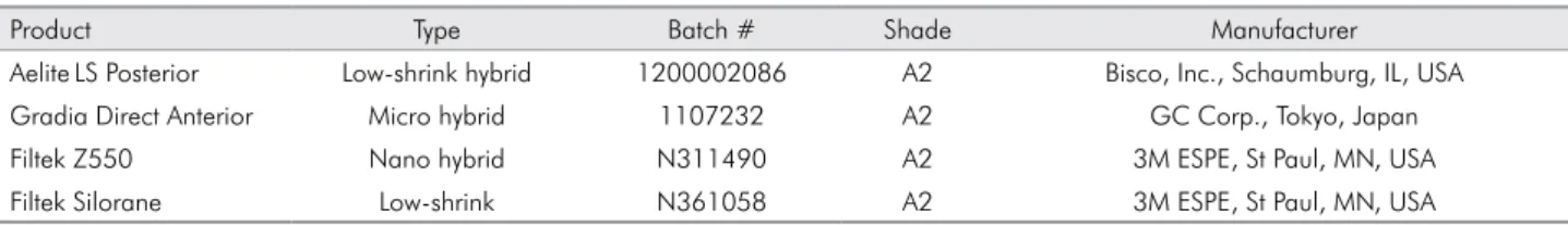
Table 1. Composite Resins Used in the Current Study.
Product Type Batch # Shade Manufacturer
AeliteLS Posterior Low-shrink hybrid 1200002086 A2 Bisco, Inc., Schaumburg, IL, USA
Gradia Direct Anterior Micro hybrid 1107232 A2 GC Corp., Tokyo, Japan
Filtek Z550 Nano hybrid N311490 A2 3M ESPE, St Paul, MN, USA
a thin layer of Clearil SE Bond. Asilicone tube (0.8 mm
internal diameter × 0.5 mm height) was placed on the bonded area and light-cured. The adhesive resin was
light-cured, and then the tubes were illed with com
-posite resin and polymerized as described in Table 2.
Specimens were stored in distilled water at 37°C for 1 week before the microshear bond test was performed.
Microshear Bond Strengths and Failure Analysis
The bond strength was tested with a universal
test-ing machine (LRX, Lloyd Instruments, Ametek Inc., Leicester, UK) at a crosshead speed of 1.0 mm/min.
Load at debonding was recorded in Newtons (N). The microshear bond strength was calculated by dividing
the load at debonding by the bonded area (mm2). For
failure mode analysis, the debonded area was examined
by stereomicroscopy at 40 × magniication. The failure mode was classiied as adhesive (if failure occurred at
the interface), cohesive (if failure affected at least parts
of the substrate or the repair composite), or mixed.17
Statistical Analysis
The Shapiro-Wilk test was used to verify the normal
distribution of the data. Then,one-way analysis of
vari-ance (ANOVA) was performed in a completely
random-ized design, Ŷij = μ + αi + eij, where Ŷij is the observation
value (MPa), μ is the overall mean, αi is the effect of the
treatment (composite or light source), and eijis the
resid-ual error. The Tukey multiple range test was utilized to
separate these differences. All computational work was performed in MINITAB (Minitab V. 13.20, 2000).
Chi-squared (χ2) analysis was applied to analyze
whether the failure mode statistically depended on the composite resin or light source. If a relationship
was identiied, then contingency coeficients (%) for
each contingency table were calculated to determine the degree of association between the failure mode and the composite resin or light source. The Z-test
was utilized to determine any further association.
Results
Results of the microshear bond strength tests are summarized in Table 3. The Filtek Z550 group
showed the highest bond strength values when the
QTH source was used for polymerization, followed
by the PAC and LED sources, albeit without
statis-tical signiicance among the subgroups. Similarly,
in the Gradia Direct Anterior group, the QTH and LED sources produced the highest and lowest bond strength values, respectively, but the difference
between them was not signiicant. All three light
sources produced similar bond strength values for the Aelite group. PAC resulted in the lowest bond strength values in the Filtek Silorane group among
the light sources used (p < 0.001).
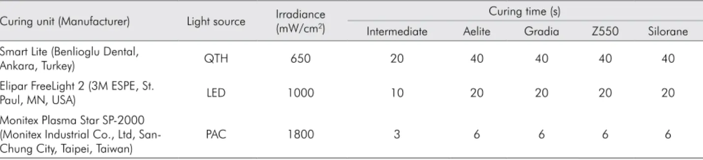
Table 2. Light-Curing Units and Curing Times Used in the Current Study.
Curing unit (Manufacturer) Light source Irradiance (mW/cm2)
Curing time (s)
Intermediate Aelite Gradia Z550 Silorane
Smart Lite (Benlioglu Dental,
Ankara, Turkey) QTH 650 20 40 40 40 40
Elipar FreeLight 2 (3M ESPE, St.
Paul, MN, USA) LED 1000 10 20 20 20 20
Monitex Plasma Star SP-2000 (Monitex Industrial Co., Ltd, San-Chung City, Taipei, Taiwan)
PAC 1800 3 6 6 6 6
Table 3. Mean and Standard Deviation Values of Micros-hear Bond Strength(MPa)for Each Group.
Group QTH LED PAC p-value
Z550 23.51A,a (6.72)
18.74AB,a (4.99)
22.00A,a (4.03)
0.112
Aelite 19.79A,a (3.43)
19.97AB,a (4.73)
17.60A,a (6.10)
0.492
Silorane 23.10A,a (5.24)
24.09A,a (4.96)
8.49B,b (7.06)
< 0.001
Gradia 21.72A,a (3.06)
17.10B,a (5.19)
19.85A,a (5.25)
0.098
p-value 0.263 0.022 < 0.001
No signiicant difference was found when com -paring the bond strength values of different
compos-ite resins polymerized with QTH. The bond strength values of LED-polymerized Filtek Silorane and Gradia Direct Anterior were signiicantly different from each
other (p = 0.022). Polymerization with PAC caused
the lowest bond strength values for Filtek Silorane
among the groups (p < 0.001).
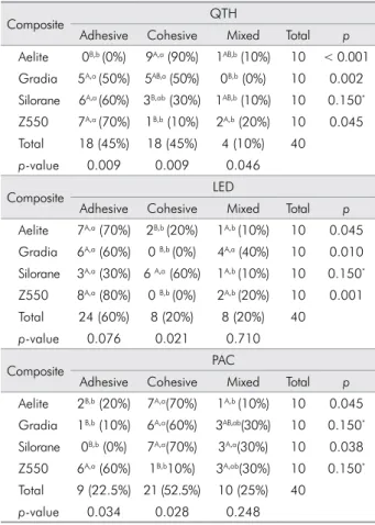
Table 4 shows the distribution of failure modes for all groups. For the Aelite group, the QTH and PAC sub-groups showed increased numbers of cohesive failures
(90% and 70%, respectively), and the LED subgroup showed an increased number of adhesive failures (70%). For the Gradia group, polymerization with QTH did not result in a signiicant difference in the number of adhesive or cohesive failures; however, polymerization
with LED or PAC resulted in an increased number of
adhesive or cohesive failures, respectively (both 60%). For the Filtek Silorane group, polymerization with
QTH resulted in an increased number of adhesive
fail-ures (60%), whereas polymerization with LED or PAC
resulted in an increased number of cohesive failures
(60% or 70%, respectively). For Filtek Z550, the number
of adhesive failures was greater than the number of cohesive or mixed failures in all subgroups.
When the failure modes of all specimens were evaluated, the QTH source exhibited the same
num-bers of adhesive and cohesive failures (45%). Adhesive
failures were more frequent when the LED source
was used (60%), and cohesive failures were more fre
-quent when the PAC source was used (52.5%).
Discussion
As an alternative to total replacement, the repair of existing restorations can enhance the longevity of
dental restorations.18 However, many factors affect the
durability of composite-to-composite bond repair.13,14,15
This study aimed to evaluate the effect of different light sources on the reparability of one silorane-based and three dimethacrylate-based composite resins.
Polym-erization with different LCUs resulted in similar bond
strength values for methacrylate-based composite res-ins, but differing values for the silorane-based com-posite resin. Thus, the null hypothesis was rejected. These results may be helpful for choosing an LCU for repairing composite restorations in everyday
sit-uations, wherein it is extremely dificult for the den -tist to know whether the restoration to be repaired is methacrylate- or silorane-based.
Specimens in this study were aged with thermal
cycling. Aging is common for composite repair tests17,19
because repairs usually become necessary months or years after a restoration is placed. The thermal cycling-induced temperature alterations may affect the com-posite-to-composite repair strength by decreasing the number of unreacted double bonds on the surface or
within the composite.16 The success of the repair
pro-cess is affected by various factors, such as the inter-mediary or repair material used, and the time after
repair.20 To focus on the effect of the light source, a
uni-form repair process was chosen. The Clearil Repair Kit was used for repair, and the aged composite sam -ples were repaired with the same substrate.
Table 4. Distribution of Failure Modes
Composite QTH
Adhesive Cohesive Mixed Total p
Aelite 0B,b (0%) 9A,a (90%) 1AB,b (10%) 10 < 0.001 Gradia 5A,a (50%) 5AB,a (50%) 0B,b (0%) 10 0.002 Silorane 6A,a (60%) 3B,ab (30%) 1AB,b (10%) 10 0.150* Z550 7A,a (70%) 1B,b (10%) 2A,b (20%) 10 0.045 Total 18 (45%) 18 (45%) 4 (10%) 40
p-value 0.009 0.009 0.046
Composite LED
Adhesive Cohesive Mixed Total p
Aelite 7A,a (70%) 2B,b (20%) 1A,b (10%) 10 0.045 Gradia 6A,a (60%) 0 B,b (0%) 4A,a (40%) 10 0.010 Silorane 3A,a (30%) 6 A,a (60%) 1A,b (10%) 10 0.150* Z550 8A,a (80%) 0 B,b (0%) 2A,b (20%) 10 0.001 Total 24 (60%) 8 (20%) 8 (20%) 40
p-value 0.076 0.021 0.710
Composite PAC
Adhesive Cohesive Mixed Total p
Aelite 2B,b (20%) 7A,a(70%) 1A,b (10%) 10 0.045 Gradia 1B,b (10%) 6A,a(60%) 3AB,ab(30%) 10 0.150* Silorane 0B,b (0%) 7A,a(70%) 3A,a(30%) 10 0.038 Z550 6A,a (60%) 1B,b10%) 3A,ab(30%) 10 0.150* Total 9 (22.5%) 21 (52.5%) 10 (25%) 40
p-value 0.034 0.028 0.248
Using different LCUs, resin composite thicknesses, and light exposure distances may change the physi-cal properties and mechaniphysi-cal behavior of restorative
materials.21 Depth of cure and microhardness tests
have been widely used to assess the relative degree of
cure of resins and, thus, the eficiency of light sources. Rode et al.21 reported no difference in microhardness
up to 2 mm thickness when LED exposure distances
of 0 and 3 mm were used. In Abate et al.,22 the distance
between the light source and the composite surface did not affect the hardness results when a QTH LCU was used. A common clinical recommendation for
the position of the LCU tip is 1 mm from the resin.23
Thus, this guideline was applied in the current study. The microshear bond strength test was accompa-nied by an analysis of failure modes. Similar bond strength values for repair were observed for the com-posite resins when the QTH was used, although their
failure modes were signiicantly different. Using the LED resulted in signiicantly different bond strength
values, but the numbers of adhesive and mixed fail-ures of the groups were similar. It can be concluded that groups with similar bond strength values do not necessarily fail in the same way. Observed dif-ferences in the failure mode among the composite resins might be explained by their differences in
lexural strength and elastic modulus, which have
been suggested to affect the bond strength and
fail-ure modes of repaired restorations.24
The PAC LCU saved time, while still achieving
suficient bond strength for methacrylate-based com -posite resins. The observed lower bond strength of
PAC-polymerized Filtek Silorane may be attributed
to the chemistry of this composite. Curing with PAC may not be adequate for all restorations because
rapid polymerization may hinder the development
of optimal properties in some materials.25 Kim et al.26
suggested a relationship between bond strength and
subsurface polymerization. Guiraldo et al.27 found that Filtek P90 did not present eficient polymerization in its deepest layers when polymerized with a QTH
LCU (900 mW/cm2). They reported that the degree
of subsurface polymerization is greater for methac -rylate- than for silorane-based composites, and they recommended increasing the exposure time or using LCUs with greater irradiance to obtain better results
for silorane-based composites. Accordingly, the lower bond strength values and greater number of cohesive failures observed in the Filtek Silorane group may
be attributed to the rapid polymerization method of
PAC, despite its high irradiance.
No previous study has compared the repair bond strength values among different composite resins
polymerized with different LCUs. However, the effect
of LCUs on dentin bond strength was investigated
in several studies, with conlicting results.28,29,30 For
instance, D’Alpino et al.28 concluded that different
LCUs inluence the restoration bond strength, whereas
Amaral et al.29 reported that the LCU and method do
not signiicantly affect bond strength. Khosla et al.30
evaluated the effects of QTH and LED on the shear bond strengths of silorane- and bis-GMA–based
com-posite resins. The type of LCU did not signiicantly
affect the shear bond strength for bis-GMA–based composite resins when the total-etch technique was used, but the QTH showed the best results for curing silorane-based composites. Nevertheless, the results of these previous studies cannot be compared directly with the data obtained in the present study because those studies evaluated the bond strength to dentine.
In the current study, only one intermediate agent
and one polymerization mode were tested. Further
research involving the use of different intermediate
agents and multiple combinations of polymerization
modes is warranted.
Conclusions
In view of the methodology used and the results
obtained, it can be concluded that:
1. Currently available LCUs (QTH, LED, and PAC)
promote similar bond strength values in the re-pair of dimethacrylate-based composites.
2. In the repair of the silorane-based composite,
PAC gave the lowest bond strength values; thus, using a QTH or LED device may be the best op-tion for this type of composite.
3. In many cases, dentists do not know the
1. Manhart J, Chen H, Hamm G, Hickel R. Buonocore Memorial
Lecture. Review of the clinical survival of direct and indirect
restorations in posterior teeth of the permanent dentition.
Oper Dent. 2004 Sep-Oct;29(5):481-508.
2. Hickel R, Manhart J. Longevity of restorations in pos
-terior teeth and reasons for failure. J Adhes Dent. 2001 Spring;3(1):45-64.
3. Gordan VV, Garvan CW, Blaser PK, Mondragon E, Mjor IA. A long-term evaluation of alternative treatments to replacement
of resin-based composite restorations: results of a seven-year study. J Am Dent Assoc. 2009 Dec;140(12):1476-84.
4. Foitzik M, Attin T. Filling revision--possibilities and execu
-tion. Schweiz Monatsschr Zahnmed. 2004;114(10):1003-11.
5. Peris AR, Mitsui FH, Amaral CM, Ambrosano GM, Pimenta LA. The effect of composite type on microhardness when
using quartz-tungsten-halogen (QTH) or LED lights. Oper Dent. 2005 Sep-Oct;30(5):649-54.
6. Caldas DB, Almeida JB, Correr-Sobrinho L, Sinhoreti MA, Consani S. Influence of curing tip distance on resin
com-posite Knoop hardness number, using three different light curing units. Oper Dent. 2003 May-Jun;28(3):315-20.
7. Barghi N, Berry T, Hatton C. Evaluating intensity output
of curing lights in private dental offices. J Am Dent Assoc. 1994 Jul;125(7):992-6.
8. Franco EB, Santos PA, Mondelli RF. The effect of different
light-curing units on tensile strength and microhardness
of a composite resin. J Appl Oral Sci. 2007 Dec;15(6):470-4.
9. Oesterle LJ, Newman SM, Shellhart WC. Rapid curing of
bonding composite with a xenon plasma arc light. Am J Or
-thod Dentofacial Orthop. 2001 Jun;119(6):610-6.
10. Jandt KD, Mills RW, Blackwell GB, Ashworth SH. Depth of cure and compressive strength of dental composites cured with blue
light emitting diodes (LEDs). Dent Mater. 2000 Jan;16(1):41-7.
11. Peutzfeldt A, Sahafi A, Asmussen E. Characterization of resin
composites polymerized with plasma arc curing units. Dent Mater. 2000 Nov;16(5):330-6.
12. Campbell IM. Introduction to synthetic polymers. Oxford: Science Publications; 1994. 213 p.
13. Tezvergil A, Lassila LV, Vallittu PK. Composite-composite
repair bond strength: effect of different adhesion primers. J Dent. 2003 Nov;31(8):521-5.
14. Rathke A, Tymina Y, Haller B. Effect of different surface treatments on the composite-composite repair bond strength.
Clin Oral Investig. 2009 Set;13(3):317-23.
15. Bonstein T, Garlapo D, Donarummo J Jr, Bush PJ. Evaluation
of varied repair protocols applied to aged composite resin. J Adhes Dent. 2005 Spring;7(1):41-9.
16. Ozcan M, Barbosa SH, Melo RM, Galhano GA, Bottino MA. Effect of surface conditioning methods on the microtensile bond strength of resin composite to composite after aging
conditions. Dent Mater. 2007 Oct;23(10):1276-82.
17. Wiegand A, Stawarczyk B, Buchalla W, Tauböck TT, Özcan
M, Attin T. Repair of silorane composite--using the same
substrate or a methacrylate-based composite?. Dent Mater.
2012 Mar;28(3):e19-25.
18. Opdam NJ, Bronkhorst EM, Loomans BA, Huysmans MC. Longevity of repaired restorations: a practice based study. J Dent. 2012 Oct;40(10):829-35.
19. Rinastiti M, Ozcan M, Siswomihardjo W, Busscher HJ. Ef -fects of surface conditioning on repair bond strengths of non-aged and aged microhybrid, nanohybrid, and nanofilled
composite resins. Clin Oral Investig. 2011 Oct;15(5):625-33.
20. Shahdad SA, Kennedy JG. Bond strength of repaired anterior
composite resins: an in vitro study. J Dent. 1998 Nov;26(8):685-94.
21. Rode KM, Kawano Y, Turbino ML. Evaluation of curing light
distance on resin composite microhardness and polymeriza
-tion. Oper Dent. 2007 Nov-Dec;32(6):571-8.
22. Abate PF, Zahra VN, Macchi RL. Effect of photopolymeriza
-tion variables on composite hardness. J Prosthet Dent. 2001 Dec;86(6):632-5.
23. Sobrinho LC, Lima AA, Consani S, Sinhoreti MA, Knowles
JC. Influence of curing tip distance on composite Knoop hardness values. Braz Dent J. 2000;11(1):11-7.
24. Boaro LC, Goncalves F, Guimaraes TC, Ferracane JL, Versluis
A, Braga RR. Polymerization stress, shrinkage and elastic
modulus of current low-shrinkage restorative composites.
Dental Mater. 2010 Dec;26(12):1144-50.
25. Deb S, Sehmi H. A comparative study of the properties of
dental resin composites polymerized with plasma and halo
-gen light. Dental Mater. 2003 Sep;19(6):517-22.
26. Kim JS, Choi YH, Cho BH, Son HH, Lee IB, Um CM, et al. Effect of light-cure time of adhesive resin on the thickness of the oxygen-inhibited layer and the microtensile bond
strength to dentin. J Biomed Mater Res B Appl Biomater. 2006 Jul;78(1):115-23.
27. Guiraldo RD, Consani S, Consani RL, Berger SB, Mendes WB, Sinhoreti MA, et al. Comparison of silorane and methacry-late-based composite resins on the curing light transmission.
Braz Dent J. 2010;21(6):538-42.
28. D’Alpino PH, Wang L, Rueggeberg FA, Svizero NR, Pereira JC, Pashley DH, et al. Bond strength of resin-based resto
-rations polymerized with different light-curing sources. J Adhes Dent. 2006 Oct;8(5):293-8.
29. Amaral CM, Peris AR, Ambrosano GM, Swift EJ Jr, Pimenta LA. The effect of light-curing source and mode on
micro-tensile bond strength to bovine dentin. J Adhes Dent. 2006 Feb;8(1):41-5.
30. Khosla M, Malhotra N, Mala K. An in vitro evaluation of shear bond strength of silorane and bis-GMA resin-based
composite using different curing units. J Conserv Dent. 2012;15(3):278-82.