ABSTRACT: Although the inluence of diabetes on salivary glands is well studied, it still presents conlicting re -sults. In this work, the regulation of the phosphofructokinase-1 enzyme (PFK-1) was studied utilizing the salivary glands of rats. Diabetes was induced by a single intraperitoneal injection of streptozotocin (60 mg/Kg of body weight) in rats (180-200 g). The animals were killed 30 days after the induction of diabetes and the submandibular and parotid salivary glands were used. Hyperglycemia was evaluated by blood sugar determination. The distri-bution of PFK-1 between the soluble and cytoskeleton fractions, the phosphate content of PFK-1, the content of fructose-2,6-bisphosphate and the activity of the PFK-2 enzyme were determined. The calculated relative glandular weight showed a higher value for the parotid gland in comparison with the control, but not for the submandibular gland. The activity of PFK-1 expressed per gland showed no variation between diabetic and control animals. How-ever, considering the speciic activity, the soluble enzyme presented a value 50% higher than that of the control and the cytoskeleton bound form increased by 84% compared to the control. For the parotid gland, no difference in the speciic activity between diabetic and control animals was observed. On the other hand, the activity per gland of the soluble enzyme increased in the diabetic animals. The phosphate content of PFK-1 increased in the submandibular and parotid glands of diabetic rats. Both the content of fructose-2,6-bisphosphate and the active form of PFK-2 were reduced in the diabetic glands. In conclusion, the increase in the activity of PFK-1 observed in the salivary glands of rats with streptozotocin-induced diabetes does not seem to be due to its modulator fructose-2,6-bisphosphate.
DESCRIPTORS: Diabetes mellitus; Phosphofructokinase-1; Salivary glands; Parotid gland; Submandibular gland. RESUMO: Apesar de existirem muitos estudos sobre a inluência do diabetes nas glândulas salivares, esses apre -sentam resultados conlitantes. Neste estudo, a regulação da enzima fosfofrutoquinase-1 (PFK-1) foi estudada utilizando-se glândulas salivares de ratos. O diabetes foi induzido por uma única injeção intraperitonial de estrep-tozotocina (60 mg/kg peso corporal) em ratos (180-200 g). Os animais foram sacriicados 30 dias após a indução do diabetes e utilizaram-se as glândulas submandibular e parótida. A hiperglicemia foi avaliada por determinação da glicemia sanguínea. A distribuição da 1 entre frações solúvel e ligada, concentração de fosfato na PFK-1, concentração de frutose-2,6-bisfosfato e a atividade da enzima PFK-2 foram determinadas. O cálculo do peso glandular relativo mostrou um aumento na glândula parótida de ratos diabéticos comparados ao controle, o que não ocorreu na glândula submandibular. A atividade da PFK-1 expressa por glândula não mostrou variação entre animais diabético e controle. Contudo, considerando a atividade especíica, a fração solúvel da enzima mostrou aumento de 50% com relação ao controle e a fração ligada ao citoesqueleto um aumento de 84% com relação ao controle. Na glândula parótida não foi observada diferença na atividade especíica entre os grupos diabético e controle. Por outro lado, a atividade por glândula da fração solúvel aumentou nos animais diabéticos. A concen-tração de fosfato da PFK-1 aumentou nas glândulas submandibular e parótida nos animais diabéticos. Tanto a concentração de frutose-2,6-bisfosfato quanto a forma ativa da PFK-2 mostraram redução nas glândulas salivares. Concluindo, o aumento na atividade da PFK-1 observado nas glândulas salivares de ratos com diabetes induzida por estreptozotocina não parece ser modulado pela frutose-2,6-bisfosfato.
DESCRITORES: Diabetes mellitus; Fosfofrutoquinase-1; Glândulas salivares; Glândula parótida; Glândula sub-mandibular.
* PhDs, Professors; **Chemist – Oral Biology Research Center, School of Dentistry, University of São Paulo.
Activity, distribution and regulation of phosphofructokinase in
salivary gland of rats with streptozotocin-induced diabetes
Atividade, distribuição e regulação da fosfofrutoquinase
em glândulas salivares de ratos com diabetes induzido por
estreptozotocina
José Nicolau*
INTRODUCTION
Diabetes mellitus is a metabolic disease that affects many organs and systems, including the oral cavity. Some investigations have described the effects of experimental diabetes induced either by streptozotocin or alloxan in the structure and functions of the salivary glands of animals2,24. It has been reported that diabetes decreases norepi-nephrine content, the density of adrenergic recep-tor and receprecep-tor-adenyl cyclase coupling in parotid glands14, as well as salivary secretion closely asso-ciated with the lowered susceptibility of the mus-carinic receptors30. Recently it was reported that isolated parotid gland from streptozotocin-induced diabetic rats presented a dose-dependent decrease in amylase release in response to noradrenaline when compared to control parotid gland17.
The role of PFK-1 in the regulation of glycolysis has been established for a variety of cell types in various animals29. The activity of this enzyme is controlled by multiple positive and negative allo-steric factors such as ATP, ADP, AMP, fructose-2,6-bisphosphate (Fru-2,6-P2) and citrate. Fru-2,6-P2 is a potent stimulator of PFK-1 and has been detected in all mammalian tissues. The concentration of Fru-2,6-P2 is the result of a balance between the activity of PFK-2/FBP-2 as kinase and as phos-phatase. Similar to other glycolytic enzymes PFK-1 may be covalently modified by phosphorylation/ dephosphorylation as a form of regulation18.
In the submandibular gland, PFK-1 was found to be synergistically regulated by ATP, fructose-6-phosphate and Fru-2,6-P2.28 In adult rat sub-mandibular gland, Fru-2,6-P2 relieves PFK-1 from inhibition by ATP11. In previous publications9,21 we have found that PFK-1 in submandibular glands of rats treated with the β-adrenergic agonist iso-proterenol has its activity and kinetics properties altered.
Considering that energy is important for the secretory functions of salivary glands, the purpose of the present investigation was to examine, in a short-term experiment, the activity, distribution and regulation of PFK-1 in the submandibular and parotid salivary glands of streptozotocin-induced diabetic rats.
MATERIAL AND METHODS
AnimalsThirty-two male rats of the Wistar strain weigh-ing 180-200 g were housed individually in plastic
cages with free access to water and food through-out the experimental period. The animals were ran-domly divided into control and diabetic groups. Diabetes was induced by a single intraperitoneal injection of streptozotocin (STZ) (Sigma-Aldrich Corporation, St. Louis, USA) dissolved in 0.01 mol/ L citrate buffer (Sigma-Aldrich Corporation, St. Louis, USA), pH 4.5 (60 mg/Kg of body weight) in overnight fasted rats. The control animals received only the citrate buffer. The rats were killed 30 days after the induction of diabetes.
All animals were handled in accordance to the guideline of Ethical Principles in Experiments with Animals approved by the “Colégio Brasileiro de Experimentação Animal”.
Tissue preparation
The animals were killed always between 9:00 and 11:00 a.m. to minimize circadian variations. The salivary glands were immediately removed, cleaned from the adherent tissue and frozen be-tween aluminium tongs previously cooled in dry ice and stored at –80°C until analyzed.
Analysis Blood glucose
It was monitored using the blood from the tail vein through the method of Somogy modified by Nelson19 (1944). Animals were considered diabetic with blood glucose levels exceeding 19.5 mM.
Determination of the soluble and cytoskeleton bound PFK-1
The soluble and particulated PFK-1 was sepa-rated as follows: The frozen tissue was homog-enized with 0.15 mol/L sucrose solution (Sigma-Aldrich Corporation, St. Louis, USA) containing 1 mol/L DTT (Sigma-Aldrich Corporation, St. Louis, USA) and 20 mol/L NaF (Sigma-Aldrich Corporation, St. Louis, USA), pH 7.5, in a glass homogenizer with a Teflon pestle. The homog-enate was centrifuged at 100 x g for 5 minutes and the sediment was discarded. The superna-tant was centrifuged at 27,750 x g for 20 min-utes. The enzymatic activity of the supernatant of this centrifugation was considered the soluble enzyme. The enzymatic activity of the sediment was considered the cytoskeleton bound PFK-1, as described elsewhere23.
NADH (Sigma-Aldrich Corporation, St. Louis, USA) at 340 nm in a system coupled to aldolase, triosephosphate isomerase and glycerophosphate dehydrogenase at two pHs: At pH 6.9, the enzyme exhibits typical allosteric kinetics and, at pH 8.2, it presents the maximum activity in a medium (Sig-ma-Aldrich Corporation, St. Louis, USA) containing 50 mol/L Hepes buffer, 10 mol/L KCl, 6.5 mol/L MgCl2, 1 mol/L NH4Cl, 5 mol/L KH2PO4, 0.1 mol/L AMP, 0.3 mol/L NADH, 0.5 U/ml aldolase, 0.5 U/ ml GDH, 5 U/ml TIM, 0.1 mol/L Fru-6-P, 0.3 mol/ L G-6-P and 1.5 mol/L ATP.
For the determination of the total and active (nonphosphorylated) form of PFK-2, according to the method described by Bartrons et al.4 (1983), the enzyme fructose-6-phosphotransferase purified from potato tubers20 was employed. Fructose-2,6-bisphosphate was determined by the ability of this metabolite to activate the enzyme PPi: fructose-6-phosphate-1-transferase purified from potato tubers20.
The phosphate content of PFK-1 purified from both salivary glands was determined based upon the methods described by Hasegawa et al.12 (1982).
Protein was estimated by the method of Lowry et al.15 (1951), using the bovine serum albumin as a standard.
Data were submitted to Student’s t test compar-ing the diabetic and control groups. Differences were accepted as statistically significant at p ≤ 0.05.
RESULTS
The diabetic animals lost weight along the ex-perimental period. The calculated relative glandular weight (RGW) of the submandibular gland (RGW = glandular weight x 100/body weight) showed no significant difference between diabetic and control rats (0.067 ± 0.005 and 0.070 ± 0.007, respective-ly), while for the parotid gland the RGW was statis-tically higher for the diabetic group (0.060 ± 0.001 and 0.030 ± 0.007 respectively for the diabetic and control groups).
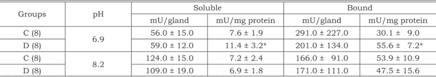
Table 1 shows the data for the submandibular gland. Expressed as mU/gland no difference was observed comparing the glands from the diabetic and control animals for both soluble and particu-lated enzyme. However, considering the activity per mg of protein (specific activity) it was seen that the soluble enzyme increased by 50% and the bound, by 84% compared to the control, when the activity was determined in conditions to present allosteric properties.
For the parotid gland (Table 2) there was an increase in the activity of the soluble form of
PFK-TABLE 1 - Activity of the soluble and citoskeleton bound fractions of PFK-1 of the submandibular salivary glands of rats with streptozotocin-induced diabetes (D) and of control animals (C) determined at pH 6.9 and at pH 8.2.
Groups pH Soluble Bound
mU/gland mU/mg protein mU/gland mU/mg protein
C (8)
6.9 56.0 ± 15.0 7.6 ± 1.9 291.0 ± 227.0 30.1 ± 9.0
D (8) 59.0 ± 12.0 11.4 ± 3.2* 201.0 ± 134.0 55.6 ± 7.2*
C (8)
8.2 124.0 ± 15.0 7.2 ± 2.4 166.0 ± 91.0 53.9 ± 10.9
D (8) 109.0 ± 19.0 6.9 ± 1.8 171.0 ± 111.0 47.5 ± 15.6
Mean ± SD. In parenthesis is the number of rats. The asterisk means statistically significant by Student’s t test comparing the diabetic and control animals (p < 0.05).
TABLE 2 - Activity of the soluble and cytoskeleton bound fractions of PFK-1 of the parotid salivary gland of rats with streptozotocin-induced diabetes (D) and of control animals (C) determined at pH 6.9 and at pH 8.2.
Groups pH Soluble Bound
mU/gland mU/mg protein mU/gland mU/mg protein
C (8)
6.9 24.0 ± 5.0 6.4 ± 1.6 36.0 ± 11.0 18.6 ± 5.0
D (8) 33.0 ± 6.0* 5.7 ± 1.1 31.0 ± 8.0 19.3 ± 5.0
C (8)
8.2 156.0 ± 51.0 35.1 ± 9.9 56.0 ± 16.0 34.4 ± 7.7
D (8) 102.0 ± 39.0* 36.4 ± 8.2 44.0 ± 12.0 28.1 ± 8.9
1 determined at pH 6.9 (37%), and a reduction at pH 8.2 (35%) expressed per gland.
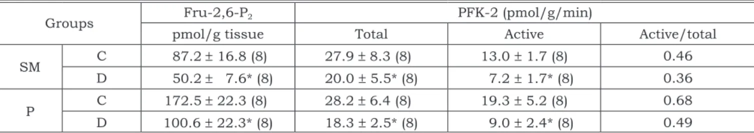
The content of Fru-2,6-P2 was reduced in the submandibular (42.5%) and parotid glands (41.7%) (Table 3). Similarly, the active form of PFK-2 showed reductions of about 44.6% in the submandibular and of about 53.2% in the parotid glands. The total activity also showed a reduction either in submandibular (28.3%) or parotid (35.1%) glands (Table 3).
The phosphate content of PFK-1 was significant-ly higher in the diabetic group than in the control rats for the submandibular gland (respectively 1.59 and 0.95 mol P/mol PFK-1) and for the parotid gland (respectively 1.07 and 0.74 mol P/mol PFK-1).
DISCUSSION
The results of the calculated RGW showed no variation for the submandibular gland. However, a higher value was found for the parotid gland of the diabetic animals, indicating an enlargement in comparison with the control animals, a fact that has already been reported3.
The role of PFK-1 in the regulation of metabo-lism is well known. The rate of the reaction cata-lyzed by PFK-1 is important to control the flow of glycolysis. The activity of this enzyme may be controlled by a variety of modulators as well as by phosphorylation/dephosphorylation. Our results for the submandibular gland show an increase in the specific activity of the enzyme determined at pH 6.9 for both forms, soluble (50%) and bound to the cytoskeleton (84%). No differences were ob-served either for the soluble or particulate enzyme in the activity determined at pH 8.2.
Contrary to the findings for the submandibular gland, the specific activity of the enzyme showed no variation for the parotid gland. However, con-sidering the determination performed at pH 6.9 in
which the enzyme presents allosteric properties, the mean activity per gland was higher for the dia-betic animals in the soluble fraction and lower than the control animals when determined at pH 8.2.
The diabetic rats consume more food than the control animals (hyperfagia)3. This increase in con-sumption may lead to an increase in the mastica-tion and an increase in the consumpmastica-tion of ATP from the glycolitic pathway. PFK-1 is a key enzyme of this metabolic pathway, and the increase in ac-tivity may be a consequence of the increase in the glycolitic pathway. The differences in the results of the submandibular and parotid glands may be due to metabolic differences between the two glands. While in the SM gland the metabolism is predomi-nantly anaerobic, the consumption of glucose may be higher than in the P gland, considering that its metabolism is predominantly aerobic22 and the gly-clolitic pathway is more efficient. Under allosteric control, the increase in the activity observed in the SM gland shows only an increase in the activation of the PFK-1, but not an increase of the total content and the total activity of PFK-1 in the SM gland. On the other hand, in the P gland, the lack of increase in the specific activity may have led to an increase of the RGW while, under allosteric regulations, the specific activity did not increase, but the total activ-ity did. Without this regulation, we can not explain the decrease of the activity of PFK-1.
Many studies have shown that the induction of diabetes may lead to different results of PFK-1 activity in several tissues. Thus, for the liver27, adi-pocytes26, enterocytes25, and heart atria8, reduced enzymatic activities were reported. The responses of the enzymatic activity to induced diabetes in animals were shown to be non-significant in lung1 and heart ventricle8. On the other hand, in rat peritoneal macrophage cells, PFK-1 activity was increased in cells from diabetic rats compared with those from normal rats7.
TABLE 3 - The activity of PFK-2 (active form and total) and the content of Fru-2,6-P2 in submandibular (SM) and parotid (P) glands of rats with streptozotocin-induced diabetes (D) and of control animals (C).
Groups Fru-2,6-P2 PFK-2 (pmol/g/min)
pmol/g tissue Total Active Active/total
SM C 87.2 ± 16.8 (8) 27.9 ± 8.3 (8) 13.0 ± 1.7 (8) 0.46
D 50.2 ± 7.6* (8) 20.0 ± 5.5* (8) 7.2 ± 1.7* (8) 0.36
P C 172.5 ± 22.3 (8) 28.2 ± 6.4 (8) 19.3 ± 5.2 (8) 0.68 D 100.6 ± 22.3* (8) 18.3 ± 2.5* (8) 9.0 ± 2.4* (8) 0.49
To examine if the modulator Fru-2,6-P2 influ-enced the activity of PFK-1, we have determined the content of this metabolite and the activity of PFK-2. The steady state concentration of Fru-2,6-P2 is de-termined by the balance between the activity ratio of the kinase and bisphosphatase of the bifunc-tional enzyme (PFK-2/FBPase). In liver, Fru-2,6-P2 is described as a molecule involved in the balance between glycolytic and gluconeogenic pathways, a potent allosteric activator of the glycolytic enzyme PFK-1 and an inhibitor of the gluconeogenic enzyme Fru-1,6-bisphosphatase. Fructose-1,6-bisphospha-tase is also present in the submandibular salivary glands of rats, however its activity is very low and does not vary with fasting, suggesting that the glu-coneogenic process is not operating in this tissue22. Thus, in salivary glands, Fru-2,6-P2 is probably implicated only in the activation of PFK-1.
The content of the metabolite was reduced by 42.5% for the submandibular gland and by 41.7% for the parotid gland. Reductions in the concentra-tion of Fru-2,6-P2 were also described in the atria region of the heart8, rat small intestine, entero-cytes25 and liver10 from diabetic animals. However, in macrophage cells, an increased level of this me-tabolite in diabetes was reported. A reduction in the mean activity of PFK-2 was observed mainly for the active (non-phosphorylated) form in submandibular (44.5%) and in parotid glands (53.4%). It has been pointed out that the reduced activity of PFK-1 ob-served in some tissues of diabetic animals was due to the reduction in the metabolite Fru-2,6-P2.16 On the other hand, in enterocytes isolated from dia-betic rats, there was a significant decrease in the level of Fru-2,6-P2, but not in the activity of PFK-125. In our work both a decrease in Fru-2,6-P2 and in the activity of PFK-2 in the diabetic animals was not accompanied by a reduction in the activity of PFK-1, suggesting that the diabetic state (decrease of insulin, hyperglicemia) reduces the activity of PFK-2 and the content of Fru-2,6-P2, but not the activity of PFK-1 in salivary gland of diabetic rats. In contrast, we have found higher specific activity of PFK-1 for the submandibular gland when com-pared with that of the control animals, and no dif-ference in the parotid gland between experimental and control animals. In view of these results, we
are led to conclude that the activation of PFK-1 in the submandibular salivary gland of diabetic rats was not due to the metabolite Fru-2,6-P2, but to another factor not known so far.
The results of the present study on the state of phosphorylation for the enzyme PFK-1 of the submandibular and parotid salivary glands of con-trol animals are within the values reported in the literature for the liver and muscle6,13. Working with skeletal muscle from mice, Bazaes et al.5 (1982) reported higher phosphate content and about 30% lower PFK-1 activity in diabetic mice than in control animals. In the present report the values obtained for phosphate content in submandibular salivary glands from diabetic animals were higher than those of the control animals, and the specific activity was also higher than in control animals. In previous papers we have found that the spe-cific activity of PFK-1 in the submandibular gland from rats injected with isoproterenol and killed 12 hours after the injection was higher than in control animals9, while the state of phosphorylation of the enzyme was also higher than in control animals23. Increasing the state of phosphorylation, we have found a reduction in the activity of PFK-1 in sub-mandibular gland from rats injected with three doses of isoproterenol23.
The results of this and of previous investiga-tions have led us to suggest that for the salivary glands, the phosphorylation of PFK-1 has dual activation. Initially, the activity of the enzyme is activated by an increased phosphorylation state. However, a higher state of phosphorylation inhibits the enzyme activity.
CONCLUSION
In conclusion, the increase in the activity of PFK-1 observed in the salivary gland of rats with streptozotocin-induced diabetes was not due to its modulator fructose-2,6-bisphosphate.
ACKNOWLEDGEMENTS
This work was supported by the “Fundação de Amparo à Pesquisa do Estado de São Paulo” (FAPESP 98/07169-9 and 99/03768-8).
REFERENCES
1. Abuelgassim AO, Salem AM, Khoja SM. Allosteric control of 6-phosphofructo-1-kinase from rat lung. Comp Biochem Physiol B 1992;101(1-2):135-8.
3. Anderson LC, Johnson DA. Effect of alloxan diabetes on rat parotid gland and saliva. Comp Biochem Physiol B 1981;70B:725-0.
4. Bartrons R, Hue L, Van Schaftingen E, Hers HG. Hormonal control of fructose 2,6-bisphosphate concentration in iso-lated rat hepatocytes. Biochem J 1983;214(3):829-37. 5. Bazaes SE, Foe LG, Kemp RG. Phosphate content of
mus-cle phosphofructokinase in the genetically diabetic mouse (C57BL/KsJ). Arch Biochem Biophys 1982;218(2):483-7. 6. Brand IA, Soling HD. Metabolite-controlled phosphorylation
of phosphofructokinase in rat hepatocytes. Eur J Biochem 1982;122(1):175-81.
7. Bustos R, Moreno-Aurioles VR, Conde M, Montano R, So-brino F. Streptozotocin-induced diabetes increases fructose 2,6-bisphosphate levels and glucose metabolism in rat mac-rophages. Biochem Med Metab Biol 1993;50(3):254-64. 8. Dunaway GA, Kasten TP, Naqui D. Insulin-mediated
regu-lation of heart atrial and ventricular 6-phosphofructo-1-kinase. J Biol Chem 1986;261(17):7831-3.
9. Ferreira FD, Nicolau J. Changes in glucose metabolism in submandibular salivary glands of rats after isoproterenol or incisor-tooth amputation. Arch Oral Biol 1987;32(7):499-503.
10. Gil J, Carreras J, Bartrons R. Effects of diabetes on fructose 2, 6-P2, glucose 1, 6-P2 and 6-phosphofruc-to 2-kinase in rat liver. Biochem Biophys Res Commun 1986;136(2):498-503.
11. Hamano E, Yamazaki T, Saito M, Kawashima H, Ozeki T, Furuyama S. Comparison of phosphofructokinases in submandibular glands of immature and adult rats. Comp Biochem Physiol B 1989;94(4):697-701.
12. Hasegawa H, Parniak M, Kaufman S. Determination of the phosphate content of purified proteins. Anal Biochem 1982;120(2):360-4.
13. Hussey CR, Liddle PF, Ardron D, Kellett GL. The isola-tion and characterizaisola-tion of differentially phosphorylated fractions of phosphofructokinase from rabbit skeletal mus-cle. Eur J Biochem 1977;80(2):497-506.
14. Komabayashi T, Ikeda T, Suda K, Izawa T. Beta-ad-renergic receptors and adenylate cyclase activity in the parotid acinar cells from acute streptozotocin-induced dia-betic rats. Res Commun Mol Pathol Pharmacol 2000;107(3-4):311-22.
15. Lowry OH, Rosebrough NS, Farr AL, Randall RJ. Pro-tein measurement with the folin phenol reagent. J Biol Chem 1951;193:265-75.
16. Madsen KL, Ariano D, Fedorak RN. Vanadate treat-ment rapidly improves glucose transport and activates 6-phosphofructo-1-kinase in diabetic rat intestine. Diabeto-logia 1995;38(4):403-12.
17. Mahay S, Adeghate E, Lindley MZ, Rolph CE, Singh J. Streptozotocin-induced type 1 diabetes mellitus alters the
morphology, secretory function and acyl lipid contents in the isolated rat parotid salivary gland. Mol Cell Biochem 2004;261(1-2):175-81.
18. Mahrenholz AM, Lan L, Mansour TE. Phosphorylation of heart phosphofructokinase by Ca2+/calmodulin protein
kinase. Biochem Biophys Res Commun 1991;174(3):1255-9.
19. Nelson N. Photometric adaptation of the Somogyi method for the determination of glucose. J Biol Chem 1944;153:375-8.
20. Nicolau J, de Souza DN, Martins HR. Pilocarpine-in-duced increases in the activity of 6-phosphofructo-2-kinase and the fructose-2,6-bisphosphate content of rat salivary glands. Arch Oral Biol 1992;37(6):483-7.
21. Nicolau J, Nunez-Burgos GB. Phosphofructokinase-1 in the nuclear and cytoplasmic fraction of the submandibu-lar salivary glands from isoproterenol-treated rats: Km and isoenzymes. Med Sci Res 1991;19:381-3.
22. Nicolau J, Sassaki KT. Metabolism of carbohy-drate in the major salivary glands of rats. Arch Oral Biol 1976;21(11):659-61.
23. Nicolau J, Souza DN, Nunez-Burgos G. Regulation of phosphofructokinase-1 on submandibular salivary glands of rats after isoproterenol administration. Arch Physiol Bio-chem 2000;108(5):437-43.
24. Reuterving CO, Hagg E, Henriksson R, Holm J. Sali-vary glands in long-term alloxan-diabetic rats. A quanti-tative light and electron-microscopic study. Acta Pathol Microbiol Immunol Scand [A] 1987;95(3):131-6.
25. Rossi I, Sanchez-Arias JA, Feliu JE. Effect of strepto-zotocin diabetes on the glycolytic flux and on fructose 2,6-bisphosphate levels in isolated rat enterocytes. Metabolism 1990;39(8):882-5.
26. Sobrino F, Saggerson ED. Measurements of gly-colytic flux rate in brown adipocytes. Effects of insulin, noradrenaline and streptozotocin diabetes. Biochem Int 1989;19(2):325-32.
27. Sochor M, Baquer NZ, Ball MR, McLean P. Regula-tion of enzymes of glucose metabolism and lipogenesis in diabetic rat liver by thyroid hormones. Biochem Int 1987;15(3):619-27.
28. Sugiya H, Fujita Y, Fukushima E, Yamazaki T, Fu-ruyama S. Fructose 2,6-bisphosphate-dependent regula-tion of phosphofructokinase in rat submandibular gland. Int J Biochem 1988;20(3):237-41.
29. Uyeda K. Phosphofructokinase. Adv Enzymol Relat Areas Mol Biol 1979;48:193-244.
30. Watanabe M, Yamagishi-Wang H, Kawaguchi M. Low-ered susceptibility of muscarinic receptor involved in sali-vary secretion of streptozotocin-induced diabetic rats. Jpn J Pharmacol 2001;87(2):117-24.