227
Correspondence: Ulisses Alexandre Croti
Hospital de Base – FAMERP – Avenida Brigadeiro Faria Lima, 5544. São José do Rio Preto, SP. CEP 15090-000
Fone (Fax): (17) 3201-5025 / 9772-6560. E-mail: [email protected]
Ulisses Alexandre CROTI, Domingo Marcolino BRAILE, Sírio HASSEM SOBRINHO, Airton Camacho MOSCARDINI
Braz J Cardiovasc Surg 2006; 21(2): 227-228
CLINICAL-SURGICAL CORRELATION
RBCCV 44205-823
Aneurisma da artéria pulmonar pós-estenose valvar pulmonar
Pulmonary artery aneurysm post pulmonary valve
stenosis
CLINICAL DATA
The case of a 2-year 5-month white Caucasian infant weighing 13.6 kg and measuring 95 cm high from José Bonifácio, São Paulo is reported. The infant had been symptomatic for the previous three months with dyspnea at normal effort. The patient was in a good general state, acyanotic, eupneic, ruddy, hydrated and afebrile. The heartbeat was regular with normal sounds and a fixed doubling of the second click, systolic murmur +/6+ at a pulmonary focus with trembling, irradiating to the left armpit. Auscultation of the lungs was symmetrical without adventitious murmurs. The liver was 1 cm from the right costal border. The pulses were symmetrical, without signs of edema and the peripheral oxygen saturation was 99%.
Article received in April, 2006 Article accepted in June, 2006
Case 4/2006
228
CROTI, UA ET AL - Pulmonary artery aneurysm post pulmonary valve stenosis - Case 4/2006
Braz J Cardiovasc Surg 2006; 21(2): 227-228
ELECTROCARDIOGRAM
An electrocardiogram evidenced sinusal rhythm with a heart beat of 125 bpm. The QRS complex was +110º, PR interval 0.16s, QRS duration 0.08s. There were signs of right ventricle overload without changes in the ventricular repolarization.
RADIOGRAM
The radiogram showed visceral situs solitus. The heart area was increased to the detriment of the right chambers with a cardiothoracic index of 0.60. The pulmonary arch was protuberant (Figure 1).
ECHOCARDIOGRAM
An echocardiogram showed situs solitus and levocardia. The venoatrial, atrioventricular and ventriculo-arterial connections were all concordant. The pulmonary artery appeared saccular and aneurysmatic with turbulent flow compatible with severe pulmonary stenosis. The maximum instantaneous gradient was 65 mmHg. An ostium secundum-type interatrial shunt (IAS) of 12 mm was evidenced. The pulmonary valvar annulus was 16 mm with a slightly thickened bi-cuspid pulmonary valve opening in a dome-shape with significant dilation of the pulmonary artery to 33 mm.
DIAGNOSIS
The diagnosis is obvious from the echocardiogram. It is important to remember that the child presented with clinical signs and symptoms only over the previous three months, probably due to the dilation of the pulmonary artery, which would compress the left pulmonary artery. This formation was facilitated by the presence of the large IAS, which provided a flow from the left chamber to the right with important hemodynamic implications. The association of pulmonary stenosis with IAS makes us think of Noonan Syndrome [1], a diagnosis not confirmed in this patient.
SURGERY
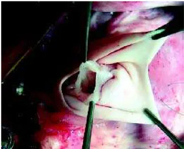
A median transsternal thoracotomy was performed with the establishment of cardiopulmonary bypass. The cannulae were introduced into the aorta and both vena cavae. Hypothermia at 34 ºC was used together with intermittent anterograde sanguineous cardioplegia at 4 ºC. After opening the pericardium, a severe aneurysmatic dilation of the pulmonary artery (Figure 2) was observed and when opened lengthwise a bi-cuspid pulmonary valve was observed, which was thin but with apparently normal movement (Figure 3). A pulmonary valvar commissurotomy was performed and the aneurysmatic wall was resected. The ostium secundum-type IAS was repaired with a bovine pericardium patch sutured using 5-0 polypropylene thread. The perfusion time was 45 minutes and the myocardial ischemia time was 30 minutes. The patient evolved well and was released from hospital on the 5th postoperative day.
REFERENCE
1. Burch M, Sharland M, Shinebourne E, Smith G, Patton M, McKenna W. Cardiologic abnormalities in Noonan syndrome: phenotypic diagnosis and echocardiographic assessment of 118 patients. J Am Coll Cardiol. 1993;22(4):1189-92.
Fig. 2 – Appearance of the aneurysmatic pulmonary artery