252
1. Pediatric Cardiovascular Surgery Service of São José do Rio Preto – Hospital de Base – São José do Rio Preto Medical School – São José do Rio Preto, SP, Brasil.
2. Pediatric Radiology Service of the Hospital de Base - São José do Rio Preto Medical School and Ultra-X Diagnóstico por Imagem of São José do Rio Preto - São José do Rio Preto, SP, Brasil.
Ulisses Alexandre CROTI1, Domingo Marcolino BRAILE1, Arthur Soares SOUZA JR2, Antônio Soares SOUZA2 Rev Bras Cir Cardiovasc 2009; 24(2): 252-255 CLINICAL-SURGICAL CORRELATION
RBCCV 44205-1086
Conexão anômala parcial de veias pulmonares com comunicação interventricular: a rara e possível
dificuldade na definição anatômica de isomerismo atrial esquerdo
Partial anomalous pulmonary venous connection
with interventricular communication: the rare and
possible difficulty on the anatomic definition of left
atrial isomerism
CLINICAL DATA
9-month-old male child, 7 kg, born in Fronteira, MG. Born at full-term with 3.3 kg. On the 10th day of life heart murmur was noted, but referred to the pediatric cardiologist with 5 months due to fatigue while breastfeeding, excessive sweating and inappropriate weight gain. With the clinics indicating congenital heart disease with pulmonary hyperflow and congestive heart failure (CHF), chest X-ray, electrocardiogram and echocardiogram were requested, which were complemented by multi-detector computed tomography (MDCT).
Under use of digital and diuretic, on the physical examination the patient presented in good general condition, afebrile, acyanotic and with tachydyspnea. Ictus
cordis positioned to the right, regular heart rhythm with
two clicks, ejection systolic murmur ++/4+ in right middle sternal border. Pulmonary auscultation with sparse bullous rales. Flaccid abdomen, liver at 3 cm from the left costal margin. Pulses were palpable and symmetrical in the four members.
ELECTROCARDIOGRAM
Sinus rhythm, heart rate 150 beats/min. PR interval of 80ms, QRS 80ms and QT 200ms. Negative P wave in D1, aVL indicating situs inversus totalis. Possible right ventricle overload (Figure 1).
Correspondence address: Ulisses Alexandre Croti
Hospital de Base – FAMERP – Avenida Brigadeiro Faria Lima, 5544 – São José do Rio Preto, SP, Brazil – CEP 15090-000.
E-mail: [email protected]
Article received on May 5th, 2009 Article accepted on June 1st, 2009
253 CROTI, UA ET AL - Partial anomalous pulmonary venous connection
with interventricular communication: the rare and possible difficulty on the anatomic definition of left atrial isomerism
Rev Bras Cir Cardiovasc 2009; 24(2): 252-255
RADIOGRAM
Slight increase of the cardiac silhouette with apex to the right, cardiothoracic index of 0.71. Signs of increased pulmonary vascular network with characteristics of hyperflow. There is compression on the right side wall of the trachea distal portion, suggesting aortic arch to the right. Liver in horizontal position (Figure 2A). In profile, it is noted increased of the anterior ventricular cavity (contact greater than 2/3 to the sternum), and in the pulmonary hila, two epibronchial arteries and bronchi with the same length (Figure 2B).
ECHOCARDIOGRAM AND TOMOGRAPHY
Echocardiogram diagnosed situs inversus totalis in dextrocardia, interatrial communication (IAC) ostium
secundum of small size, wide ventricular septal defect (VSD),
mild pulmonary valve stenosis by thickening of the valves
Fig. 2 - Chest radiography: (A) Front in PA with dextrocardia and increased cardiac area. (B) Left profile with two epibronchial arteries and bronchi of the same length
A
B
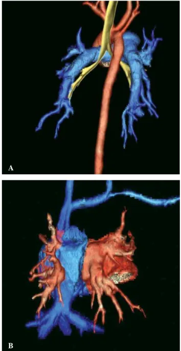
Fig. 3 - Multi-detector computed tomography after 3D reconstruction: (A) Posterior view with descending aorta to the right, symmetrical and epibronchial pulmonary arteries. Bronchi also symmetrical originating two similar lobe bronchi, all with characteristics anatomically left. (B) Rear view with superior vena cava to the left, supra-hepatic veins draining into a common vein that, in its turn flows into the atrium to the left and two venous pulmonary trunks draining into each of the atriums
A
254
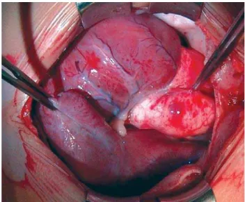
Fig. 4 – Atrium positioned to the left being pulled by the forceps and with external anatomical characteristics that resemble the right atrium
Fig. 5 – Atrium positioned to the left, opened, where vena cava are connected. It is noted abnormal orifices of pulmonary veins that drain into the left lung. It is also noted smooth internal appearance that indicates atrium anatomically left
and commissural fusion, dilated azygos vein draining into the superior vena cava (SVC) located to the left near the right atrium (RA) also positioned to the left. Presence of vein abnormally draining into the posterior region of the RA positioned to the left, which suggests such veins are left pulmonary veins, showing partial anomalous connection of left pulmonary veins (PAPVC) in RA. At Doppler, there was mean gradient between the right ventricle and pulmonary trunk (PT) of 24 mmHg and no significant gradient across the VSD.
MDCT, in addition to PAPVC, concluded that there was left atrial isomerism, with the pulmonary veins that drained the lung to the left and the SVC flowing into the atrium at left, which showed characteristics of left atrium (LA), and the pulmonary veins that drained the lung to the right flowing into the atrium at right also with characteristics of LA. The inferior vena cava (IVC) continued as azygous vein, which drained into the SVC positioned to the left. There was also a IAC and the aortic arch was to the right (Figures 3A and 3B).
DIAGNOSIS
The clinical presentation of CHF in the presence of PAPVC and IAC were dominant and sufficient to indicate the surgical correction, however, conflicting data respect to the situs was evident on the physical examination, electrocardiogram and echocardiogram, suggesting situs inversus totalis. A radiography with bronchi of the same length and liver in horizontal position favored the left atrial isomerism, according the findings of MDCT that showed two bronchi with left configuration, symmetrical, sealed; two epibronchial pulmonary arteries and both the bilobulated lungs, in addition to the presence of polysplenia and interruption of the IVC, which is an extremely reliable data of left atrial isomerism. Also, the fact of having two left pulmonary veins draining to the atrium positioned to the left, pointed to the left atrial isomerism, which was confirmed during the operation.
It is important to emphasize that the external aspect of the atrial appendage is not the best model of right or left morphology, and the presence of trabeculations on the atrial inferior wall should be valorized in order to characterize such atrium as right or the absence of them in order to designate an atrium as left [1 ].
OPERATION
With the surgeon positioned to the left of the patient, as previously described for patients with situs inversus
totalis [2], after transsternal median thoracotomy and
opening the pericardial sac, it was possible to note the external characteristics of the heart, with difficult definition respect to whether the atrium positioned to left was anatomically right or left (Figure 4).
Regardless of external aspect, the operation consisted
of making circular purse-string sutures on the aorta using 5-0 polypropylene yarn and 6-0 polydioxanone yarn on the IVC and supra-hepatic veins. Heparinization with 4 mg/kg and introduction of cannulas of diameters appropriate for the weight.
Conventional cardiopulmonary bypass was initiated, hypothermia at 25ºC, aortic clamping, opening of the atrium positioned to left. Two pulmonary veins from the left lung were found draining into the aforementioned atrium (one highest pulmonary vein and near the great IVC and the CROTI, UA ET AL - Partial anomalous pulmonary venous connection
with interventricular communication: the rare and possible difficulty on the anatomic definition of left atrial isomerism
255
REFERENCES
1. Sharma S, Devine W, Anderson RH, Zuberbuhler JR. The determination of atrial arrangement by examination of appendage morphology in 1842 heart specimens. Br Heart J. 1988;60(3):227-31.
2. Croti UA, Braile DM, Kozak MF, Kozak ACLFBM. Correlação clínico-cirúrgica. Caso 1/2005 – Serviço de Cirurgia Cardíaca Pediátrica – Hospital de Base da Faculdade Estadual de Medicina de São José do Rio Preto. Rev Bras Cir Cardiovasc. 2005;20(1):94-5.
other near the interatrial septum and more medially in relation to the position of the IVC and the supra-hepatic veins). The inner atrial wall was smooth, without trabeculations, suggesting indeed we were facing a left atrial isomerism (Figure 5).
Small IAC was then noted in high position in the interatrial septum. All oval fossa was resected and it was found that there was possibility to redirect the flow of all the pulmonary veins to the mitral valve.
The PT - positioned to the right and anteriorly - was opened earlier and the pulmonary valve was completely normal, with three valves, without thickening or fusions and with diameter appropriate for the child’s weight. The right ventricle outflow tract, positioned to the right, also was absolutely normal and with appropriate caliber. The TP was sutured with 5-0 polypropylene yarn.
The IAC was muscular of right ventricle outflow tract. To avoid any risk of blockage, eight U points using 6-0 polypropylene were passed and anchored in bovine pericardium patches, all distant from the edges and superficially on the right ventricle wall, which was above and to the right. The IAC was closed with a large bovine pericardium patch as usual. The tricuspid valve needed three points anchoring the valve to the anterior septum in order to maintain it totally competent and without stenosis. The flow redirection of all pulmonary veins was possible with a large bovine pericardium patch, which was sutured from the highest edge of the interatrial septum, placing it inferiorly to the IVC and anteriorly to the pulmonary vein that drained the left lung of such a way that allowed the direction of the pulmonary veins flow to the mitral valve and the blood from the IVC and supra-hepatic to the tricuspid valve.
The atrium positioned to the left was closed with 5-0 polypropylene after appropriate withdrawal of air from left and right cavities. The perfusion time was 111 minutes and myocardial ischemia, 82 minutes.
The evolution in the Intensive Care Unit was normal, with inotropic drugs within the first 48 hours. The hospital discharge occurred on the 9th postoperative day under use
of digital and diuretic. The echocardiogram performed 3 months after the operation - despite the technical difficulties - showed normal physiology recovered without residual defects.
ACKNOWLEDGMENT
To Prof. Dr. Vera Demarchi Aiello, for the guidelines and discussion of the case.
CROTI, UA ET AL - Partial anomalous pulmonary venous connection with interventricular communication: the rare and possible difficulty on the anatomic definition of left atrial isomerism