422
1. Pediatric Cardiovascular Surgery Service of São José do Rio Preto – Hospital de Base – Faculdade de Medicina de São José do Rio Preto.
Maria Fernanda Ferrari Balthazar JACOB1, Carlos Henrique DE MARCHI1, Ulisses Alexandre CROTI1, Domingo Marcolino BRAILE1
Rev Bras Cir Cardiovasc 2009; 24(3): 422-424
RBCCV 44205-1113
Defeito do septo atrioventricular associado à tetralogia de Fallot em paciente com síndrome de
Down
Atrioventricular septal defect with tetralogy of
Fallot in patient with Down’s syndrome
Mailing address: Ulisses Alexandre Croti
Hospital de Base – FAMERP – Avenida Brigadeiro Faria Lima, 5544. ZIP Code: 15090-000 – São José do Rio Preto, SP, Brazil. Phone (Fax): 17 - 3201 5025 / 3222 6450 / 9772 6560. E-mail: [email protected]
Article received on August 6th, 2009
Article accepted on September 8th, 2009
CLINICAL-SURGICAL CORRELATION
CLINICAL DATA
Female infant; born on term with 2.6 kg, normal delivery. Mother; fourth pregnancy, denied the use of medication during pregnancy. Presented respiratory discomfort at birth, evolving with neonatal infection and use of antibiotic therapy for 29 days.
At two months, she was referred to our Service for evaluation with use of digoxin, furosemide and captopril.
After confirming the diagnostic, the therapy with beta blocking agents was started with scheduled clinical follow-ups every three months, due to the absence of cardiac failure. At two years-old, there was an aggravation of the symptoms and hypoxemic crisis, resulting in the child’s referral to surgical treatment.
The physical examination presented satisfactory general conditions, cyanotic ++/4+, normotensive and with adequate weight and growth. The normal positioned ictus cordis, hyperphonetic single second sound and ejective systolic murmur +++/6+. Normal abdomen. Extremities well perfused, present and symmetric pulsation.
423
JACOB, MFFB ET AL - Atrioventricular septal defect with tetralogy of Fallot in patient with Down’s syndrome
Rev Bras Cir Cardiovasc 2009; 24(3): 422-424
ECHOCARDIOGRAM
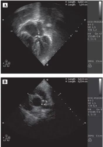
Situs solitus in levocardia. Presence of atrial septal defect (ASD): one ostium secundum of 1.8 mm and another ostium primum of 6 mm; ventricular septal defect (VSD) of 10.4 mm and single atrioventricular valve, presenting total defect of atrium-ventricular septum type C of Rastelli. Simultaneously, there was present stenosis in the right ventricle outlet and enlargement of the left ventricle outlet, pulmonary valvar stenosis with 6.1 mm of valvar ring and overriding of the aorta in less than 50%, typical of tetralogy of Fallot. The systolic peak gradient between the right ventricle and the pulmonary trunk was of 74 mmHg, the pulmonary arteries measured on the left 7.6 mm and on the right 6.1 mm and the diameter of the pulmonary artery trunk 5.3 mm (Figure 2).
DIAGNOSTIC
The final disgnostic was confirmed by the echocardiogram as being total atrium-ventricular septal defect with tetralogy of Fallot. Therefore, it was chosen the maintenance of the clinical treatment, since there were no early evidences of cardiac insufficiency or cyanosis predominance [1].
Through the clinical treatment the hypoxemic crisis were avoided, although there were signs of right ventricle dysfunction, in addition to polycitemia with all the inherent risks to it.
This situation indicated the moment for operation. The corrective surgical procedure demanded caution regarding the possibility of atrium-ventricular valve insufficiency and residual shunts, ill-prognostic factors and with higher possibility for the need of reoperation [2,3]. ELECTROCARDIOGRAM
Sinusal rhythm with anterior superior divisional blocking, frequency 100 beats/min.
SAQRSbetween - 90º and - 180º, PR 200 ms, QRS 80 ms. Axis deviated to the right with atrial and right ventricular overload.
RADIOGRAM
Visceral Situs solitus in levocardia. Enlarged cardia area with prevalence of right chambers and cardiothoracic index of 0.70. Enlarged pulmonary vascular trama (Figure 1).
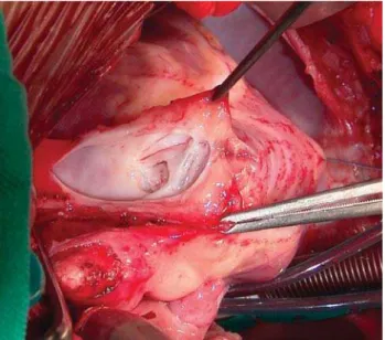
Fig. 3 – Aspect of the heart immediately after opening the sternum. The pulmonary trunk is hypoplastic and the right atrium and right ventricle are distended.
Fig. 2 – A: Echocardiogram in apical cut, four chambers in systolic cycle evidencing dilation of the right chambers with hypertrophy of the right ventricle and the diameters of the atrial septal defect ostium primum: (+) of 6 mm and the entricular septal defect (X) of 10 mm. B: Echocardiogram in parasternal cut, short axis at the level of the base vessels showing mild dilation of the ascending Ao and stenosis of the pulmonary trunk with its diameters in the medium segment (+) and distal (X) of 6.1 and 5.3 mm respectively. AE: left atrium, AD: right atrium, VE: left ventricle, VD: right ventricle, Ao: aorta, TP: pulmonary trunk, CIA: atrial septal defect, CIV: ventricular septal defect
A
424
REFERENCES
1. Hoohenkerk GJ, Schoof PH, Bruggemans EF, Rijlaarsdam M, Hazekamp MG. 28 years’ experience with transatrial-transpulmonary repair of atrioventricular septal defect with tetralogy of Fallot. Ann Thorac Surg. 2008;85(5):1686-90.
2. Brancaccio G, Michielon G, Filippelli S, Perri G, Di Carlo D, Iorio FS, et al. Transannular patching is a valid alternative for tetralogy of Fallot and complete atrioventricular septal defect repair. J Thorac Cardiovasc Surg. 2009;137(4):919-23.
3. Formigari R, Di Donato RM, Gargiulo G, Di Carlo D, Feltri C, Picchio FM, et al. Better surgical prognosis for patients with complete atrioventricular septal defect and Down’s syndrome. Ann Thorac Surg. 2004;78(2):666-72.
Fig. 4 – Pulmonary trunk opened with a dysplastic pulmonary valve. The small diameter of the pulmonary valvar ring and the commissural fusion are prominent.
Fig. 5 – Enlargement of the right ventricle outlet tract with bovine pericardium patch.
OPERATION
Sternotomy, heparine administration, insertion of cannulas in the aorta and vena cava, extracorporeal circulation aid (ECC) with hypothermia at 26ºC. Aortic clamping with hypothermic (4ºC) anterograde blood cardioplegia, intermittently each 20 minutes (Figure 3).
After opening the right atrium the single atrium-ventricular valve was reached. The pulmonary trunk was hypoplastic and the right ventricle outlet with stenotic pulmonary valvar ring (Figure 4).
The technique opted was the anterior opening of the pulmonary trunk, pulmonary valvar ring and right ventricle outlet tract, performing broad resection and enlargement with bovine pericardium patch (Figure 5).
The total atrium-ventricular septal defect was corrected with the double patch technique, closing the ventricular septal defect and the atrial septal defect with independent bovine pericardium patches. This approach was carefully handled in order to avoid the obstruction of the left ventricle outlet, leaving the patch satisfactorily in this area. The cleft of the left atrium-ventricular valve was occluded with separate stitches of polypropylene 6-0.
The disconnection of the ECC was supported with the aid of inotropics, after 148 minutes of ECC aid and 119 minutes of myocardial ischemia.
The patient was held in the Intensive Care Unit for 6 days, having a satisfactory clinical evolution, although presenting mild pericardium effusion, which was reverted by the oral administration of diuretic and corticosteroid drugs.
The patient was released from hospital on the 15thday
of postoperative using digoxin and diuretic drugs. Followed-up in the medical office two months ago, she is presently acyanotic, not complaining about fatigue, with echocardiogram showing moderate to important pulmonary valvar insufficiency, which is due to the treatment applied in the right ventricle outlet, only using the implantation of bovine pericardium patch without valve or valved tube [2].
JACOB, MFFB ET AL - Atrioventricular septal defect with tetralogy of Fallot in patient with Down’s syndrome