RBCCV 44205-1461 DOI: 10.5935/1678-9741.20130032
Comparação entre a Dopplermetria e o luxo livre da artéria torácica interna de cães com e sem o uso
de noradrenalina
Comparison between the Doppler lowmetry and
the free low of dog's internal thoracic artery with
and without use of norepinephrine
Alessandro Bordinhão
11. Masters in Principles of Surgery / Evangelical Hospital of Curitiba, Specialist of the Brazilian Society of Cardiovascular Surgery, Cardiovascular Surgeon at Santa Casa de Presidente Prudente, Curitiba, PR, Brazil.
Work performed at the Evangelical Hospital of Curitiba, Curitiba, Paraná, Brazil.
Correspondence address: Alessandro Bordinhão
Al. Augusto Stellfeld 1908 - Bigorrilho - Curitiba, Paraná, Brazil – Zip code: 80730-150.
E-mail: alessandrobordinhao@uol.com.br
Article received on September 25th, 2012 Article accepted on November 13th, 2012
Abstract
Objective: This work aims to study comparatively the free
low and the Doppler lowmetry of the internal thoracic artery in anesthetized dogs, with and without continuous intravenous administration of norepinephrine.
Methods: The sample was made up of ten mongrel dogs, which dissected the left and right internal thoracic arteries and evaluated your stream; irst, by Doppler lowmetry and then by free low. The mean arterial pressure and the diameter of the arteries at the beginning of the procedure were registered. The worklow checks by two methods occurred in three times: zero time, 10 and 25 minutes. After the irst check in time zero, the continuous infusion of norepinephrine in the right atrium; other checks were made in the same way that the irst time, to 10 and 25 minutes, in the same arteries and by two methods, each one in his artery, noting the results, as well as the corresponding average blood pressure.
Results: The results of the lowscan, between Doppler lowmetry and free low, there were similar; being the irst, zero times, ten and twenty-ive minutes, respectively, 183, 230.1 and 237 ml/min compared to seconds, 168.6, 226.8 and 226.4 ml/min (P=0.285). The mean arterial pressures of three times
and the average diameter of the arteries, showed no statistically signiicant differences between the methods, so did not inluence on the comparison of the results.
Conclusion: The evaluations, both from Doppler lowmetry and free low, were similar in three times checked.
Descriptors: Mammary arteries. Ultrasonography, Doppler. Continuous low. Norepinephrine. Dogs.
Resumo
Objetivo: Este trabalho objetiva estudar comparativamente o luxo livre e a dopplerluxometria da artéria torácica interna de cães anestesiados com e sem a administração de noradrenalina endovenosa contínua.
ocorreram em três tempos: tempo zero, 10 e 25 minutos. Após a primeira veriicação no tempo zero, iniciou-se a infusão contínua de noradrenalina no átrio direito; as avaliações aos 10 e 25 minutos foram feitas da mesma forma que na primeira vez, nas mesmas artérias e pelos dois métodos, anotando-se os resultados, assim como a pressão arterial média correspondente. Resultados: Os resultados da veriicação de luxo, entre Dopplermetria e luxo livre, apresentaram-se similares; sendo
os primeiros, nos tempos zero, 10 e 25 minutos, respectivamente, 183, 237 e 230,1 ml/min, comparados aos segundos, 168,6, 226,8 e 226,4 ml/min (P=0,285). A média das pressões arteriais dos
três tempos e o diâmetro médio das artérias não apresentaram diferenças estatisticamente signiicativas entre os métodos, portanto, não inluenciaram na comparação dos resultados.
Conclusão: As avaliações, tanto da dopplerluxometria quanto do luxo livre, foram semelhantes nos três tempos veriicados.
Descritores: Artéria torácica interna. Ultrassonograia Doppler. Fluxo contínuo. Norepinefrina. Cães.
Abbrebiations, acronyms and symbols
INTRODUCTION
The internal thoracic artery (ITA) is used in coronary artery bypass surgery as a graft, and in the anastomosis of the coronary artery for its long patency and adequate perfusion. Compared to the saphenous vein graft, the artery is clear in 85% of patients, 10 years after surgery, while with the vein, it occurs on average in 60% of the cases [1,2]. ITA dissection is simple in most cases. Due to these characteristics, ITA is the irst-choice artery for revascularization of the anterior descending artery, the most important in the cardiovascular structure [3].
Due to the importance of ITA in CABG, we should properly evaluate its patency, especially after dissection to avoid early occlusion, assessing its low. An ITA with low low can be
used as a free graft.
The evaluation of the ITA free blood low is composed of its incision and verify it in a container at a given time. This investigation was compared to measurement by transthoracic Doppler of the same artery, in a study by Canver et al. [4] in 1991, which demonstrated adequate correlation between the methods.
Some studies used a scanning method for evaluation of the
free low of the ITA. Wendler et al. [5] in 1999, comparatively evaluated ITA low dissected with a pedicle and a skeletonized one, comparing them by the free low. Most studies of ITA aims to analyze their patency over the years and few studies have the objective to evaluate their low in the intraoperative period, as the present article does, therefore, this is our most
relevant aspect.
Experimental study in dogs, conducted by Sasajima et al. [6] demonstrated the histopathological correlation of the ITA in dogs and in humans. Tada et al. [7] have analyzed the dog’s ITA through Doppler lowmetry under the effect of inotropic and vasoactive substances and Suematsu et al. [8] defended the epicardial Doppler lowmetry as a good method for evaluating
the ITA in CABG in dogs.
This work aims to comparatively study the free low and ITA Doppler lowmetry in anesthetized dogs with and without
continuous intravenous administration of norepinephrine.
METHODS
This work was performed at the Laboratory for Experimental Surgery of the Medical Research Institute from the Graduate Program in Principles of Surgery, Evangelical University of Paraná. The study was approved by the Research Ethics Committee of the Evangelical University of Paraná (no 4586/01), according to the Declaration of Helsinki.
We followed the principles of ethics in animal
experimentation, recommended by the Brazilian College of Animal Experimentation, speciically for dogs, according to the National Research Council [9].
Animal experimentation
The sample consisted of 10 mongrel dogs (Canis familiaris), with body weight ranging from 15 kg to 25 kg,
from the kennel of Curitiba City Hall. The sample size was suficient to be statistically evaluated.
Preoperative period
The animals were fed with dog food (Nuvita® for adult dogs) and had free access to water. The dogs were kept under observation for 15 days, subjected to vaccination and treatment of parasites. They were weighed and fasted for 12 hours before surgery.
Anesthetic technique
The animal was taken to the operating room where the
cephalic vein was punctured with Venoix® nº 21 scalp, which was connected to a 500 ml bag of 0.9 % saline solution (B. Braun S/A) to drip until the end of the procedure.
The anesthesia was endovenously induced with sodium thiopental hypnosis (Thionembutal®, Abbott Lab Brazil Ltda.)
at a dose of 5 mg/kg. The solution was obtained after diluting 1 g of thiopental sodium lyophilized in 1 ml of 0.9% saline
solution, obtaining 25 mg / ml of anesthetic solution.
The animal was considered anesthetized when it was
unconscious, with the involuntary movements ceased and
no reaction to surgical management. We also performed an
orotracheal intubation by direct laryngoscopy with 7.5 mm cannula in diameter, with a ballon of cardiac tamponade (RUNCH probe).
The animal was kept in ventilation controlled by a respirator (Takaoka® model 671) using inhalation anesthesia with halothane (Halothano®, Cristália) for continuous
vaporization.
Surgical Technique
1. The animal was placed in supine position with its cranial
and caudal limbs ixed.
2. The fur from ventral thoracic region, close to the skin,
was shaved with a razor blade.
3. In antisepsis of the surgical region, the povidone-iodine solution was used (Povedine Degermante®, Darrow), and then, povidone-iodine dye solution (Povedine Tintura®, Darrow).
4. The sterile surgical drapes were positioned, delimiting
the area to be incised.
5. Median thoracic incision was performed, from the jugular notch to the xiphoid process with a electric scalpel (Wem ® model BC-160), depthened to the breastbone, where
the hemostasis was performed.
6. The sternum was sawed in the midline, with Gigli saw, and hemostasis performed with bone wax (2.5 g, Poly Suture)
and infrasternal bleeding vessels with a 4.0 USP cotton thread
(Algoil®, Cirumédica).
7. The right ITA and left ITA was dissected using
skeletonization, starting from its origin in the subclavian artery and continuing until near the xiphoid process [5], linking its branches with 3-0 USP cotton thread (Algoil®, Cirumédica.)
After dissection of the ITA, 300 mg of papaverine hydrochloride were injected into the artery wall, in order to avoid spasms.
8. The pericardium was longitudinally incised along its entire length before being ixed to the subcutaneous tissue with three 2.0 USP cotton thread (Algoil®, Cirumédica), rising it and exposing the heart.
9. Suturing was performed in tobacco pouch with braided
polyester threads (Ethibond 2.0) in the aorta and right atrium.
10. A catheter was inserted into the aortic pouch and
connected to a sphygmomanometer clock, functioning as an arterial pressure checking system.
11. A no 8 nasogastric tube was introduced in the atrial pouch, connected to a serum catheter with an infusion pump (Nutrimat II®, B. Braun) and a lask with norepinephrine
standard glucose solution consisting of 4 mg of norepinephrine
diluted in 250 ml of 5% dextrose solution (16 g/ml) [10]. The infusion has not been started yet.
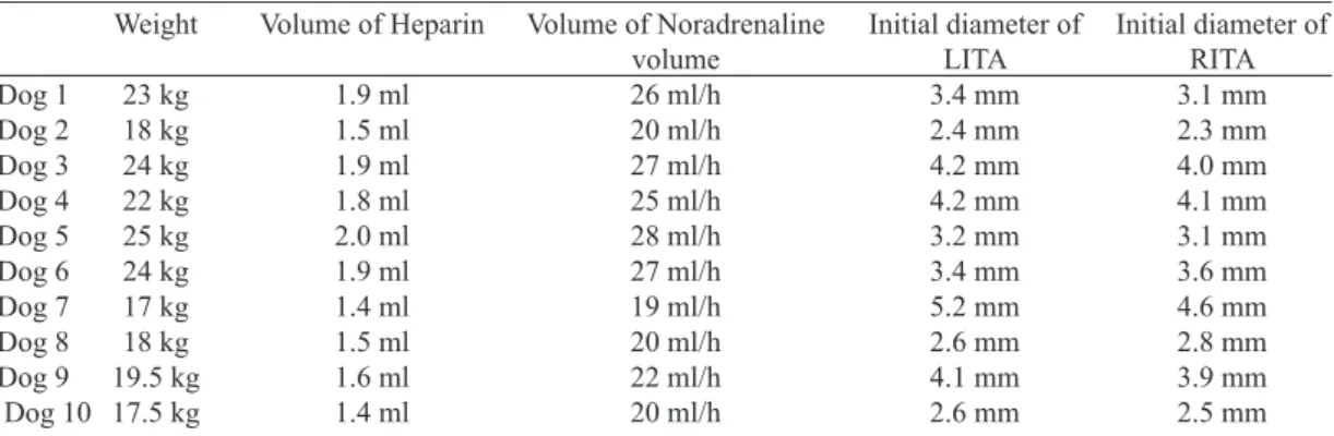
12. Heparin was intravenously injected (400 IU / kg) in the dog, with the purpose of anticoagulation. The dose of heparin injected was directly correlated to the weight (P=0.00001) (Table 1).
13. Veriication by the Doppler method (performed by a cardiologist with subspecialty in echocardiography and Doppler) was initiated by placing the transducer on one of ITAs. The irst veriication was adopted as the zero time (Vd0) assessing the diameter (Table 1) and the velocity of arterial blood low, and then calculating the arterial volume per minute. Blood pressure (Pd0) was measured by the sphygmomanometer connected to the aorta.
14. The contralateral ITA has its diameters observed (Table 1) by Doppler and then had its distal end transversely incised. The artery was dissected away from its bed and its caudal end
clamped with Bulldog forceps.
15. The clamped artery was displaced over a container and
then the clamp released. The bleeding was evaluated for 15
seconds. Blood volume was multiplied by four, deining the 1-minute low, then, we noted the mean arterial blood pressure when the veriication started (P0). The collected blood was injected with a 20 ml syringe through the dog’s venous access.
16. A continuous infusion of norepinephrine solution was
started at a constant low of 0.3 g / kg per minute, it was then calculated for each dog, according to the weight, in milliliters per hour (Table 1) standard dose [10].
17. After ten minutes of continuous infusion of
norepinephrine solution, the methods were evaluated by Doppler (VD10) and the free low (V10) in the same arteries and the same way as at time zero, verifying the mean arterial pressures corresponding to each method ( PD10 and P10).
18. After 25 minutes of infusion of norepinephrine, further measures were taken in the arteries by Doppler (Vd25) and then the free low (V25) and the blood pressure was observed in each of the two moments (Pd25) and free low (P25).
20. Initially, a four-dog pilot group underwent the experiment described above, for technical development and
to test the devices used.
21. ITA was evaluated by one and then by the other method, taking turns from one dog to the next.
Euthanasia
After the completion of the assessments, the animal was sacriiced with 40 ml of 19.1% potassium chloride, endovenously administered.
The Doppler lowmetry
The Doppler used to perform the measurements was the
AU3 Partner and the artery was examined by a 5-10MHz linear transducer, and the volume low calculated by multiplying the diameter by the average velocity of the blood low.
Variables analyzed
Variables were evaluated in three stages; zero time
corresponding to the beginning of the experiment and at two different times, 10 and 25 minutes (Tables 2 and 3):
1. The variables of Doppler evaluation were: Vd0, VD10 and Vd25, and mean arterial pressures corresponding to each veriication were; Pd0, PD10 and Pd25.
2. The free low variables were: V0, V10 and V25, and mean arterial pressures corresponding to each veriication were, respectively, P0, P10 and P25.
Statistical Analysis
The analysis was performed by the nonparametric Friedman and Kruskal-Wallis tests. Comparing the results, we adopted the null hypothesis that the low results measured by the two methods are equal versus the alternative hypothesis
Table 1. The weight of the dogs, the volume of heparin performed on each animal, the amount of noradrenaline
performed per hour and the diameter of internal thoracic arteries at zero time.
Dog 1 Dog 2 Dog 3 Dog 4 Dog 5 Dog 6 Dog 7 Dog 8 Dog 9 Dog 10 Weight 23 kg 18 kg 24 kg 22 kg 25 kg 24 kg 17 kg 18 kg 19.5 kg 17.5 kg
Volume of Heparin
1.9 ml 1.5 ml 1.9 ml 1.8 ml 2.0 ml 1.9 ml 1.4 ml 1.5 ml 1.6 ml 1.4 ml
Volume of Noradrenaline volume 26 ml/h 20 ml/h 27 ml/h 25 ml/h 28 ml/h 27 ml/h 19 ml/h 20 ml/h 22 ml/h 20 ml/h
Initial diameter of LITA 3.4 mm 2.4 mm 4.2 mm 4.2 mm 3.2 mm 3.4 mm 5.2 mm 2.6 mm 4.1 mm 2.6 mm
Initial diameter of RITA 3.1 mm 2.3 mm 4.0 mm 4.1 mm 3.1 mm 3.6 mm 4.6 mm 2.8 mm 3.9 mm 2.5 mm
LITA = left internal thoracic artery. RITA = right internal thoracic artery
Table 3. Results of Doppler lowmetry low variables(Vd0, Vd10 and Vd25) and free low variables (V0, V10 and V25).
Dog 1 Dog 2 Dog 3 Dog 4 Dog 5 Dog 6 Dog 7 Dog 8 Dog 9 Dog 10 Vd0 146 126 285 287 102 172 264 124 200 126 Vd10 198 160 357 321 166 232 280 143 330 188 Vd25 198 143 376 338 156 220 280 156 268 166 V0 132 136 240 280 130 132 236 120 200 120 V10 240 160 280 320 168 180 260 160 320 180
Note: The unit of measurement used for volume was milliliters per minute V25 248 144 320 320 160 168 272 180 280 172
Table 2. Results of blood pressures measured by the Doppler method (Pd0, Pd10, Pd25) and the free low method (P0, P10, P25). Dog 1 Dog 2 Dog 3 Dog 4 Dog 5 Dog 6 Dog 7 Dog 8 Dog 9 Dog 10 Pd0 70 80 100 100 70 100 100 80 90 90 Pd10 85 110 120 140 90 110 120 100 120 130 Pd25 90 120 120 130 80 120 130 100 110 120 P0 70 80 100 100 70 100 90 80 80 90 P10 80 120 120 140 90 110 120 110 120 130
that the results would be different. In all tests, we considered a 5% signiicance level. The variation of the diameter of the internal thoracic arteries and blood pressure were also analyzed by the same tests, as factors that inluence the result of the low between the two methods. Since the null hypothesis has P<0.05, they would be different in the two groups.
RESULTS
The experiments were uneventful. It was easy to dissect the ITA in the animals, presenting a good caliber and a good low. The average diameter of the ITAs veriied by Doppler was 3.54 mm and the free low of 3.39 mm.
No difference was observed between diameters of ITA in
the 2 methods, Doppler and free low (P=0.705).
The minimum diameter measured by the arterial Doppler method was 2.3 mm, and the free low of 2.4 mm, while the maximum diameter was 5.2 mm for the irst and, 4.6 mm for
the second method. There was a standard deviation of 0.89 for
Doppler, while for the free low of 0.76. The standard error was 0.28 for the irst and 0.24 for second method.
The values for mean arterial pressure measured by the Doppler method correspond to Pd0, PD10 and Pd25 and the free low to P0, P10 and P25 (Table 2).
The average blood pressure in the Doppler lowmetry method at zero time was 88 ± 12.29 mmHg, with a minimum of 70 mmHg and the maximum of 100 mmHg, whereas in the free-low method at the same time, the average was 86 ± 11.74 mmHg, with a minimum of 70 mmHg and a maximum of 100 mmHg.
The average blood pressure by Doppler method was 112.5 ± 17.20 mmHg at the 10- minute time, the minimum and the maximum of 85 mmHg and 122.5 mmHg, respectively, at the free low was 114 ± 17.76 mm Hg, with a minimum of 80 mmHg and a maximum of 140 mmHg. At the 25-minute time, the mean blood pressure in Doppler lowmetry method was 112 ± 16.87 mmHg, the minimum and maximum of 80 mmHg and 122.5 mmHg, respectively, the free low was 113.5 ± 16.67 mmHg, with a minimum of 80 mmHg and the maximum of 130 mmHg.
The blood pressure measured by Doppler method and the free low were compared with analyzes of nonparametric variance (Friedman). No signiicant difference was found, therefore, the pressure in both methods were similar during the three times (P=0.309).
The values found for the arterial low in the Doppler variables correspond to Vd0, Vd10 and Vd25 and for free low, V0, V10 and V25, and can be veriied in Table 3.
The mean arterial low in the Doppler lowmetry time at zero time was 183 ± 71.6 ml/min, with a minimum of 102 ml/min and a maximum of 287 ml/min, while the free low method at the same time presented an average of 168.6 ± 59.7 ml/min, with the minimum of 120 ml / min and the maximum of 280 ml/min.
The mean arterial low in Doppler lowmetry was 237.5 ± 78.7 ml/min at the 10-minute time, with a minimum of 143 ml/min and a maximum of 357 ml/min and the free low was
226.8 ± 65.2 ml/min with the minimum of 160 ml/min and
the maximum of 320 ml/min. At the 25-minute time, the mean arterial low in Dopplermetria method was 230.1 ± 82.1 ml/ min, with a minimum of 143 ml/min and a maximum of 376 ml/min and the free low was 226 4 ± 68.8 ml/min, with a minimum of 144 ml/min and a maximum of 320 ml / min.
The results revealed no signiicant difference in blood low between the methods in the times analyzed (P=0.285). Regression analysis shows that blood pressure is the determining factor for the variation in low (P=<0.00001*). By adjusted R2, approximately 30% of the variation of the low are due to the pressure variation.
DISCUSSION
The importance of this experiment was evidenced by
demonstrating similar results between the standard method
(Doppermetry) and the free low method, equaling them in daily clinical practice and allowing the free low method as an option in studies, for example, to evaluate the effect of drugs into the blood low, or to test methods of ITA dissection [11] and others. The free low is also useful in the daily assessment of ITA dissected in coronary artery bypass surgery.
The animal chosen was the adult dog for having a ITA with
good caliber and good low [12]. The artery of the dog has similar histopathological characteristics with the human’s, as demonstrated by Sasajima et al. [6]. All experimental work used the dog to the assessment of ITA low [6,7,12,13].
Norepinephrine was used in order to compare the two
methods in different blood pressures and consequently different blood low speeds, therefore, it provides other data for the comparison between Doppler lowmetry and free low [14].
It is important to note that drug was used in a constant
volume by means of an infusion pump, not showing signiicant luctuations in blood pressure during the analysis of the two types of veriication. We used the halothane inhalational anesthetic, since it was continously administered with the same quantity. The continuous inhalation anesthesia is the one which causes less oscillations in blood pressure [15]. In studies analyzed, the maintenance of anesthesia during surgery was performed in the same manner as in this experimental work [6,7,12,13].
The factors that can inluence the arterial low results
between the methods are:
1. The difference in diameters between the arteries in both
methods. The experiment showed the similarity between the diameters of arteries veriied in Doppler lowmetry and free low, being statistically signiicant (P=0.705).
REFERENCES
1. Cameron A, Davis KB, Green G, Schaff HV. Coronary bypass surgery with internal-thoracic-artery grafts: effects on survival over 15-year period. N Engl J Med. 1996;334(4):216-9.
2. Cameron AA, Green GE, Brogno DA, Thornton J. Internal thoracic artery grafts: 20-years clinical follow-up. J Am Coll Cardiol. 1995;25(1):188-92.
3. Barner HB. The internal mammary artery as a free graft. J Thorac Cardiovasc Surg. 1973;66(2):219-21.
4. Canver CC, Ricotta JJ, Bhayana JN, Fiedler RC, Mentzer RM Jr. Use of duplex imaging to assess suitability of the internal mammary artery for coronary artery surgery. J Vasc Surg. 1991;13(2):294-300.
5. Wendler O, Tscholl D, Huang Q, Schäfers HJ. Free low capacity of skeletonized versus pedicled internal thoracic artery grafts in coronary artery bypass grafts. Eur J Cardiothoracic Surg. 1999;15(3):247-50.
6. Sasajima T, Bhattacharya V, Wu MH, Shi Q, Havashida N, Sauvage LR. Morphology and histology of human and canine internal thoracic arteries. Ann Thorac Surg. 1999;68(1):143-8.
7. Tada Y, Tsuboi H, Suzuki K, Katoh T, Zempo N, Fujimura Y, et al. The response of blood low between the internal thoracic and ileocecal arteries to inotropic agents in a canine model. Surg Today. 1998;28(1):70-5.
8. Suematsu Y, Takamoto S, Ohtsukat T. Intraoperative echocardiographic imaging of coronary arteries and graft anastomoses during coronary artery bypass grafting without cardiopulmonary bypass. J Thorac Cardiovasc Surg. 2001;122(6):1147-54.
Doppler lowmetry and free low veriication. The experiment showed the similarity between the mean arterial pressure, recorded at the beginning of the Doppler lowmetry and free low, being statistically signiicant (P=0.309).
3. The decrease in blood flow due to the blood loss
during the free low veriication. It would be important if the measurement taken at the 1-minute time were corrected by performing the measurement in 15 seconds, the blood loss was lower, and then injected into the animal.
There are few studies comparing the two methods. Canver
et al. [4] separately used the two types of low veriication of the internal thoracic artery, the Doppler lowmetry in the preoperative period and free low in the intraoperative period,
demonstrating the correlation of them with a “P” statistically
signiicant, as in this work. In the study of Cagli et al. [16] performed on humans, there was a comparison between the free low in the intraoperative period with Doppler lowmetry in the pre-and postoperative period, with a smaller low in the irst method. However, there is a strong bias in this study,
for comparing a method in which the patient is anesthetized
(free low) whose blood pressure is different from a non-anesthetized patient (Doppler lowmetry), demonstrating a lower low in the irst one, and other problems such as the manipulation of the dissection be present in only one method (free low).
Authors as Tada et al. [7] used the free low to assess the inotropic action of substances on the arterial low, while Choi and Lee [17] and Wendler et al. [5] used to compare the free low of the dissected ITA with and without the pedicle, demonstrating that both methods are reliable. Hata et al. [18] demonstrated that ITA even with low under 20 ml/min can be used in CABG, increasing the low in the coronary artery with severe obstruction, similar to the ITA with greater low.
The low veriication by Doppler can identify factors of early occlusion of arterial or venous graft in the coronary artery, as the low low in the coronary bed after the anastomosis. This method is more comprehensive than the free low. Van der Meulen et al. [19] used the intraoperative Doppler to verify blood low speed in six internal thoracic artery before and after anastomosed to the anterior descending coronary artery. Suematsu et al. [8] used the Doppler lowmetry compared to histological examination in dogs and then compared to angiography, in humans, in evaluating the low of anastomoses graft to the coronary arteries, showing good correlation between the methods (P<0.001) [8 ]. Doppler would be an excellent evalution method of ITA in situ and after the graft
anastomosis in CABG [20,21].
The free low is currently used on a smaller scale than the Doppler and angiography, but it is still an eficient method in different papers [22,23]. Its easy and simple applicability, no need for a device or an expert in the ield, can increase the number of surveys assessing blood low, being especially
useful in countries with deicits of research inancial resources,
as in Brazil. CONCLUSION
A comparative evaluation between the free flow and
Doppler lowmetry of the ITA in anesthetized dogs with and
without administration of norepinephrine presents results
9. National Research Council. Commission on Life Sciences. Institute of Laboratory Animal Resources. Committee on Dogs. Dogs: laboratory; 1996.
10. Mosby’s Drug Consult. Norepinephrine bitartrate, drug resume. St Louis: Mosby; 2002.
11. Mannacio V, Di Tommaso L, De Amicis V, Stassano P, Vosa C. Randomized flow capacity comparison of skeletonized and pedicled left internal mammary artery. Ann Thorac Surg. 2011;91(1):24-30.
12. Lee CN, Orzulak TA, Schaff HV, Kaye MP. Flow capacity of the canine internal mammary artery. J Thorac Cardiovasc Surg. 1986;91(3):405-10.
13. Gitter R, Anderson JM Jr, Jett GK. Inluence of milrinone and norepinephrine on blood low in canine internal mammary artery grafts. Ann Thorac Surg. 1996;61(5):1367-71.
14. Shimizu T, Hirayama T, Suesada H, Ikeda K, Ito S, Ishimaru S. Effect of low competition on internal thoracic artery graft: postoperative velocimetric and angiographic study. J Thorac Cardiovasc Surg. 2000;120(3):459-65.
15. Massone F. Anestesiologia veterinária: farmacologia e técnicas. 3ª ed. Rio de Janeiro:Guanabara Koogan;1999. p.225.
16. Cagli K, Emir M, Kunt A, Ergun K, Muharrem T, Murat T. Evaluation of low characteristics of the left internal thoracic artery graft: perioperative color Doppler ultrasonography versus intraoperative free-bleeding technique. Tex Heart Inst J. 2004;31(4):376-81.
17. Choi JB, Lee SY. Skeletonized and pedicled internal thoracic artery grafts: effects on free low during bypass. Ann Thorac Surg. 1996;61(3):909-13.
18. Hata M, Shiono M, Orime Y, Hata H, Yagi S, Yamamoto T, et al. Should use of the internal thoracic artery be avoided under conditions of low free flow? Postoperative hemodynamic assessment using pulsed Doppler echocardiography. Jpn Circ J. 1999;63(7):533-6.
19. Van der Meulen J, van Son JA, van Asten WN, Skotnicki SH, Lacquet LK. Intraoperative Doppler spectrum analysis of blood low in the internal mammary artery used for myocardial revascularization. Thorac Cardiovasc Surg. 1991;39(5):281-3.
20. Louagie YA, Haxhe JP, Jamart J, Buche M, Schoevaerdts JC. Intraoperative assessment of coronary artery bypass grafts using a pulsed Doppler lowmeter. Ann Thorac Surg. 1994;58(3):742-9.
21. Forte AJV, Lobo Filho JG, Leitão MCA. Padrão ecocardiográico do luxo da artéria torácica interna esquerda após revascularização do miocárdio com enxerto composto. Arq Bras Cardiol. 2007;89(5):e163-4.
22. Une D, Shimizu S, Kamiya A, Kawada T, Shishido T, Sugimachi M. Both skeletonized and pedicled internal thoracic arteries supply adequate graft flow after coronary artery bypass grafting even during intense sympathoexcitation. J Physiol Sci. 2010;60(6):407-13.