Relationship of Exercise Capacity and Left
Ventricular Dimensions in Patients with a
Normal Ejection Fraction. An Exploratory
Study
Markus Meyer*☯, Rachel K. McEntee☯, Iwan Nyotowidjojo, Guoxiang Chu, Martin
M. LeWinter
Division of Cardiology, University of Vermont College of Medicine, Burlington, Vermont, United States of America
☯These authors contributed equally to this work. *[email protected]
Abstract
Objectives
Extreme endurance exercise is known to be associated with an enlargement of the left ven-tricular (LV) chamber, whereas inactivity results in inverse changes. It is unknown if these dimensional relationships exist in patients.
Methods
We analyzed the relationship of exercise capacity and LV dimension in a cohort of sequen-tial patients with a normal ejection fraction undergoing stress echocardiography. In a total of 137 studies the following questions were addressed: (a) is there a difference in LV dimen-sions of patients with an excellent exercise capacity versus patients with a poor exercise ca-pacity, (b) how is LV dimension and exercise capacity affected by LV wall thickness and (c) how do LV dimensions of patients who are unable to walk on a treadmill compare to the above groups.
Results
Patients with a poor exercise capacity or who are unable to physically exercise have a 34 percent smaller LV cavity size when compared to patients with an excellent exercise capaci-ty (p<0.001). This reduction in LV chamber size is associated with concentric LV
hypertro-phy and a reciprocal increase in resting heart rate. In addition, cardiac output reserve is further blunted by chronotropic incompetence and a tachycardia-induced LV volume reduc-tion. In conclusion the relationship of exercise capacity and cardiac dimensions described in extreme athletes also applies to patients. Our exploratory analysis suggests that patients who cannot sufficiently exercise have small LV cavities.
a11111
OPEN ACCESS
Citation:Meyer M, McEntee RK, Nyotowidjojo I, Chu G, LeWinter MM (2015) Relationship of Exercise Capacity and Left Ventricular Dimensions in Patients with a Normal Ejection Fraction. An Exploratory Study. PLoS ONE 10(3): e0119432. doi:10.1371/ journal.pone.0119432
Academic Editor:Dinender K Singla, University of Central Florida, UNITED STATES
Received:September 4, 2014
Accepted:January 13, 2015
Published:March 10, 2015
Copyright:© 2015 Meyer et al. This is an open access article distributed under the terms of the Creative Commons Attribution License, which permits unrestricted use, distribution, and reproduction in any medium, provided the original author and source are credited.
Data Availability Statement:All relevant data are within the paper.
Funding:This research was supported by an American Heart Association Scientist Development Grant 0730056N (MM), NIH grants 1R21HL94807-01 (MM) and NIH Institutional Training Grant T32 HL07647 (MML). The funders had no role in study design, data collection and analysis, decision to publish, or preparation of the manuscript.
INTRODUCTION
In response to external demands the myocardium undergoes adaptive changes. This process has been termed cardiac plasticity and changes at the cellular and macroscopic levels have been documented under various environmental conditions [1]. The most common and best under-stood cardiac adaptation is concentric left ventricular remodeling, which most commonly de-velops in response to pressure overload due to hypertension and aortic stenosis [2,3].
The adaptive changes that occur with endurance exercise and deconditioning are not as well understood but have been described in the exercise physiology literature [4–6]. Adaptive eccen-tric remodeling with left veneccen-tricular chamber enlargement is well documented in endurance athletes such as long-distance cyclists [4]. Extreme physical inactivity induced by bed rest or zero-gravity conditions in healthy subjects has been demonstrated to rapidly reduce left ven-tricular chamber volumes and mass [5,6]. It is unknown if analogous exercise phenotypes are found in a general clinical population with less extreme physical activity and inactivity.
Changes in left ventricular mass and volume have been documented in select clinical popula-tions that can be assumed to have a reduced exercise capacity, namely morbidly obese patients [7] and patients with severe pulmonary diseases [8–10]. In morbidly obese patients with and without heart failure a tendency towards eccentric left ventricular remodeling in the group of pa-tients with heart failure symptoms was explained by an increase in the cardiac workload [7]. Sev-eral studies describe small left ventricular chamber dimensions in patients with COPD [8–10] and patients with end-stage pulmonary arterial hypertension are reported to develop left ventric-ular atrophy, that is probably the result of a left ventricventric-ular workload reduction [11,12]. All adap-tive changes in the left ventricular dimensions volume have to be considered in the context of physiological aging, which is generally accompanied by smaller LV chamber volumes [13].
Although population or disease-based studies provide interesting structural insights into the remodeling of the left ventricle they typically do not directly relate left ventricular dimen-sions to exercise capacity, which is the primary objective of this study. Based on the exercise physiology findings and a large patient study that confirmed that poor exercise capacity is asso-ciated with diastolic dysfunction [14] it could be speculated that exercise capacity should also be reflected in the left ventricular dimensions. We hypothesized that poor exercise capacity in patients with a normal ejection fraction should be associated with a smaller left ventricle and chamber volumes, irrespective of the underlying cause or clinical presentation. Because this ini-tial analysis suggested that an increase in left ventricular wall thickness significantly contribut-ed to our findings we also explorcontribut-ed the impact of left ventricular wall thickness on left
ventricular volumes and exercise capacity.
METHODS
The primary objective of this exploratory study was to determine if there are principal associations between exercise capacity and left ventricular dimensions in the population of adult patients re-ferred for clinical stress testing. A secondary objective was to explore the effects of wall thickness on LV volumes and exercise capacity. To study these relationships we retrospectively analyzed a sequential cohort of patients undergoing Bruce-protocol based stress-echocardiography [15,16]. We also evaluated the left ventricular structure of patients who are unable to perform treadmill ex-ercise and therefore underwent pharmacological echocardiographic stress testing.
Patient Population
shortness of breath, coronary artery disease, and other (percentages of each shown in Tables
1–3). Exclusion criteria were EF<50%, baseline or stress induced wall motional abnormalities, clinically significant (moderate) valvular disease and poor echocardiographic image quality. The database was screened in reverse chronological order according to the date of study. The following patient characteristics were tabulated: test indication, cardiovascular risk factors, rea-son for test termination, gender, age, pre-test blood pressure and heart rate, body weight and height, medications, exercise duration, estimated metabolic equivalents of tasks (METs) and NYHA functional class. The University of Vermont Institutional Review Board has reviewed and approved this study. The data analysis was anonymous and no consent was required. A total of 137 studies were included in the analysis.
Group Comparisons
In order to gain insight into the relationship between exercise capacity and LV chamber dimen-sions we selected groups of patients based on the following additional criteria:
Poor exercise capacity versus excellent exercise capacity. We first compared LV dimen-sions of patients with poor exercise capacity, defined as the ability to exercise less than 5 min-utes on a standard Bruce treadmill protocol with patients with excellent exercise capacity, defined as the ability to exercise more than 15 minutes on the protocol. For both groups 20 studies were included in the analysis.
Normal LV wall thickness versus increased LV wall thickness. Since our initial analysis re-vealed a highly significant difference in wall-thickness at the extremes of exercise capacity we explored the effects of wall-thickness on LV volumes and exercise capacity. This analysis was performed in patients that were reported to have a normal exercise capacity on the clinical stress test interpretation. Inclusion criteria with either anormalseptal wall thickness, (9mm in women and10mm in men, n = 28), ormore than a mild increasein wall thickness, (>12mm regardless of gender, n = 21) [17]. All patients in this group reached more than 85% of the maximum predicted heart rate.
Inability to exercise. At our institution, dobutamine stress echocardiography is only per-formed in patients who are unable to walk on a treadmill. We hypothesized that this group of patients might represent a further physiologic and/or anatomic extreme of the poor exercise group. 48 patients were analyzed in this group.
Echocardiography
(AWT) in the parasternal short axis view. AWT was calculated by subtracting the endocardial radius from the epicardial radius derived from the short axis LV area calculation (A = r2π).
Stress Test
Patients on the treadmill underwent a standardized Bruce treadmill protocol [15]. Images were obtained both at rest and at peak exercise following guideline recommendations [16]. In pa-tients who underwent pharmacologic echocardiographic stress testing, intravenous dobuta-mine was delivered by an infusion pump starting at 5 mcg/kg/min for 3 minutes. The infusion rate was stepwise increased to 10, 20 and then 40 mcg/kg every 3 minutes until at least 85% of the maximum predicted heart rate was reached [16]. Images were captured at baseline, inter-mediate rate steps and at peak heart rate.
Data Analysis and Inter-observer Variability
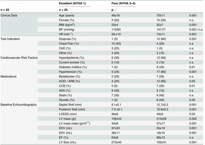
A power analysis using preliminary data of patients who exercised less than 5 minutes or more than 15 minutes revealed 80% power to detect a difference with group sizes of 20 using Table 1. Excellent versus Poor Exercise Capacity.
Excellent (NYHA 1) Poor (NYHA 3–4)
n = 20 n = 20 p<
Clinical Data Age (years) 46±16 70±11 0.001
Female (%) 4 (20) 10 (50) n.s.
BMI (kg/m2) 25±4 32±7 0.001
BP (mmHg) 115/83 141/77 0.001/ n.s.
HR (min-1) 59-±10 73±11 0.001
Test Indication Dyspnea (%) 1 (5) 12 (60) 0.001
Chest Pain (%) 10 (50) 4 (20) n.s.
CAD (%) 5 (25) 1 (5) n.s.
Other (%) 4 (20) 3 (15) n.s.
Cardiovascular Risk Factors Hyperlipidemia (%) 6 (30) 12 (60) n.s.
Current smoker (%) 2 (10) 2 (10) n.s.
Diabetes mellitus (%) 1 (5) 9 (45) 0.01
Hypertension (%) 5 (25) 17 (85) 0.001
Medications Betablocker (%) 5 (25) 7 (35) n.s.
ACEI / ARB (%) 4 (20) 12 (60) 0.05
CCB (%) 0 (0) 7 (35) 0.01
ASA (%) 9 (45) 3 (15) n.s.
Statin (%) 7 (35) 9 (45) n.s.
Diuretic (%) 1 (5) 8 (40) 0.05
Baseline Echocardiography Septal Wall (mm) 8.1±0.1 12.7±0.3 0.001
Posterior Wall (mm) 7.2-±0.1 10.6±0.2 0.001
LVEDD (mm) 56±8 49±9 0.05
LV mass (gr) 156±43 215±59 0.005
LV mass index (gr/m2.7) 34±8 57±17 0.001
EDV (mL) 97±20 55±18 0.001
ESV (mL) 36±11 18±10 0.001
EF (%) 63±8 68±13 n.s.
LV Size (mL) 215±43 152±41 0.001
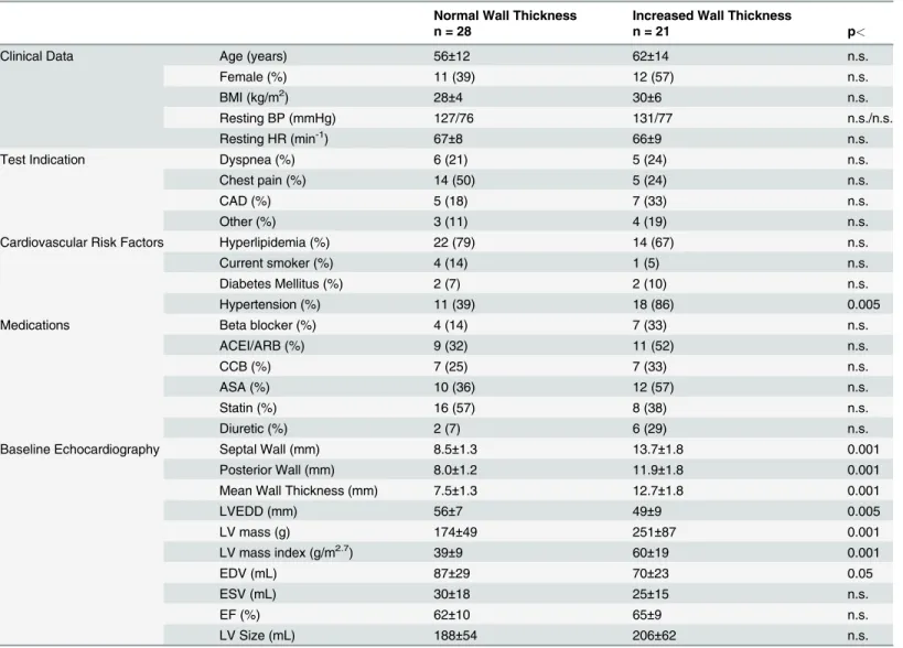
2-sample t-tests with a two-tailed 5% Type I error level. Accordingly, we aimed to enroll at least 20 patients per group. LV end-diastolic and end-systolic volumes from the dobutamine stress test (rest and peak stress) and treadmill stress test (rest and peak stress) were plotted against the respective heart rates. The stress-induced change in LV volume was calculated in each patient and expressed as the change in LV volume (in ml) over a change in heart rate of 50 beats per minute. Between and within group comparisons were performed using Student’s t-tests. A two-sided Fisher’s exact test was employed for categorical data. P-values<.05 were considered to be statistically significant. Values are reported as mean ± standard deviation. An analysis of inter-observer variability of LV volumes resulted in a good correlation, with r = 0.88 and a typical error (standard deviation of the differences divided by the square root of 2) of 5 mL (95% CI, 6 to 18 mL). Statistical analysis was performed using IBM SPSS 18.0.0 software. Table 2. Normal versus Increased LV Wall Thickness.
Normal Wall Thickness Increased Wall Thickness
n = 28 n = 21 p<
Clinical Data Age (years) 56±12 62±14 n.s.
Female (%) 11 (39) 12 (57) n.s.
BMI (kg/m2) 28±4 30±6 n.s.
Resting BP (mmHg) 127/76 131/77 n.s./n.s.
Resting HR (min-1) 67±8 66±9 n.s.
Test Indication Dyspnea (%) 6 (21) 5 (24) n.s.
Chest pain (%) 14 (50) 5 (24) n.s.
CAD (%) 5 (18) 7 (33) n.s.
Other (%) 3 (11) 4 (19) n.s.
Cardiovascular Risk Factors Hyperlipidemia (%) 22 (79) 14 (67) n.s.
Current smoker (%) 4 (14) 1 (5) n.s.
Diabetes Mellitus (%) 2 (7) 2 (10) n.s.
Hypertension (%) 11 (39) 18 (86) 0.005
Medications Beta blocker (%) 4 (14) 7 (33) n.s.
ACEI/ARB (%) 9 (32) 11 (52) n.s.
CCB (%) 7 (25) 7 (33) n.s.
ASA (%) 10 (36) 12 (57) n.s.
Statin (%) 16 (57) 8 (38) n.s.
Diuretic (%) 2 (7) 6 (29) n.s.
Baseline Echocardiography Septal Wall (mm) 8.5±1.3 13.7±1.8 0.001
Posterior Wall (mm) 8.0±1.2 11.9±1.8 0.001
Mean Wall Thickness (mm) 7.5±1.3 12.7±1.8 0.001
LVEDD (mm) 56±7 49±9 0.005
LV mass (g) 174±49 251±87 0.001
LV mass index (g/m2.7) 39±9 60±19 0.001
EDV (mL) 87±29 70±23 0.05
ESV (mL) 30±18 25±15 n.s.
EF (%) 62±10 65±9 n.s.
LV Size (mL) 188±54 206±62 n.s.
RESULTS
Exercise Capacity and Left Ventricular Dimensions
Patients in the poor exercise capacity group were able to exercise an average of 3 minutes and 39 seconds, whereas patients in the excellent exercise capacity group exercised for 16 minutes and 25 seconds. The estimated metabolic equivalent of tasks (METs) was 5±1METs in the poor exercise capacity group and 16±1METs in the excellent exercise capacity group. This translates to NYHA functional classes 3–4 and 1, respectively.
There were multiple significant differences in the baseline characteristics between groups as shown inTable 1. The poor exercise capacity group was older, had a higher body mass index and a significantly higher blood pressure. In addition, resting heart rate was significantly higher in the group with poor exercise capacity (difference of 14 beats per minute, p<.001). There was also a significant difference in the test indications; 60 percent of the patients with poor exercise capacity had dyspnea as the test indication, whereas this was only the case in 5 percent in pa-tients with excellent exercise capacity (p<.001). Finally patients with poor exercise capacity had higher rates of hypertension and diabetes mellitus and were more likely to take ACE/ ARB’s, calcium channel blockers and diuretics (p<.05 for all). Notably, there was no difference in the use of beta receptor antagonists between the groups.
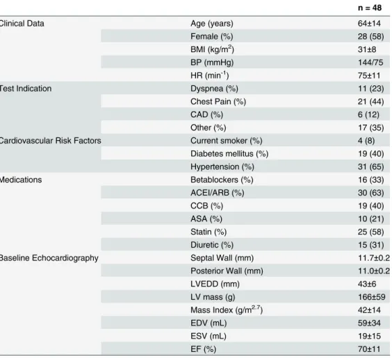
Table 3. Unable to Walk on Treadmill.
n = 48
Clinical Data Age (years) 64±14
Female (%) 28 (58)
BMI (kg/m2) 31±8
BP (mmHg) 144/75
HR (min-1) 75±11
Test Indication Dyspnea (%) 11 (23)
Chest Pain (%) 21 (44)
CAD (%) 6 (12)
Other (%) 17 (35)
Cardiovascular Risk Factors Current smoker (%) 4 (8)
Diabetes mellitus (%) 19 (40)
Hypertension (%) 31 (65)
Medications Betablockers (%) 16 (33)
ACEI/ARB (%) 30 (63)
CCB (%) 19 (40)
ASA (%) 10 (21)
Statin (%) 25 (58)
Diuretic (%) 15 (31)
Baseline Echocardiography Septal Wall (mm) 11.7±0.2
Posterior Wall (mm) 11.0±0.2
LVEDD (mm) 43±6
LV mass (g) 166±59
Mass Index (g/m2.7) 42±14
EDV (mL) 59±34
ESV (mL) 19±15
EF (%) 70±11
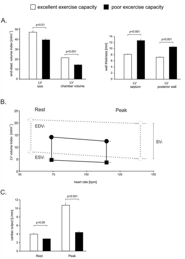
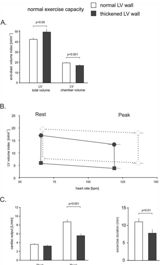
There were significant differences in cardiac dimensions as shown inTable 1andFig. 2. In the group with a poor exercise capacity, the non-indexed and indexed LV end-diastolic cham-ber volume was 44 and 34 percent smaller (both p<.001), and the septal and posterior LV wall was increased by 57 percent and 48 percent respectively (both p<.001), to suggest concentric remodeling. Accordingly, LV mass was significantly higher in patients with a poor exercise ca-pacity, whereas the external LV dimensions were smaller. Total LV size, which includes the myocardium as shown inFig. 1, that was 30 percent (non-indexed) and 16 percent (indexed) smaller (p<.001, p<.01).
In addition to the anatomic differences, physiologic differences were noted between the two groups. Despite a similar use of beta receptor antagonists patients with an excellent exercise ca-pacity increased their heart rate by 99±24 bpm compared to 50±15 bpm in patients with very poor exercise capacity (p<. 001). The volumetric assessments also allowed us to estimate cardi-ac output. As shown inFig. 2C, patients with an excellent exercise capacity nearly tripled their cardiac output with exercise whereas patients with a poor exercise capacity only increased their cardiac output by about 50% (p<.001). This marked difference was mainly driven by higher resting heart rates, reduced chronotropic response and additional reductions in end-diastolic volume at peak exercise.
The Effect of LV Wall Thickness on Volumes and Exercise
We compared patients with a normal septal wall thickness to patients with a more than mild increase in septal wall thickness. All patients reached the target heart rate and were clinically judged as having a normal exercise capacity. The resulting groups were similar in age, body mass index, resting blood pressure and heart rate as shown inTable 2. Also, there were no sig-nificant difference in test indications and medications. However, patients with an increased wall thickness had higher rates of hypertension (p<.005).
Fig 1. LV size and LV cavity volumetric assessment from the apical 4-chamber view.
Fig 2. LV Volume and heart rate relationship in patients with a normal EF in patients with poor and excellent exercise capacity.Panel A
demonstrates the differences in resting total LV size and LV chamber volume. The septal and posterior wall thickness was significantly increased in patients with poor exercise capacity. Panel B demonstrates the relationship of heart rate and indexed LV chamber volume at rest and with peak exercise. Circles: end-diastolic volumes, squares: peak systolic volumes. The vertical lines equal the stroke volume index. Panel C depicts cardiac outputs at rest and peak exercise in both groups. Error bars±SE.
In patients with an increased septal wall thickness the posterior and average LV wall thick-ness was increased by 48 percent and 69 percent respectively (both p<.001), whereas the LV end-diastolic diameter was reduced by 13 percent (all p<.005). Consistent with this finding, the indexed and non-indexed LV chamber volume was reduced by 13 percent and 19 percent (both p<.05). In patients with increased myocardial thickness the non-indexed and indexed LV mass were increased by 44 percent and 54 percent (both p<.001). The indexed total LV size, which includes the myocardium, was 17 percent larger in patients with an increased LV wall thickness as shown inFig. 3A(p<.05).
Patients with a normal wall thickness increased their heart rate with exercise by 90±14 bpm, whereas subjects with increased wall thickness could only increase their heart rate by 67±14 bpm (p<.001). In patients with a normal wall thickness, the left ventricular end-diastolic volume did not significantly change with exercise, whereas a heart rate-dependent volume loss was observed in the group with a thickened myocardium, as shown inFig. 3B. This change is manifest in a steeper slope of the line connecting end-diastolic volumes at rest and at peak exercise (-20±4mL/ 50bpm versus -7±2mL/50bpm, p<.05). Together, these variables significantly impaired cardiac output reserve in the group with an increased LV wall thickness. As a result, these patients in-creased their cardiac output by only 77 percent versus 250 percent in patients with a normal wall thickness (p<.01) as shown inFig. 3C. There were also significant differences in exercise duration; patients with increased wall thickness exercised an average of 7 minutes and 53 seconds, whereas patients with a normal wall thickness exercised an average 11 minutes and 5 seconds (p<.01). Similarly, exercise capacity expressed in METs was 9±3 METs and 12±3 METs, respectively (p<.01). This confirmed that both groups were within the limits of a normal exercise capacity.
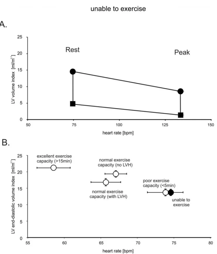
Inability to Exercise on a Treadmill and Cardiac Dimensions
Pharmacological stress tests are performed in patients who cannot physically exercise. We hy-pothesized that irrespective of the reasons that prevented treadmill exercise this inability should be reflected in the LV phenotype. As shown inTable 3, these patients have similar base-line characteristics to the patients in the poor exercise capacity group. They are older, have an increased body mass index, hypertension, features of concentric LV remodeling and higher resting heart rates.
Administration of dobutamine resulted in a pronounced diastolic and systolic volume loss, with near cavity obliteration at peak stress as shown inFig. 4A. As a result of this pronounced reduction in the LV chamber size, cardiac output increased by only 34 percent. In patients who met left ventricular hypertrophy (LVH) partition values (>48g/m2.7in men,>44g/m2.7in women) we found a more pronounced end-diastolic volume loss. In patients with LVH, the slope of the volume loss was-28±5mL/50bpm compared to-13±5mL/50bpm in patients with-out LVH (p<.01). The same principal association was confirmed using our septal thickness cri-teria (p<.01).
Importantly, when directly compared to the other groups, left ventricular end-diastolic vol-ume was identical to the group with a poor exercise capacity, as shown inFig. 4B. Thus, in comparison to patients with a normal or excellent exercise capacity, patients who cannot suffi-ciently exercise also appear to have smaller LV cavity volumes.
DISCUSSION
Exercise Capacity and LV Dimensions
exercise capacity were found to have smaller left ventricular cavities and higher resting heart rates. In this observational study we can only speculate about cause-and-effect as we do not know if the observed reduction in chamber volume is in response to a sedentary lifestyle, aging or a preexisting phenotype associated with a low exercise capacity. However, rapid changes in left ventricular dimensions have been documented in healthy subjects exposed to bedrest or zero gravity conditions [5,6]. It appears therefore very likely that the observed differences in chamber volume are the result of an adaptive process of which hypertension-induced concen-tric remodeling appears to play an important role.
Our principal finding of a reduced LV cavity size was confirmed in the analysis of a diverse group of patients who were deemed unable to walk on a treadmill and were therefore referred for pharmacological stress testing. This particular result suggests that it may not matterwhy
patients are unable to exercise; inactivity, for whatever reason, is associated with a small LV chamber size. Although small LV cavity sizes are typically encountered in patient with hyper-tension-induced concentric remodeling, as seen in this study, LV volume reductions are also reported in situations where the LV workload is severely reduced [12]. In our second analysis we explored the specific impact of wall thickness on LV volumes in patients who were consid-ered to have a normal exercise capacity. This data suggests that patients with a seemingly nor-mal exercise capacity but an increase in LV wall thickness are better able to maintain their LV chamber volume by an increase in the external dimensions of the LV. The finding that an in-creased LV wall thickness is still associated with a reduced cardiac output reserve and a lower exercise capacity mirrors changes seen in athletes, where individuals who performed non-endurance strength exercise such as weightlifting tend to have thicker left ventricular walls [19,20]. Pluim et al. compiled the literature on athletes in a meta-analysis examining this con-cept and conclude that although there seems to exist an“endurance trained heart”and a
“strength trained heart,”adapted to handle high volume loads versus pressure loads respective-ly, this concept is not absolute but exists on a continuum [21].
The assessment of LV chamber volumes using the biplane method of discs is a recom-mended component of a transthoracic echocardiogram [17]. Because the measurement of the LV volume integrates both LV chamber size and the presence of concentric remodeling, it may be able to predict exercise capacity. After appropriate partition values have been further de-fined, an LV-dimension based“fitness-estimate”may present an additional opportunity to dis-cuss the beneficial effects of physical exercise with patients as it is well established that physical fitness portends prognostic information. A recent longitudinal study examining the relation-ship between fitness level in middle age and subsequent hospitalizations for heart failure or myocardial infarction concludes that low levels of fitness in early middle aged were associated with increased rates of hospitalization for heart failure later in life [22]. In support of this no-tion Fujimoto et al. reported that one year of walking based exercise training in initially seden-tary seniors, culminating in about 30 minutes of exercise per day, increases cardiovascular fitness and cardiac output primarily through decreasing peripheral vascular resistance. Howev-er, they also document a significant decrease in resting heart rate and increase in stroke volume index [23]. Importantly, the same investigators demonstrated that older individuals who exer-cise throughout their lives maintain normal LV dimensions [24], which supports our assertion, that differences in cardiac phenotype observed in this analysis are predominately maladaptive. In addition, structured exercise is so far the only intervention that appears to provide a clinical
The vertical lines equal the stroke volume index. Panel C depicts cardiac output and exercise duration at rest and with exercise in both groups. Error bars±SE.
Fig 4. LV volume and heart rate relationship in patients with normal ejection fraction who are unable to walk on a treadmill.Panel A demonstrates the relationship of heart rate and LV volume index at rest and with peak exercise. Both systolic and diastolic chambers volumes decline substantially between rest and peak exercise which results in near LV cavity obliteration in systole. Circles: end-diastolic volumes, squares: peak systolic volumes. The vertical lines equal the stroke volume index. Panel B summarizes the relationship of heart rate and end-diastolic LV volume of all groups studied. Error bars±SE.
benefit in patients with heart failure and preserved ejection fraction, which tends to be associat-ed with smaller LV cavity volumes and concentric remodeling [25,26].
Variables Limiting Cardiac Reserve
As cardiac output is the product of heart rate and stroke volume, it is essential to discuss the impact of these variables on our findings.
Heart rate. Despite the fact that heart rate is by far the most important contributor towards cardiac output reserve in healthy individuals [27], relatively little attention has been given to the effects of heart rate on ventricular size, pump function and exercise capacity in patients. Our data demonstrate that resting heart rates are elevated in patients with a poor exercise ca-pacity. This alone will inherently reduce cardiac output reserve. In addition these patients have chronotropic incompetence, or an inability to increase the peak heart rate sufficiently with ex-ercise. This is a well-established observation in patients with heart failure with preserved ejection fraction (HFpEF) and an attempt has been made at correcting this deficit with rate-adaptive pacemaker therapy [28]. However, this trial was stopped prematurely and no informa-tion is available if the already enrolled patients derived a clinical benefit.
Chamber volume. Stroke volumes are generated by changes in chamber volume throughout the cardiac cycle and a smaller cavity size is typically associated with a smaller stroke volume [5,6]. This also implies that patients with smaller left ventricular cavities will more quickly develop a increased contractility-induced cavity obliteration. In resting patients, the stroke volume reduc-ing effect of a smaller ventricular cavity appears to be compensated by an increase in restreduc-ing heart rate but functions to reduce cardiac reserve during exercise as discussed above. Considering the di-mensional restraints, only a significant exercise-induced increase in diastolic chamber volume (which conceivably could be accomplished by a shape change from an ellipsoidal to a spherical LV geometry) could improve stroke volumes since the normal pericardium precludes significant LV expansion [29]. This effect may underlay the exercise-induced 13 percent increase in LV cavity size in healthy subjects who underwent nuclear imaging during bicycle exercise [27].
impaired left ventricular filling in the elderly and patients with diastolic dysfunction will also influence the LV volume in less predictable ways [38]. Tachycardia-induced reductions in left ventricular filling pressures could reduce the filling of the LV withpacing-induced or
pharma-cological tachycardia[35]. However, this later mechanism is clearly not playing a role with
physical exercise where LV filling pressures are known to disproportionally increase in patients with diastolic dysfunction [39].
Limitations
Similar to athletic and sudden-deconditioning studies, this exploratory analysis can only be used to provide initial insights into the relationship between left ventricular dimension and fit-ness at the extremes of the patient population that requires confirmation from larger prospec-tive studies. Ideally such a study should include an assessment of diastolic function, which currently is not routinely assessed during stress echocardiography.
Summary
In this exploratory analysis we found that the relationship between exercise capacity and LV chamber size observed in athletes and deconditioned subjects is also seen in patients. Patients with a poor exercise capacity or who are unable to exercise on a treadmill tend to have smaller and concentrically remodeled left ventricles with a reduced cavity volume. Thickening of the myo-cardium appears to result in an additional exercise-induced reduction in LV chamber volume. This volume reduction is potentiated by a higher resting heart rate and chronotropic insufficiency which both limit cardiac reserve. This is information is depicted in our heart rate-volume dia-grams that provide a straight-forward visualization of all variables relevant to cardiac output.
Author Contributions
Conceived and designed the experiments: MM MML. Performed the experiments: RKM IN GC MM. Analyzed the data: RKM IN MM. Wrote the paper: RKM MML MM.
REFERENCES
1. Hill JA, Olson EN. Cardiac Plasticity. N Engl J Med. 2008; 358: 1370–1380. doi:10.1056/ NEJMra072139PMID:18367740
2. Linzbach AJ. Heart failure from the point of view of quantitative anatomy. J Am Coll Cardiol. 1960; 5: 370–382.
3. Gaasch WH, Zile MR. Left Ventricular Structural Remodeling in Health and Disease With Special Em-phasis on Volume, Mass, and Geometry J Am Coll Cardiol. 2011; 58: 1733–1740. doi:10.1016/j.jacc. 2011.07.022PMID:21996383
4. Abergel E, Chatellier G, Hagege AA, Oblak A, Linhart A, Ducardonnet A, et al. Serial left ventricular ad-aptations in world-class professional cyclists: implications for disease screening and follow-up. J Am Coll Cardiol. 2004; 44: 144–149. PMID:15234423
5. Levine BD, Zuckerman JH, Pawelczyk JA. Cardiac atrophy after bed-rest deconditioning: a nonneural mechanism for orthostatic intolerance. Circulation. 1997; 96: 517–525. PMID:9244220
6. Perhonen MA, Franco F, Lane LD, Buckey JC, Blomqvist CG, Zerwekh JE, et al. Cardiac atrophy after bed rest and spaceflight. J Appl Physiol. 2001; 91: 645–653. PMID:11457776
7. Alpert MA, Boyd ET, Kelly DL. Effect of weight loss on cardiac chamber size, wall thickness and left ventricular function in morbid obesity. J Am Coll Cardiol. 1985; 55: 783–786.
8. Jörgensen K, Houltz E, Westfelt U, Nilsson F, Scherstén H, Ricksten SE. Effects of lung volume reduc-tion surgery on left ventricular diastolic filling and dimensions in patients with severe emphysema. Chest. 2003; 124: 1863–1870. PMID:14605061
10. Watz H, Waschki B, Meyer T, Kretschmar G, Kirsten A, Claussen M, et al. Decreasing Cardiac Cham-ber Sizes and Associated Heart Dysfunction in COPD: Role of Hyperinflation. Chest. 2010; 138: 32–38. doi:10.1378/chest.09-2810PMID:20190002
11. Manders E, Bogaard HJ, Handoko ML, van de Veerdonk MC, Keogh A, Westerhof N, et al. Contractile dysfunction of left ventricular cardiomyocytes in patients with pulmonary arterial hypertension. J Am Coll Cardiol. 2014; 64: 28–37. doi:10.1016/j.jacc.2014.04.031PMID:24998125
12. Meyer M. Left Ventricular Atrophy in Pulmonary Arterial Hypertension. J Am Coll Cardiol. 2014; 64: 38–40. doi:10.1016/j.jacc.2014.04.027PMID:24998126
13. Cheng S, Fernandes VR, Bluemke DA, McClelland RL, Kronmal RA, Lima JA. Age-related left ventricu-lar remodeling and associated risk for cardiovascuventricu-lar outcomes the multi-ethnic study of atherosclero-sis. Circ Cardiovasc Imaging. 2009; 2: 191–198. doi:10.1161/CIRCIMAGING.108.819938PMID: 19808592
14. Grewal J, McCully RB, Kane GC, Lam C, Pellikka PA. Left ventricular function and exercise capacity. JAMA. 2009; 301: 286–294. doi:10.1001/jama.2008.1022PMID:19155455
15. Fletcher GF, Ades PA, Kligfield P, Arena R, Balady GJ, Bittner VA, et al. AHA Scientific Statement. Ex-ercise Standards for Testing and Training. A Scientific Statement From the American Heart Associa-tion. CirculaAssocia-tion. 2013; 128: 873–934. doi:10.1161/CIR.0b013e31829b5b44PMID:23877260
16. Pellikka PA, Nagueh SF, Elhendy AA, Kuehl CA, Sawada SG. American Society of Echocardiography recommendations for performance, interpretation, and application of stress echocardiography. J Am Soc Echocardiogr. 2007; 20: 1021–1041. PMID:17765820
17. Lang RM, Bieri g M, Devereux RB, Flachskampf FA, Foster E, Pellikka PA, Picard MH, et al. Chamber Quantification Writing Group; American Society of Echocardiography’s Guidelines and Standards Committee; European Association of Echocardiography. J Am Soc Echocardiogr. 2005;
18:1440–1463. PMID:16376782
18. Sawada SG, Segar DS, Ryan T, Brown SE, Dohan AM, Williams R, et al. Echocardiographic detection of coronary artery disease during dobutamine infusion. Circulation. 1991; 83: 1605–1614. PMID: 1673646
19. Pelliccia A, Maron BJ, Spataro A, Proschan MA, Spirito P. The upper limit of physiologic cardiac hyper-trophy in highly trained elite athletes. N Engl J Med. 1991; 324: 295–301. PMID:1824720
20. Baggish AL, Wang F, Weiner RB, Elinoff JM, Tournoux F, Boland A, et al. Training-specific changes in cardiac structure and function: a prospective and longitudinal assessment of competitive athletes. J Appl Physiol. 2008; 104: 1121–1128. PMID:18096751
21. Pluim BM, Zwinderman AH, van der Laarse A, van der Wall EE. The Athlete’s Heart. A Meta-Analysis of Cardiac Structure and Function. Circulation. 2000; 101: 336–344. PMID:10645932
22. Berry JD, Pandey A, Gao A, Leonard D, Farzaneh-Far R, Ayers C, et al. Physical Fitness and Risk for Heart Failure and Coronary Artery Disease. Circulation: Circ Heart Fail. 2013; 6: 627–634. PMID: 23677924
23. Fujimoto N, Prasad A, Hastings JL, Arbab-Zadeh A, Bhella PS, Shibata S, et al. Cardiovascular Effects of 1 Year of Progressive and Vigorous Exercise Training in Previously Sedentary Individuals Older Than 65 Years of Age. Circulation. 2010; 122: 1797–1805.
24. Arbab-Zadeh A, Dijk E, Prasad A, Fu Q, Torres P, Zhang R, et al. Effect of aging and physical activity on left ventricular compliance. Circulation. 2004; 110: 1799–1805. PMID:15364801
25. Kitzman DW, Brubaker PH, Morgan TM, Stewart KP, Little WC. Exercise training in older patients with heart failure and preserved ejection fraction: a randomized, controlled, single-blind trial. Circ Heart Fail. 2010; 3: 659–667. doi:10.1161/CIRCHEARTFAILURE.110.958785PMID:20852060
26. Udelson JE. Heart failure with preserved ejection fraction. Circulation. 2011; 124: e540–3. doi:10. 1161/CIRCULATIONAHA.111.071696PMID:22105201
27. Higginbotham MB, Morris KG, Williams RS, McHale PA, Coleman RD, Cobb FR. Regulation of stroke volume during submaximal and maximal upright exercise in normal man. Circ Res. 1986; 58: 281–291. PMID:3948345
28. Kass DA, Kitzman DW, Alvarez GE. The Restoration of Chronotropic Competence in Heart Failure Pa-tients with Normal Ejection Fraction (RESET) Study: Rationale and Design RESET Trial. J Card Fail. 2010; 16: 17–24. doi:10.1016/j.cardfail.2009.08.008PMID:20123314
29. LeWinter MM, Tischler MD. Pericardial Diseases. In: Bonow RO et al editors. Braunwald’s Heart Dis-ease: A textbook of cardiovascular medicine. 9th ed. Philadelphia; Elsevier Saunders; 2011; 1651–1671.
31. Kitzman DW, Higginbotham MB, Cobb FR, Sheikh KH, Sullivan MJ. Exercise intolerance in patients with heart failure and preserved left ventricular systolic function: failure of the Frank-Starling mecha-nism. J Am Coll Cardiol. 1991; 17: 1065–1072. PMID:2007704
32. Borlaug BA, Melenovsky V, Russell SD, Kessler K, Pacak K, Becker LC, et al. Impaired chronotropic and vasodilator reserves limit exercise capacity in patients with heart failure and a preserved ejection fraction. Circulation. 2006; 114: 2138–2147. PMID:17088459
33. Liu CP, Ting CT, Lawrence W, Maughan WL, Chang MS, Kass DA. Diminished contractile response to increased heart rate in intact human left ventricular hypertrophy. Systolic versus diastolic determinants. Circulation. 1993; 88: 1893–1906. PMID:8403335
34. Kasner M, Westermann D, Steendijk P, Dröse S, Poller W, Schultheiss HP, et al. Role of left ventricular stiffness in heart failure with normal ejection fraction. Circulation. 2008; 117: 2051–2060. doi:10.1161/ CIRCULATIONAHA.107.716886PMID:18413502
35. Wachter R, Schmidt-Schweda S, Westermann D, Post H, Edelmann F, Kasner M, et al. Blunted fre-quency: dependent upregulation of cardiac output is related to impaired relaxation in diastolic heart fail-ure. Eur Heart J. 2009; 30: 3027–3036. doi:10.1093/eurheartj/ehp341PMID:19720638
36. Zile MR, Baicu CF, Gaasch WH. Diastolic heart failure—abnormalities in active relaxation and passive stiffness of the left ventricle. N Engl J Med. 2004; 350: 1953–1959. PMID:15128895
37. Selby DE, Palmer BM, LeWinter MM, Meyer M. Tachycardia-induced diastolic dysfunction and resting tone in myocardium from patients with a normal ejection fraction. J Am Coll Cardiol. 2011; 58: 147–154. doi:10.1016/j.jacc.2010.10.069PMID:21718911
38. Lakatta EG, Levy D. Arterial and cardiac aging: major shareholders in cardiovascular disease enter-prises: Part I: aging arteries: a“set up”for vascular disease. Circulation. 2003; 107: 139–146. PMID: 12515756