Comparison of different techniques for mouse transgenesis
Joana Paula de Assunção Almeida
Dissertation for Master degree in Medical Microbiology
Comparison of different techniques of transgenesis
Joana Paula de Assunção Almeida
Master in Medical Microbiology
External advisor: Rui Costa (Champalimaud Foundation)
External Co-advisor: Susana Lima (Champalimaud Foundation)
Internal advisor: Ricardo Parreira (IHMT/UNL)
Hosting Institution: Neurobiology of Action, Champalimaud Foundation
Acknowledgements
To start, I must thank the Champalimaud Neuroscience Programme (CNP), which I consider my scientific family, for having hosted me just after I finished veterinary school and for contributing to both my professional and personal development. A special thanks to Costa lab, where I learnt all the experimental techniques, for the great work environment, and for stimulating discussions. In particular, a very special thanks to Ana Nóvoa, Ana Vaz, Fatuel Tecuapetla and Joaquim Alves da Silva for their enormous contribution to this thesis.
Lastly, my major acknowledgement, mixed with a great admiration and esteem, to my supervisors Rui Costa and Susana Lima for their time, patience, teaching and
Abstract
Resumo
O uso de ratinhos transgénicos em neurociências aumentou consideravelmente nos últimos anos devido ao crescente interesse em compreender o cérebro e a necessidade de solucionar situações clínicas do foro neurológico e psiquiátrico. Para esse efeito, diferentes métodos de produção de animais transgénicos têm sido testados.
O objectivo desta tese foi comparar métodos de integração aleatória de um transgene no genoma de ratinhos em termos de eficiência, estabilidade da integração do transgene, número de animais e de horas de trabalho necessárias para cada método. Assim, foi comparado o método mais utilizado - microinjecção pronuclear (PNMI) - com duas outras técnicas cujo desempenho foi considerado promissor – a transferência génica através dos testículos por electroporação e transfecção por lentivírus in vivo. As três técnicas foram realizadas usando um gene repórter sob o controlo de um promotor constitutivo, e depois reproduzidas usando um gene de interesse de modo a permitir obtenção de um animal capaz de ser usado em experimentação laboratorial.
O transgene de interesse utilizado codifica uma proteína de fusão correspondendo a uma variante da rodopsina (channelrhodopsin) fundida à proteína enhanced yellow
Index
Index of Tables... vii
Index of Figures ... vii
Introduction ...1
1.DNA pronuclear microinjection ...6
Material and Methods ...6
Results and Discussion ...9
2. Proof of principle for in vivo DNA injection in mice testis ... 11
Material and Methods ... 11
Results and Discussion ... 12
3. In vivo DNA injection in mice testis ... 13
Material and methods ... 13
Results and Discussion ... 15
4.Infection of seminiferous tubule cells with lentiviruses ... 16
Proof of principle - Testing lentiviral transfection ... 16
Material and Methods ... 16
Lentivirus transduction in vivo ... 16
Results and Discussion ... 17
5.Application of the techniques ... 19
5.1.Pronuclear Microinjection ... 20
Material and Methods ... 20
Results and Discussion ... 22
5.2.Testis Electroporation ... 24
Material and Methods ... 24
Results and Discussion ... 24
5.3.Viral injection ... 26
6.Adeno-associated Virus in vivo injection ... 27
Material and Methods ... 28
Results and Discussion ... 29
7. Conclusion ... 30
References ... 34
Appendix I: CMV-GFP-WPRE-polyA ... 40
Appendix II: TH-ChR2-EYFP ... 41
Index of Tables
Table 1: Comparison of obtained values with reference parameters for C57BL6/J mice.9
Table 2: Pronuclear microinjection parameters ...9
Table 3: Intratesticular injection and electroporation of pCAGGS-GFP ... 12
Table 4: Results from testis electroporation with CMV-GFP sequence ... 15
Table 5: Lentiviral transfection results ... 17
Table 6: Results from TH-ChR2-EYFP pronuclear microinjection ... 22
Table 7: Results of testis electroporation using TH-ChR2-EYFP sequence ... 24
Table 8: Number of transgenic mice/total offspring for each technique ... 31
Index of Figures Figure 1: Schematic map of the plasmid containing the CMV-EYFP sequence ...6
Figure 2: Pronuclear microinjection.. ...7
Figure 3: 16,5 dpc embryos after pronuclear microinjection of CMV-GFP.. ... 10
Figure 4: Electroporation procedure... 14
Figure 5: in vivo Lentiviral injection.. ... 17
Figure 6: Schematic map of the plasmid containing the TH-ChR2-EYFP sequence ... 19
Figure 7: PCR analysis of transgenic animals and respective offspring.. ... 22
Figure 8: Characterization of ChR2-EYFP expression in Ventral tegumental area in transgenic mice generated by PNMI. ... 23
Figure 9: PCR analysis of offspring of the electroporated males that remained fertile.. 24
Figure 10: Characterization of ChR2-EYFP expression in Ventral tegumental area in transgenic mice generated by Testes Electroporation.. ... 25
Figura 11: schematic representation of the plasmid carrying a AAV double floxed ChR2-EYFP ... 27
Introduction
A “transgenic animal” is classically defined as an animal that has a foreign gene(s) stably incorporated into its genome through human intervention (3). Transgenesis has become one of the most important tools in medical research since it allows us to manipulate an animal´s genome enabling the construction of disease models and the study of tissues and/or organs’ functions in vivo. Mice, in particular, are a powerful animal model. Firstly, because mice are mammals and are consequently
phylogenetically close to humans; and secondly because mice are easier to manipulate genetically when compared to other mammals.
The use of transgenic mice is increasing in all fields of research, particularly in the field of neurosciences due to the urgent need to solve neurological medical conditions, which, in turn, depend on the generation of animal models of those conditions.
There are two different approaches for the production of genetically modified animals: gene targeting and the random integration of the transgene in the genome. Gene targeting is the most reliable technique since it guarantees that the gene of interest is inserted in the right place in the genome of a targeted animal host. It also prevents undesired results that can happen when the gene lands in the middle of a coding
sequence, which can cause a mutation. It permits the so-called “knock in” (insertion) or “knock out” (deletion or interruption) of genes and consequently a consequent gain or a loss of function. However, the cloning strategies used and the maintenance of an
embryonic stem-cell(ES cells) culture are very labour-intensive and expensive procedures. For this reason and because it is beyond the scope of this thesis, gene targeting techniques will not be further explored.
For the random integration of the transgene in the genome, different methodologies have been tested during the last decade. Despite the fact that integration of foreign DNA into the genome in a stable form that can be passed onto successive generations,
2
The first method developed for random integration of a transgene is pronuclear microinjection, which was developed in the 1980s by Gordon and other investigators (6;14) and is still the most widely used method for transgenic production. However, it comes with the disadvantage of requiring expensive equipment and highly skilled personnel, and the rate of gene integration is relatively low. Also, the number of animals needed to obtain a good founder population is large, raising ethical concerns. In lieu of this, alternative methods that overcome these problems have been developed.
Sperm-mediated gene transfer (SMGT) or testis-mediated gene transfer (TMGT) includes several potential methods that were extensively explored in the last two decades (44). Both experimental approaches are based on the idea of introducing exogenous DNA in the oocyte by the most natural means - the sperm. The advantage of these methods over the pronuclear microinjection is the possibility of producing a founder male that can generate large numbers of descendants using a relatively simple procedure. Many experiments have been carried out, but the obtained results are frequently controversial and hard to replicate (11).
The use of liposomes as vehicles in gene transfer experiments, for instance, either for direct lipofection of the sperm or direct injection in testis has also been tried. However, in 1991 Bachiller showed that after sperm lipofection, despite the fact that DNA transfer into sperm was very efficient, the generation of stable transgenic mice by this method was not attained (4). The direct injection of cationic liposomes in the testis did not show promising results in mice, as integration of exogenous DNA molecules in the cell’s genome was not met with success (8; 25). Finally, low ratios of offspring carrying the transgene with absence of expression characterized most experimental endeavours (1; 54).
Currently, the most promising TMGT strategy is in vivo testis electroporation, which consists of injection of a DNA construct in the testis, which are then subjected to
electric pulses that destabilize the membrane of spermatogonial cells, and allow DNA to enter. This method was first described in 1997 (37) and since then hundreds of
3
constraints on amounts and sizes of the DNA used, and no immunogenicity is expected (38) since testis are immunological privileged organs. However, the generation of transgenic animal using this method had never actually been reported until 2008 when a group of investigators from New Dehli was able to successfully produce transgenic pups from 94% of male mice electroporated with transgenes (33). If this could be replicated, then it would reveal itself as a powerful tool, since each electroporated male provides a valuable resource for continuous production of transgenic founders.
The use of viral vectors in transgenesis is another field that has been widely explored because it embodies another powerful approach to transfer genetic material into cells. The most important aspects when considering which virus to use are its tropism, efficiency in terms of off-targeted cell infection, carrying capacity of the viral vector to be used, and the levels of gene expression. Adenovirus, adeno-associated virus and retrovirus are those most frequently used as vectors.
Adenoviruses were used for gene transfer into testis in vivo and it is consensual that it was indeed effective for infection of different types of cells including sertoli and leydig cells but not germ cells (5; 26; 27). Adeno-associated viruses (AAV), which are non-pathogenic members of the Parvoviridae family, mediate long-term gene expression in both dividing and non-dividing cell types (46). However, the small size of their viral capsid limits to up to approximately 5kb (12) the DNA packaging capacity of the viral particles. Also, only 10% of the exogenous DNA integrates into the host genome, with the remaining 90% remaining in an episomal form (48), which is a limitation for the production of transgenic animals. The retroviruses require mitotic cell division for transduction, and can permanently integrate exogenous DNA into the genome of the infected cell (34). The disadvantages of this class of viruses include low production yields, random integration (15), and reduced packaging size (8-10kb). Lentiviruses are a subclass of retrovirus that can transduce both dividing and non-dividing cells. As
4
The ultimate goal of this thesis involves the comparison of different methods of random integration of a transgene in the genome in terms of efficiency (number of transgenic animals obtained per procedure) and stability of transgene integration. A technique is considered successful when there is a stable integration of the transgene in the genome, which occurs when the gene is transmitted to the offspring and expressed in biological levels resulting in a characteristic phenotype. The costs, the number of animals required, and the labour intensity of each technique will also be taken in account. In accordance to the discussion above, we decided to compare the most used method – pronuclear microinjection – with the two other techniques that seemed promising to us, TMGT by electroporation and in vivo lentiviral transfection.
We first tested all the techniques using a reporter gene – coding for the green fluorescent protein (GFP), the expression of which was driven by a constitutive promoter, the cytomegalovirus (CMV) promoter, in order to compare the number of animals expressing GFP in all cells. Then, the techniques were reproduced using a specific promoter for dopamine (DA) producing neurons, [the tyrosine hydroxylase (TH) gene promoter (45)] and a gene coding for a protein of interest. The final transgene encodes for a fusion protein called channelrhodopsin-enhanced yellow fluorescent (ChR2-EYFP) in dopaminergic neurons, resulting in a transgenic animal suitable for use in specific experiments of interest for work done in our lab. The
channelrhodopsin is a light-gated ion channel originating from microalgae, and has been largely used in optogenetics studies. ChR2 absorbs light with an approximate
wavelength of 450nm, inducing a conformational change in the protein that translates into the opening of a nonspecific pore for cations, conducting H+, Na+, K+, and Ca2+ ions. When inserting this protein in neurons, the entrance of the cations into the cell leads to membrane depolarization which, in turn, can generate an action potential. The fusion of the ChR2 with a reporter protein allows its identification and localization in the brain. This is one of the most useful tools in neurosciences nowadays since it makes possible to tag neuronal populations in vivo and monitoring of their activity (2;30) or stimulate specific populations of neurons, and correlate neuronal activity with
5
The variant of ChR2 most used in optogenetic studies has been codon optimized for mammalian expression (humanized form) and subsequently improved through
engineering to make it more suitable for neuroscience applications (31;55). However, several difficulties were found in the production of good transgenic mice lines carrying this optimized protein, as very low levels of expression were observed in all the possible founders. We hypothesized that this modification (the mammalian codon optimization) could be the cause of the low levels of expression as resulting from gene siliencing effects. Thus, we decided to apply the referred techniques to generate a transgenic mouse using a wild type version of the ChR2-EYFP driven by a tyrosine hydroxilase promoter. Other hypothesis for the poor expression of ChR2 is that the accumulation of this protein in the membrane of the cells impedes its normal function, resulting in cellular death.
The transgenic animals we aimed to construct would express this light channel only in the dopaminergic cells, which are involved in some of the most important biological functions in mammals; for example: learning (13), habit formation (19), and motor function (10). We hypothesized that optogenetic tools would make possible to activate specifically this group of neurons, while simultaneously observing the behaviour of a freely moving animal (49). These are very important tools in basic neuroscience
6
Figure 1: Schematic map of the plasmid containing the CMV-EYFP
sequence
1.
DNA pronuclear microinjection
Material and Methods
Plasmid DNA
Plasmid DNA was prepared by inserting the coding sequence for GFP and the Woodchuck hepatitis virus posttranscriptional regulatory element (WPRE) sequences in a pcDNA3 vector (Invitrogen) using KpnI/NheI restriction sites. Final sequence of the obtained
recombinant construct was confirmed by direct sequencing (see Appendix I). The fragment injected (CMV-GFP-WPRE-polyA) was obtained from the original plasmid (Figure 1)
using MluI/PvuII restriction enzymes and cleaned after gel band extraction and purification using a Quiagen gel extraction Kit. The WPRE has been known for its effect on enhancing the expression of an exogenous cDNA by stabilization of mRNA and the facilitation of the mRNA transport from the nucleus to the cytoplasm (41). The polyA tail in the 3’ terminus is also an important component of the construct due to its role in the mRNA activation by cytoplasmatic polyadenylation (52).
For microinjection, the purified fragment was diluted in a microinjection buffer to a 2ng/µ l concentration, which is the optimal concentration for transgene integration (9,39).
Animals and microinjection
7
5 groups of C57BL6/J females 7-8 weeks old were superovulated by intraperitoneal injection of 5 units of pregnant mare’s serum gonadotropin (PMSG) followed by 5 units human chorionic gonadotropin (hCG) 48 h later and then mated with C57BL6/J males (9).
The following morning females were checked for the presence of vaginal plug and euthanized using CO2. Fertilized embryos were collected from females’ oviducts and
placed in a hyaluronidase solution for 5 minutes at maximum in order to remove cumulus cells. Embryos were washed 4 times in a warm manipulation medium (M2) and 6 times in incubation medium (M16) covered with mineral oil to prevent from rapid air and temperature exchanges. Embryos were incubated for some hours up to
microinjection at 37⁰C in a 5% CO2 incubator. For microinjection, embryos were
moved again into warm M2 medium where they were washed 4 more times and then placed in the microinjection chamber, in a M2 drop covered with mineral oil. One pronucleus was injected until a visual swelling occurred as seen in Figure 2. Embryos were washed in M16 medium 6 times prior to an overnight incubation.
8
Embryos transfer into pseudopregnant females
After injection, embryos that survived were collected and washed 4 times in warm M2 medium and introduced, in both oviducts of a pseudopregnant NMRI female.
Pseudopregnant females are obtained after mating with vasectomized mice, in order to make the uterus more receptive and suitable for embryo implantation due to hormonal changes. NMRI strain was chosen for embryos transfer due to high fertility rates and good maternity skills (43).
Females were anesthetized with a ketamine/ xylazine mixture, the oviducts exposed dorsally and an average of 30 embryos were bilaterally distributed inside.
Embryo phenotype analysis
Pregnant females were euthanized using CO2 when embryos reached 16.5 days post
coitum1 (dpc) stage and embryos were collected to phosphate buffer saline (PBS) and screened for ubiquitous expression of GFP under a fluorescent stereoscope (Zeiss Stereo Lumar V12).
1
9
Results and Discussion
When comparing our experimental parameters with the one published in the literature of reference, some of the fluctuations are displayed in the data from Table 1.
Table 1: Comparison of obtained values with reference values for C57BL6/J mice
Obtained values (%) Reference values (%)
# Zygotes / female (superovulation efficiency) 17.7 20-50 (39)
% Injectable Zygotes / Total (male performance) 52.7 90 (39)
% Eggs transferred/Inj (Eggs survival) 62.7 70-72 (3)
% Born/ Inj 4.3 5-9 (3)
% Transgenics/Litter 31.3 20 (39)
Experimental results showed less efficiency in superovulation which despite females being within the best reproductive age (6-8 weeks of age), can be explained by different husbandry conditions within facilities, or by the experimenter. At the same time, the percentage of injectable zygotes was also below the reference parameters. This measure reflects male’s performance, but since the number of plugs was acceptable, this may suggest a lower quality of the sperm, possibly due to over usage of the males. By non-injectable zygotes, we refer to dead non-fertilized zygotes, as well as to zygotes with 3 or even 4 pronuclei instead of two, which can be caused by polyspermia, meaning fertilization of the same zygote by two or three spermatozoa, which make those zygotes non-viable. In optimal conditions the percentage of non-injectable zygotes is not
expected to be higher than 10% of the total number. Table 2 shows the yields of each microinjection session.
Table 2: Pronuclear microinjection parameters
Microinjection # Females # Zygotes Transfer # Embryos 16,5dpc
Total Injectable # Embryos Total # Tg
1 7 137 31 17 0 0
2 7 70 35 22 1 1
3 8 90 70 32 0 0
4 10 148 60 80 4 1
5 8 263 177 120 11 3
10
These results probably also reflect a certain degree of inexperience by the manipulator. As a matter of fact, pronuclear microinjection and embryo transfer are highly
demanding techniques and are usually performed by trained microinjectionists. However, the percentage of transgenics per injection is slightly higher than the ones usually reported by transgenic facilities, and the number of transgenic pups in each litter is around 20% which is also the efficiency rate usually obtained by other
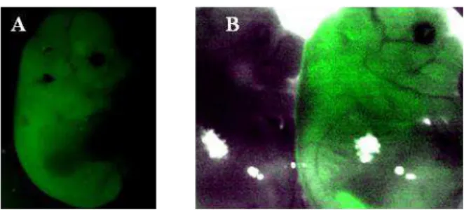
microinjectionists. This effect frequently affects the experimental results of technicians starting pronuclear microinjection, since there is a tendency to inject a higher amount of DNA solution than that necessary in order to better visualize the increase of pronuclear volume. In turn, this results in higher mortality of zygotes after injection, most probably due to the changes in cytoplasm concentration, for example. However, higher numbers of transgenic animals per litter are also obtained. After using 40 females, 5 transgenic animals were obtained. A picture of the transgenic animals and the comparison of transgenic and non transgenic is shown below in Figure 3:
A B
11
2. Proof of principle for
in vivo
DNA injection in mice testis
Material and Methods
Before going forward with the planned experimental work, we wanted to reproduce the methods described in Suveera & Subeer’s (33) and evaluate its reproducibility in our facility, since it was the first time that this method was described with positive results. The results are shown below.
To test the technique 5 C57BL6/J male mice were injected and electroporated with a circular plasmid containing GFP under the control of the CMV enhancer/ß-actin promoter (pCAGGS-GFP) which leads to a ubiquitous expression in eukaryotic cells.
12
Results and Discussion
As seen in Table 3, transfection of testicular cells occurred in all the testes submitted to DNA injection and electroporation, although the proportion of transfected cells or transfected cell type were not evaluated. This data allowed us to predict that
spermatogonia, as the other type of cells, would a priori integrate the injected DNA, allowing the generation of transgenic sperm, and consequently, a transgenic offspring.
Significant differences were not seen within the five conditions tested, so a decision was made to proceed with a complete study with the conditions used in the reference study (33).
Table 3: Intratesticular injection and electroporation of pCAGGS-GFP
25µl DNA (0,5µg/µl) 8 pulses 40 V, 50 msec
1 sec interval
25µl DNA (1µg/µl)
8 pulses 40V, 50msec 1 sec interval
25µl DNA (1µg/µl) 8 pulses
50V, 50msec
1 sec interval
25µl DNA (1µg/µl) 8 pulses
40V, 100msec
1 sec interval
Rete testis
25µl DNA (1µg/µl) 8 pulses, 40V, 50msec 1 sec interval
13
3.
In vivo
DNA injection in mice testis
Material and methods
Animals and surgery
For this procedure, five 30-40 days old C57BL6/J males were used. Mice were
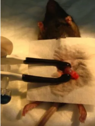
anesthetised with a ketamine and xylazine mixture and both testes exposed by a cut in the lower abdominal area. Using a 30-gauge needle a puncture was made in the tunica albuginea to facilitate the insertion of the glass micropipette.
For this experiment we used the same construct used for pronuclear microinjection (CMV-GFP-WPRE-polyA), and the same fragment, extracted from an agarose gel after plasmid restriction with the same restriction enzymes. The DNA solution used (where the CMV-GFP-WPRE-polyA DNA fragment was at a concentration of 0,5µg/µ l, also contained a 0,04% dye called Trypan Blue which allowed us to observe the localization of the solution during the injection. About 20µl of DNA solution (≈10µg of DNA) was injected into the intertubular space in three different directions to ensure its maximum spread.
Only one of the testis was submitted to this procedure, and the contralateral one was removed. This ensured that all the sperm produced by the animal came from the electroporated testis and allows a better comparison within animals.
Electroporation (Figure 4)
14 Breeding and phenotype analysis
Once the animal had recovered from the surgery for one week, it was bred with
C57BL6/J females over at least 35 days, which is the period of time taken for a cycle of spermatogenesis. In the females, the presence of plugs was registered because it allowed inference about males’ reproductive fitness. Females were euthanized at 16,5 dpc and embryos collected and screened for green fluorescence under a stereoscope (Zeiss Stereo Lumar.V12).
15
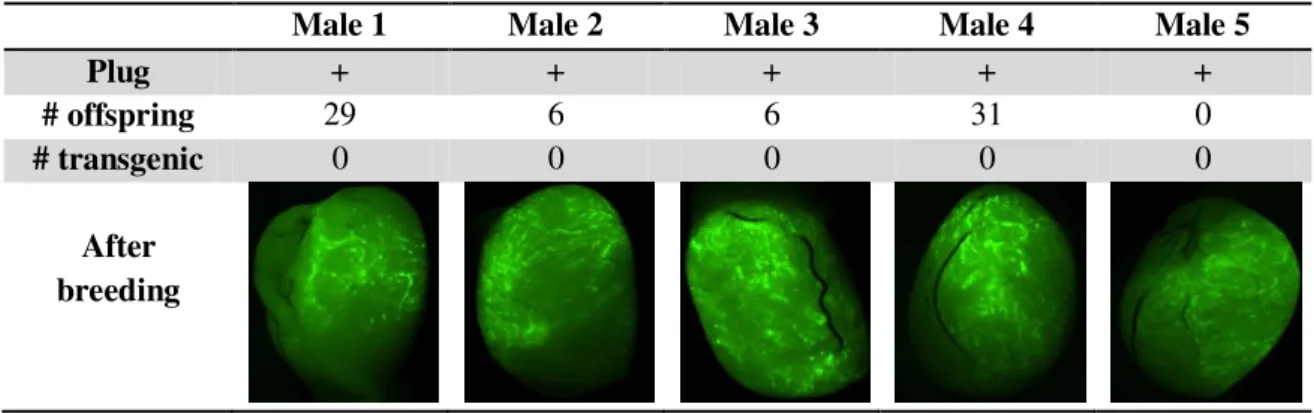
Results and Discussion
According to the results shown in Table 4, testis electroporation appeared to result in successful cell transfection with the formerly injected DNA fragment. However, there was not enough data to verify if the transfected cells are spermatogonia or what their proportion was.
Table 4: Results from testis electroporation with CMV-GFP sequence
Male 1 Male 2 Male 3 Male 4 Male 5
Plug + + + + +
# offspring 29 6 6 31 0
# transgenic 0 0 0 0 0
After breeding
16
4.
Infection of seminiferous tubule cells with lentiviruses
Proof of principle - Testing lentiviral transfection
Before performing the experimental protocol in testes, the transfection efficiency of the viral vectors was verified by cell transduction. The virus used was a GFP-carrying lentivirus obtained from a commercial stock from Meditecno (GFP Lentivirus Control LTV-300), while the cells used in the transfection experiments were the 293 LTV Cell Line. Cell transfection was performed according to the protocol provided by the
company (see Appendix III). After transfection, the cells were kept for 72 hours at 37ºC under a CO2 atmosphere, and at this moment green cells were observed under a
microscope, confirming the efficiency of the used lentiviral transduction approach (data not shown).
Material and Methods
Lentivirus transduction in vivo
Five C57BL6/J males age 30-40 days old were injected via rete testis with 10µ l of Dulbecco modified Eagle medium supplemented with 10% fetal calf serum
(DMEM/FCS) containing lentiviral particles at a concentration of 1x10⁴ IU/µ l (32). Males were anesthetized with a ketamine and xylazine mixture, and the non-injected testis was removed in order to avoid the dilution of the final sperm. Males were used 6 weeks after the surgery for mating with BL6 females. Three breeding pairs were set up for each male.
17
Results and Discussion
As demonstrated by the results shown in Table 5, only two of the 5 males (40%) submitted to this technique remained fertile. This result suggested that either the
injection of the lentivirus in the rete testis damaged the functionality of the structure due to a physical trauma, as these are small and sensitive tubules. The damage may also have arisen from any inflammatory process caused by the virus itself since it is not expected that the animal carries antibodies against lentivirus, once it is housed in microbiologically controlled environment.
Table 5: Lentiviral transfection results
Male 1 Male 2 Male 3 Male 4 Male 5
Presence of plug Yes Yes No No No
# Pups 11 17 0 0 0
# Transgenic 0 0 0 0 0
Also, by observing testis under a fluorescent stereoscope (Figure 5), it was noted that the spread of infection was very limited as demonstrated by the lack of fluorescence in the testes. The green color observed in the pictures B and D of Figure 5 is due to autofluorescence process and does not correspond to GFP expression. This situation was also confirmed as a result of the analysis of testis slices, where no green cells were observed.
A B C D
Figure 5: in vivo Lentiviral injection. A) Testes injected with lentiviral vector of GFP seen with transmitted light channel. B) The same testes as in A seen with GFP channel showing the absence of GFP expression but with considerable autofluorescence. C)
18
In the proof of principle experiment, good transfection efficiencies were obtained due to the use of reagents that enhance the transduction efficiency. These reagents are
19
5.
Application of the techniques
After testing the previous techniques using a reporter gene, we decided to repeat the same experiments using the gene of interest. The chosen gene was the light-gated ion channel channelrhodopsin-2 fused with a fluorescent protein, under the control of a promoter for dopaminergic cells.
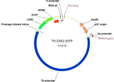
Specifically, the DNA fragment used includes the rat tyrosine hydroxilase (TH) promoter to drive the expression of the wild type ChR2 fused with the reporter protein enhanced yellow fluorescent protein - ChR2-EYFP. A heterologous intron was also cloned between the promoter and the gene in order to increase the expression efficiency of the gene (40). A WPRE and polyA tail were also cloned downstream to the previous sequences (final sequence in Appendix II). The final result displayed in Figure 6. The DNA fragment used in the transgenesis experiments was obtained by cutting the plasmid with HindIII and PvuI restriction enzymes (NEBiolabs) followed by its purification with QIAquick Gel Extraction Kit (Quiagen).
After the cloning steps, the previously mentioned techniques were applied to obtain founders of the desired transgenic line, as described below.
20
5.1.
Pronuclear Microinjection
Material and Methods
After the fragment was prepared, pronuclear microinjection was performed following the same methodology described above. In this case, the offspring were screened for the presence of the transgene after the weaning age (≥ 21 days).
PCR analysis
The DNA was extracted and purified from a piece of tail tip biopsies using Extract-N-Amp Tissue PCR Kit (Sigma-Aldrich). The animals were screened by polymerase chain reaction (PCR) analysis using GFP universal primers (THGFP-F:
AAGTTCATCTGCACCACCG and THGFP-R: TGCTCAGGTAGTGGTTGTCG), which allows the amplification of a GFP-specific, 450bp product. The PCR reactions, carried out using a VWR Duo thermal Cycle, consisted of a thermal profile that included 35 cycles of denaturation at 94°C for 30 sec, primer annealing at 55°C for 30 sec and primer extension at 72°C for 7 min.
Perfusion
The animals were anesthetized using a ketamine and xylazine mixture. The beating heart is exposed, a needle is inserted in the left ventriculum and a cut is made in the right auricular. 20ml of saline is infused in the left ventriculum using a pump. A cut made to the right auricula to allow the blood flow. After the saline, the same amount of 4% paraformaldehyde (PFA) was infused. The head of the animal was severed, and the brain removed from the cranial bones using forceps. The brain was kept overnight in 4% PFA, in the cold (4ºC), and then processed for histology.
Histology and immunohistochemistry
21
minutes each were performed with fresh PBS. Secondary antibodies (Alexa Fluor 594 Goat anti – mouse) were then added in PBS-T 0,4% at a dilution of 1/1000 and
22
Results and Discussion
Three of the five obtained animals showed the presence of the transgene in the genome as seen in Table 6. One of these 3 animals (F0) died after birth and the remaining two were crossed with wild-type animals.
Table 6: Results from TH-ChR2-EYFP pronuclear microinjection
Microinjection # Females # Zygotes Transfer # Pups
Total Injectable # Embryos Total # Tg
1 8 110 60 25 3 1
2 8 168 86 34 0 0
3 9 220 60 60 1 1
4 10 172 101 22 1 1
5 6 80 45 33 0 0
Total 41 750 352 174 5 3
The offspring was also screened by PCR for the transgene presence as shown in Figure 7. The F1 animals also showed the presence of the transgene which inferred that the F0 animals could be good founders.
Figure 7: PCR analysis of transgenic animals and respective offspring. Results of PCR using genomic DNA (gDNA) obtained from ear biopsies of progeny generated from founders 2 and 3 (F0). WT = gDNA of wild type mice (C57BL6/J animal), + = genomic DNA from an animal known to be a transgenic animal; H2O = blank without
23
The animals that revealed GFP band in the PCR were submitted to a perfusion protocol with fixative and the brain was removed and processed for histology.
Immunohistochemistry to tag both YFP and dopaminergic cells using green and red fluorescent secondary antibodies (to reveal the YFP and dopaminergic neurons, respectively) was carried as described previously.
As seen in Figure 8, the dopaminergic cells located in the ventral tegumental area (VTA) and in the substancia nigra seemed normal and healthy, but no ChR2 expression was detected in any of them. Figure 8 shows some examples selected from totality of animals observed, as the results are consistent across all animals.
24
Figure 9: PCR analysis of offspring of the electroporated males that remained fertile. Results of PCR using genomic DNA (gDNA) obtained from ear biopsies WT = gDNA of wild type mice (C57BL6/J animal), + = genomic DNA from an animal known to be a transgenic animal; H2O = blank without any DNA, only
water. This offspring was generated by mating the males with C57BL6/J females.
5.2.
Testis Electroporation
Material and Methods
The DNA preparation and the technique were performed following exactly the same methodology described above. Five 1 month old male mice were submitted to this procedure.
Results and Discussion
The offspring generated by each male was screened by PCR analysis using the same previously used primers and reaction conditions, in order to detect the presence of the transgene. Same examples of the PCR results for the offspring of each animal are depicted in Figure 9.
In Table 7, the performance of each male submitted to the electroporation protocol with the referred transgene are summarized as well as the number of animals generated and the percentage of transgenic offspring obtained.
Table 7: Results of testis electroporation using TH-ChR2-EYFP sequence
Male 1 Male 2 Male 3 Male 4 Male 5
Plug presence No Yes Yes Yes Yes
# offspring 0 29 25 16 13
25
The animals that showed a GFP band in the PCR were perfused with fixative and the brains removed. The brains were submitted to immunohistochemistry using antibodies to tag dopaminergic neurons (red fluorescence) and EYFP (ChR2; yellow-green fluorescence), exactly as described in the previous sections. The results are shown below in Figure 10.
Figure 10: Characterization of ChR2-EYFP expression in Ventral tegumental area in transgenic mice generated by Testes Electroporation. A-L) Zeiss AxioImager images of four different VTA areas from different transgenic animals showing the absence of EYFP expression in dopaminergic cells (upper panel) and the same cells after TH-staining (middle panel). Lower panel: merged images of YFP and anti-TH staining.
4X
4X
4X
10X
10X
10X
10X
10X
10X
20X
20X
26
5.3.
Viral injection
27
6.
Adeno-associated Virus
in vivo
injection
In order to test if the lack of expression of the ChR2 in the dopaminergic cells obtained with the previous techniques was due to the construct used, adeno-associated virus serotype 1 (AAV2/1) carrying a hChR2-EYFP were injected in the substancia nigra of a THCre knock in mice expressing a Cre recombinase in dopaminergic neuros – Ventral tegumental area (VTA) and substantia nigra. This virus carries the hChR2 sequence inverted and within a double pair of lox P sites as seen in Figure 11. This strategy assures that the hChR2 sequence will be specifically expressed in the THCre positive cells. The AAV-1 was injected directly in the mouse brain through stereotaxic surgery (7) and the stereotaxic coordinates were found by consulting The Mouse Brain in Stereotaxic Coordinates (42).
Figura 11: schematic representation of the plasmid carrying a AAV double floxed
28
Material and Methods
Surgery and AAV infection
A 2 month old TH-Cre animal was anesthetized with volatile isofluorane. The hair of the head was shaved and the skin cleaned with betadine and 70% ethanol. When reaching a stage 3 of anesthesia – the surgical stage (51) - the animal was placed in a stereotaxic frame (Dual Ultra Precise Stereotaxic Apparatus Kopf) and fixed with two ear bars. The coordinates used for viral injection and cannula implantation were the following relatively to Bregma: AP, –3.0 mm; DV, –3.3 mm; and ML, ± 0.5 mm. Ophthalmic ointment was applied in the eyes to avoid drying and blindness, and the animals were kept on a warm pad to avoid hypothermia.
The bone sutures of the skull were exposed using a stereoscope and two small holes made using a dental drill. A glass micropipette for virus injection was made on a Sutter puller with the following characteristics: ≥ 2mm of length and tip diameter between 20 and 50 µ m. The injection was done bilaterally using an AAV suspension at a titter of 1012 infectious particles per millilitre. The injection was done using a NanoJect II and a microinjector (Picospritzer) device, for 27 min at a flow rate of 4.6nl every 5 sec, which results in the injection of approximately 1.5µ l of total volume. After injection, I waited 15 minutes before pulling the micropipette to avoid the reflux of viral suspension. A cannula containing an optical fiber was implanted in both holes, fixed to the skull with dental acrylic, and the wound closed using Vet bond® tissue adhesive.
In vivo confirmation of viral transfection
After the animal has recovered from surgery, it was submitted to brain stimulation by connecting a laser to the implanted cannula. Stimulation consisted in shinning blue light into the brain in 5 pulses of 1mW power, 14Hz. If the recombination of the virus had occurred at the expected location in the brain, when shining a light into one hemisphere, the animal would rotate to the contralateral one. This is an indicator of the functionality of the surgery and that the animal is ready for experiments. After the injection
procedure and validation of efficient of the recombination by mice behavioural
29
Results and Discussion
As seen in Figure 12, the target area of infection was perfectly reached and
recombination occurred specifically in the substancia nigrareticulata (SNr) and ventral tegmental areas. The histological pictures also show that the cells labelled with green fluorescence are indeed the dopaminergic cells.
30
7.
Conclusion
We were able to replicate the results obtained by Majumdar, (33) and here we show that this method can successfully transfer DNA into testicular cells, as confirmed by PCR analysis. Furthermore, the obtained results have also demonstrated that transmission of the transgene to the offspring had taken place.
Regarding testis electroporation, some studies have shown that this technique has no permanent adverse effects for either testis integrity or sperm quality. Nevertheless, our results indicate that electroporation is, at least under the experimental conditions used, accompanied by damage in spermatogenesis process (18; 28; 50). In this study, and due to scarcity of the obtained data, we cannot conclude whether the infertility caused was transitory or permanent but it was possible to conclude that this method can cause infertility in 20% of the males submitted to this procedure. The possible causes for this may be the trauma caused either by the surgery, the heat generated with the
electroporation, or by the high pressure in the testis caused by the injection of the 20 µl of solution.
As far as the absence of transgenic offspring using a reporter gene is concerned it is impossible to draw any statistically sound conclusions due to the small number of animals submitted to this method. One of the downsides of the experimental approach followed is associated to the possibility that foreign genes may likely to be present inside the cells in an episomal form, when transferred by in vivo electroporation (38) which turns gene expression transient. In fact, the percentage of transfected germ line with this method was shown to decreases with time, from 1.3%-2.0% of all the germ line cells after 7 days after the electroporation to 0% 1 month after electroporation (53). Other studies also estimate that only about 5.0-10% of the epididymal sperm should be carrying the transgene (18) resulting in the high unpredictability of the results.
31
In our hands, the injection of lentiviral solution in the rete testis did not result in the generation of any transgenic animals. This could have been due to the low titter of the virus suspension used, the low spread of infection (which is an intrinsic characteristic of the virus), or lack of infection of germ line.
As a summary, when comparing the three methods tested here, the one that generated higher percentage of transgenic mice is testicular electroporation as seen in the Table 8: Number of transgenic mice/total offspring for each technique
Table 8: Number of transgenic mice/total offspring for each technique
PNMI Testes electroporation Lentiviral injection
CMV – GFP 5/ 16 0/ 72 0/28
TH-ChR2-EYFP 3/6 53/ 83 _
In terms of gene expression, the reporter gene showed good expression in all the transgenic animals generated by pronuclear microinjection. When using the testes electroporation method, the expression of GFP in the offspring was not, however, observed. In any case the small numbers of animals used does not allow us to draw any significant conclusion from the obtained data. However, in a qualitative perspective, the lentiviral injection of the reporter gene did not, result in any GFP positive animal either. Unfortunately, and again, the small number of animals used does not allow further considerations . The low titter of the virus or gene-silencing processes, like
hypermethylation for example, that has been seen in lentiviral integrants (22) can be a possible reason to justify the results obtained.
Conversely, when using the TH-ChR2-YFP construct, both testes electroporation and pronuclear microinjection generated transgenic offspring in which it was possible to detect the transgene by PCR. However, when analysing the processed slides, there was no expression of ChR2-EYFP in the dopaminergic neurons. The intrinsic characteristics of the construct could be a possible cause for the lack of expression, although the AAV injection of the same sequence resulted in expression of ChR2 inside the target cells – dopaminergic neurons. This suggests that the absence of biological activity of ChR2 in dopaminergic cells of the transgenics generated using the techniques described
32
Also, the first hypothesis postulated in the beginning of this thesis, that one possible reason for the absence of ChR2 expression would be the result of cellular death caused by the accumulation of the channel in the membrane, was not confirmed according. As a matter of fact our data, the histological slices show healthy dopaminergic cells in the transgenic animals. The second hypothesis, that the wild type version of ChR2 could result in higher levels of expression than the optimized version, does not seem to have been confirmed either, according to the data shown. However, more experiments and control animals would be necessary to achieve a proper conclusion.
The Cre-Lox systems, combined with the injection of AAV, have been found to be a good alternative for targeting a specific population of cells with a given gene. The problems associated with this method are the need to submit all the experimental animals to a surgery which, besides increasing the financial costs involved, adds more variants to the experimental system, due to users’ manipulation variability, animal recovery, and viral infection timing. However, the production of a transgenic animal that is able to express a given target protein in the right population of cells would always be of added value to researchers due to shorter experimental time-spans and increasing reproducibility of results. Even if the production of a transgenic line is very expensive, in the long term it may became cheaper if researchers take into account the cost of surgeries, viral production and expansion and labour time.
There are alternatives for trangenesis that could help to overcome these problems. One possibility is the performance of intra-cytoplasmatic sperm injection (ICSI) with pre-treated sperm, which membranes have been permeabilized to allow the introduction of exogenous DNA. This has been successfully done (21; 36), and the results obtained have been similar to those resulting from the use of traditional pronuclear
microinjection (20). Unfortunately, this method also requires expensive equipment and skilled personnel.
Another alternative is the microinjection of both bacterial artificial chromosomes
(BACs) and yeast artificial chromosomes (YACs) since they are designed in such a way that allows the integration of the gene in a specific place in the genome and the
33
34
References
1. Amaral M, Campos V, Seixas F, Cavalcanti P, Selau L, Deschamps J, Collares T (2011). Testis-mediated gene transfer in mice: comparison of transfection reagents regarding transgene transmission and testicular damage. Biol Res 44: 229-234 2. Arenkiel B., P. J. (2007). In Vivo Light-Induced Activation Expressing
Channelrhodopsin-2 of Neural Circuitry in Transgenic Mice. Neuron 54 , 205–218. 3. Auerbach, A. B. (2004). Production of functional transgenic mice by DNA
pronuclear. Acta Biochimica Polonica, Vol. 51 No. 1 , 9-31.
4. Bachiller D, Schellander K, Peli J, Rüther U (1991). Liposome-mediated DNA uptake by sperm cells. Mol Reprod Dev. Nov;30(3):194-200.
5. Blanchard KT, Boekelheide K (1997). Adenovirus-mediated gene transfer to rat testis in vivo. Biol Reprod. Feb;56(2):495-500.
6. Brinster RL and Palmiter RD. (1984-1985). Introduction of genes into the germ line of animals. Harvey Lect. 80:1-38.
7. Cetin A, Komai S, Eliava M, Seeburg PH, Osten P (2006). Stereotaxic gene delivery in the rodent brain. Nat Protoc;1(6):3166-73.
8. Chang KT, Ikeda A, Hayashi K, Furuhata Y, Banai M, Nishihara M, Ohta A, Ogawa S, Takahashi M (1999). Possible mechanism for the testis mediated gene transfer as a new method for producing transgenic animals. J Reprod Dev 45: 37-42.
9. Cho A, Haruyama N, Kulkarni A.B (2009). Generation of Transgenic Mice. Curr Protoc Cell Biol. March; chapter: Unit–19.11
35
11.Coward K., Kubota H., Parrington J. (2007). In vivo gene transfer into testis and sperm: Developments and future application. Systems biology in reproductive medicine, 53:4, 187-197.
12.Dong JY, Fan PD, Frizzell RA( 1996). Quantitative analysis of the packaging capacity of recombinant adeno-associated virus. Hum Gene Ther. Nov 10;7(17):2101-12.
13.Flagel SB, Clark JJ, Robinson TE, Mayo L, Czuj A, Willuhn I, Akers CA, Clinton SM, Phillips PE, Akil H. (2011). A selective role for dopamine in stimulus-reward learning. Nature. Jan 6;469(7328):53-7. Epub 2010 Dec 8.
14.Gordon et al. Integration and stable germ line transmission of genes injected into mouse pronuclei (1981) . Science December, 1244-1246.
15.Hacein-Bey-Abina S, et al (2003). LMO2-associated clonal T cell proliferation in two patients after gene therapy for SCID-X1. Science. Oct 17;302(5644):415-9.
16.Hamaguchi I, Woods NB, Panagopoulos I, Andersson E, Mikkola H, Fahlman C, Zufferey R, Carlsson L, Trono D, Karlsson S. (2000) Lentivirus vector gene expression during ES cell-derived hematopoietic development in vitro. J Virol. Nov;74(22):10778-84.
17.Heintz N (2001). BAC to the future: the use of bac transgenic mice for neuroscience research. Nat Rev Neurosci. Dec;2(12):861-70.
18.Hibbitt O, Coward K, Kubota H, Prathalingham N, Holt W, Kohri K, Parrington J. In vivo gene transfer by electroporation allows expression of a fluorescent transgene in hamster testis and epididymal sperm and has no adverse effects upon testicular integrity or sperm quality. (2006) Biol Reprod. Jan;74(1):95-101.
36
20.Hirabayashi M, Kato M, Amemiya K, Hochi S (2008). Direct comparison between ICSI-mediated DNA transfer and pronuclear DNA microinjection for producing transgenic rats. Exp Anim. Apr;57(2):145-8.
21.Hirabayashi M, Hochi S.(2010) Generation of transgenic rats by ooplasmic injection of sperm cells exposed to exogenous DNA. Methods Mol Biol. 2010;597:127-36. 22.Hofmann A, Kessler B, Ewerling S, Kabermann A, Brem G, Wolf E, Pfeifer A.
(2006). Epigenetic regulation of lentiviral transgene vectors in a large animal model. Mol Ther. Jan;13(1):59-66.
23.Kanatsu-Shinohara M, Toyokuni S, Shinohara T.(2004). Transgenic mice produced by retroviral transduction of male germ line stem cells in vivo. Biol Reprod.
Oct;71(4):1202-7.
24.Kentaro, Y. (2008). Mammalian testis: A target of in vivo electroporation. Develop Growth Differ, 50 , 513-515.
25.Kim JH, Jung-Ha HS, Lee HT, Chung KS (1997). Development of a positive method for male stem cell-mediated gene transfer in mouse and pig. Mol Reprod Dev; 46:515–526.
26.Kojima Y, Sasaki S, Umemoto Y, Hashimoto Y, Hayashi Y, Kohri K (2003). Effects of adenovirus mediated gene transfer to mouse testis in vivo on spermatogenesis and next generation. J Urol. Nov;170(5):2109-14.
27.Kojima Y, Hayashi Y, Kurokawa S, Mizuno K, Sasaki S, Kohri K. (2008). No evidence of germ-line transmission by adenovirus-mediated gene transfer to mouse testes. Fertil Steril.May;89(5 Suppl):1448-54.
28.Kubota H, Hayashi Y, Kubota Y, Coward K, Parrington J. Comparison of two methods of in vivo gene transfer by electroporation (2005). Fertil Steril. Apr;83 Suppl 1:1310-8.
37
30.Lima SQ, Hromádka T, Znamenskiy P, Zador AM.(2009).PINP: a new method of tagging neuronal populations for identification during in vivo electrophysiological recording. PLoS One. Jul 7.
31.Lin JY. (2011). A user's guide to channelrhodopsin variants: features, limitations and future developments. Exp Physiol. Jan;96 (1):19-25.
32.Lois C. Advanced Protocols for Animal Transgenesis. An ISTT Manual. Chapter 10: Generation of Transgenic Animals with Lentiviral Vectors. Springer 2011. 33.Majumdar, S. D. (2008). Transgenesis via permanent integration of gene in
repopulating spermatogonial cells in vivo. Nature Methods vol.5 nº.7 , 601-603. 34.Mancheño-Corvo P, Martín-Duque P. (2006). Viral gene therapy. Clin Transl
Oncol. Dec;8(12):858-67.
35.Masahito Ikawa, N. T.-Y. ( 2003). Generation of Transgenic Mice Using Lentiviral Vectors. Nature Molecular Therapy, Vol 8, Nº 4 .
36.Moisyadi S, Kaminski JM, Yanagimachi R (2009). Use of intracytoplasmic sperm injection (ICSI) to generate transgenic animals. Comp Immunol Microbiol Infect Dis. Mar;32(2):47-60.
37.Muramatsu T, Shibata O, Ryoki S, Ohmori Y, Okumura J.Foreign gene expression in the mouse testis by localized in vivo gene transfer. Biochem Biophys Res Commun. 1997 Apr 7;233(1):45-9.
38.Muramatsu T, Nakamura A, Park HM. In vivo electroporation: a powerful and convenient means of nonviral gene transfer to tissues of living animals (Review). (1998). Int J Mol Med. Jan;1(1):55-62.
39.Nagy, A. (2003). Manipulating the mouse embryo. Cold spring harbor laboratory press.
38
41.Pañeda, A. (2006). Effect of WPRE on Transgene Expression Using Different Promoters in the Context of Hydrodynamically Delivered Plasmid Vectors. Molecular Therapy , S413 - S414.
42.Paxinos G., Keith B. J. Franklin. The mouse brain in stereotaxic coordinates 3rd edition, 2008. Academic Press ISBN: 9780123694607
43.Rose C, Schwegler H, Hanke J, Yilmazer-Hanke D.M (2012). Pregnancy rates, prenatal and postnatal survival of offspring, and litter sizes after reciprocal embryo transfer in DBA/2JHd, C3H/HeNCrl and NMRI mice. Theriogenology, Volume 77, Issue 9 , Pages 1883-1893.
44.Sato, M. (2006). Direct gene delivery to murine testis as a possible means of transfection of mature sperm and epithelial cells linin epididymal ducts. Reproductive Medicine and Biology 2006 , 1-7.
45.Sawamoto K., Nakao N.,Kobayashi K., Matsushita N., Takahashi H., Kakishita K., Yamamoto A, Yoshizaki T,,Terashima T,, Murakami F, Itakura T., Okano H. (2001). Proc Natl Acad Sci U S A. May 22; 98(11): 6423–6428.
46.Schaffer DV, Koerber JT, Lim KI (2008). Molecular engineering of viral gene delivery vehicles. Annu Rev Biomed Eng;10:169-94.
47.Somia N, Verma IM.(2000) Gene therapy: trials and tribulations. Nat Rev Genet. Nov;1(2):91-9.
48.Thomas CE, Ehrhardt A, Kay MA (2003). Progress and problems with the use of viral vectors for gene therapy. Nat Rev Genet. May;4(5):346-58.
49.Tsai HC, Zhang F, Adamantidis A, Stuber GD, Bonci A, de Lecea L, Deisseroth K. (2009). Phasic firing in dopaminergic neurons is sufficient for behavioral
conditioning. Science. May 22;324 (5930):1080-4.
39
51.Van Zuphen LFM, Baumans V and Beynen AC. (2001). Principles of laboratory animal science, revised edition. Elsevier Ltd.
52.West M. F., Verrotti A.C., Salle´ s F. J., Tsirka S.E. and Strickland S. (1996). Isolation and Characterization of Two Novel,Cytoplasmically Polyadenylated, Oocyte-Specific, Mouse Maternal RNAs. Developmental Biology, 175 , 132-141. 53.Yomogida K, Yagura Y, Nishimune Y. (2002). Electroporated Transgene-Rescued
Spermatogenesis in fertile mutant mice with a sertoli cell defect. Biology of Reproduction 67, 712-717.
54.Yonezawa T, Furuhata Y, Hirabayashi K, Suzuki S, Takahashi M, Nishihara M (2001). Detection of transgene in progeny at different developmental stages following testis mediated gene transfer. Mol Reprod Dev 60: 196-201.
55.Zhang F, Wang LP, Boyden ES, Deisseroth K. (2006). Channelrhodopsin-2 and optical control of excitable cells. Nat Methods. Oct;3(10):785-92.
56.Zhao S, Ting JT, Atallah HE, Qiu L, Tan J, Gloss B, Augustine GJ, Deisseroth K, Luo M, Graybiel AM, Feng G (2011). Cell type–specific channelrhodopsin-2
40
Appendix I: CMV-GFP-WPRE-polyA
41
Appendix II: TH-ChR2-EYFP
44
ctcgccgaccactaccagcagaacacccccatcggcgacggccccgtgctgctgcccgacaaccactacctgagctaccagtccgccctgagcaaag
accccaacgagaagcgcgatcacatggtcctgctggagttcgtgaccgccgccgggatcactctcggcatggacgagctgtacaagtaatagactagt
cacatctagcgataatcaacctctggattacaaaatttgtgaaagattgactggtattcttaactatgttgctccttttacgctatgtggatacgctgctttaatgc
ctttgtatcatgctattgcttcccgtatggctttcattttctcctccttgtataaatcctggttgctgtctctttatgaggagttgtggcccgttgtcaggcaacgtgg
cgtggtgtgcactgtgtttgctgacgcaacccccactggttggggcattgccaccacctgtcagctcctttccgggactttcgctttccccctccctattgcc
acggcggaactcatcgccgcctgccttgcccgctgctggacaggggctcggctgttgggcactgacaattccgtggtgttgtcggggaaatcatcgtcct
ttccttggctgctcgcctgtgttgccacctggattctgcgcgggacgtccttctgctacgtcccttcggccctcaatccagcggaccttccttcccgcggcct
gctgccggctctgcggcctcttccgcgtcttcgccttcgccctcagacgagtcggatctccctttgggccgcctccccgcatcgctagagggccctattct
atagtgtctctagagggcccgtttaaacccgctgatcagcctcgactgtgccttctagttgccagccatctgttgtttgcccctcccccgtgccttccttgacc
ctggaaggtgccactcccactgtcctttcctaataaaatgaggaaattgcatcgcattgtctgagtaggtgtcattctattctggggggtggggtggggcag
45
Appendix III: Transduction Protocol
ViraDuctin™ Lentivirus Transduction Kit. Product Manual. Catalog number LTV-200. Cell Biolabs
I. Transduction of Adherent Cells
1. The day before transduction, trypsinize and count the cells, plating 0.2-2 x 10⁵ cells in 0.5
mL complete culture medium per well of a 24-well plate. Incubate cells at 37°C overnight.
2. On the day of transduction, thaw your lentiviral stock and dilute the lentiviral stock into
complete culture medium to a final volume of 0.5 mL in a sterile tube. Mix by inverting; do not vortex. You may prepare serial dilutions if desired.
3. Add 5 μL of ViraDuctin™ Lentivirus Transduction Reagent A (100X), mix by inverting.
Immediately add 5 μL of ViraDuctin™ Lentivirus Transduction Reagent B (100X) and mix
by inverting.
4. Incubate 30 minutes at 37°C.
5. Remove the culture medium from the cells. Apply all lentivirus/ ViraDuctin™ complexes to
cells. Refer to the literature to determine the proper MOI for your specific cell.
6. Incubate at 37°C overnight.
7. Remove the media containing virus and replace with 0.5 mL of complete culture medium.
8. Dilute the appropriate amount of ViraDuctin™ Lentivirus Transduction Reagent C (8X) to
1X with complete culture medium (for example, add 70 μL of 8X Reagent C to 490 μL of
complete culture medium).
9. To completely remove the transduction complex, remove the culture medium and replace
with 500 μL of the diluted ViraDuctin™ Lentivirus Transduction Reagent C (1X) in each
well; gently rock the plate for 30-60 seconds. IMMEDIATELY aspirate the medium
containing ViraDuctin™ Lentivirus Transduction Reagent C and replace with 0.5 ml of
complete culture medium. Wash twice with complete culture medium to remove any residue complex.
10. 48-72 hrs after transduction, proceed with desired method of detection including functional
46 II. Transduction of Suspension Cells
1. On the day of transduction, thaw your lentiviral stock and dilute the lentiviral stock into
complete culture medium to a final volume of 0.5 mL in a sterile tube. Mix by inverting; do not vortex. You may prepare serial dilutions if desired.
2. Add 5 μL of ViraDuctin™ Lentivirus Transduction Reagent A (100X), mix by
inverting. Immediately add 5 μL of ViraDuctin™ Lentivirus Transduction Reagent B
(100X) and mix by inverting.
3. Incubate 30 minutes at 37°C.
4. Pellet your suspension cells for 5 minutes at 1000 g and remove supernatant. Resuspend
cell pellet by adding lentivirus/ ViraDuctin™ complexes. Refer to the literature to
determine the proper MOI for your specific cell.
5. Incubate at 37°C overnight.
6. Centrifuge for 5 minutes at 1000 g; remove the media containing virus and replace with
0.5 ml of complete culture medium.
7. Dilute the appropriate amount of ViraDuctin™ Lentivirus Transduction Reagent C (8X)
to 1X with complete culture medium (for example, add 70 μL of 8X Reagent C to 490
μL of complete culture medium).
8. To completely remove the transduction complex, centrifuge for 5 minutes at 1000 g and
remove the supernatant. Add 500 μL of the diluted ViraDuctin™ Lentivirus
Transduction Reagent C (1X) to each well and gently rock the plate for 30-60 seconds.
9. Centrifuge for 5 minutes at 1000 g; IMMEDIATELY aspirate the medium containing
ViraDuctin™ Lentivirus Transduction Reagent C and resuspend in 0.5 ml of complete
culture medium. Repeat twice to remove any residue complex.
10.48-72 hrs after transduction, proceed with desired method of detection including