Review
Printed in Brazil - ©2016 Sociedade Brasileira de Química0103 - 5053 $6.00+0.00
*e-mail: [email protected], [email protected]
Intended and Unintended Consequences and Applications of Unnatural Interfaces:
Oligo
p
-Phenylene Ethynylene Electrolytes, Biological Cells and Biomacromolecules
Harry C. Pappas,a,b Patrick L. Donabedian,a,b Kirk S. Schanze*,c and David G. Whitten*,b
aDepartment of Nanoscience and Microsystems Engineering and bCenter for Biomedical
Engineering, Department of Chemical and Biological Engineering, University of New Mexico, 87131-1341 Albuquerque-New Mexico, United States of America
cDepartment of Chemistry, University of Florida, 32611-7200 Gainesville-Florida, United States of America
This short review focuses on an extended study of synthetic oligomericphenyleneethynylene (OPE) electrolytes that have been investigated for both their antimicrobial activity and their fluorescence sensing properties. In both cases, interfaces between these synthetic electrolytes and naturally occurring materials such as proteins, lipids, nucleic acids and cells are critical to their function or activity. The review contains a general overview with a focus at the end on three recent examples including biological and chemical sensing, induction of spore germination and fluorescent detection of amyloid protein aggregates.
Keywords: conjugated polyelectrolyte, phenylene ethynylene, oligomer, biosensing, biocide, bacteria
1. Introduction
We are writing this at a time when the scientific enterprise is facing a challenge where, as suggested by Whitesides,1 “core disciplines have drifted more towards ‘iteration’ and ‘improvement’ and away from discovery”. In this review we discuss a research project that began when we were in the course of developing biosensors based on synthetic fluorescent conjugated polyelectrolytes (CPE) and we started an orthogonal investigation of these materials as potential antimicrobial agents against vegetative bacteria and bacterial spores.2 In the initial investigations we found that a cationic CPE was a potent antimicrobial against Escherichia coli (E. coli) vegetative cells and Bacillus anthracis Sterne (B. anthracis) spores. The initial results stimulated a research program that has included not only “iteration” and “improvement” efforts but also a significant amount of “discovery” focused research. A key point we will develop is that the “discovery” focused research efforts have themselves led to important consequences and potential applications that are driven by new ideas and, above all, curiosity.
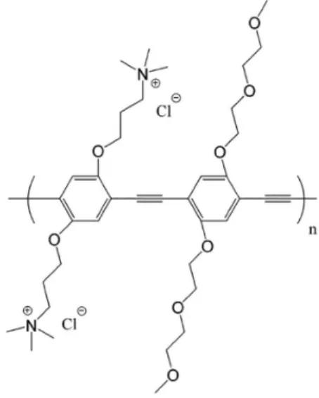
The specific focus of this review is on the properties and interfacial activity of a series of charged, conjugated
polymeric phenylene ethynylene (PPE, Figure 1). These compounds and their antimicrobial properties and reactivity have been investigated by a number of researchers in laboratories at the Universities of Florida (UF) and New Mexico (UNM) over the last ten years. As indicated above the research was initiated by an exploratory investigation of a cationic CPE with two representative bacterial species.2 The polymers exhibit a number of interesting properties in that while they are polyelectrolytes and water soluble, they are also moderately to strongly hydrophobic. The first conjugated polyelectrolyte that our research group worked with was an anionic poly(phenylene vinylene) derivative. We found that this material was fluorescent in aqueous solution and that its fluorescence was quenched by very low concentrations of neutral or cationic electron acceptors.3 In subsequent investigations it was found that these materials also could easily coat oppositely charged supports including planar surfaces and microspheres.
microspheres but to do this we turned to the use of phenylene ethynylene polyelectrolytes (PPE) proposed by Schanze and co-workers6 due to their much stronger fluorescence and our finding that they retained their fluorescence when adsorbed onto solid surfaces.5,6 For our initial probes into their antimicrobial activity we added the cationic PPE (PPE-1) in aqueous solution to aqueous suspensions of E. coli and B. anthracis Sterne spores.2 In these experiments we observed that brief exposure of either bacteria to PPE-1 in the dark did not cause much harm, but on irradiation of the suspensions with visible light absorbed by PPE-1 there was efficient killing of both strains of bacteria. It was also observed through fluorescent microscopy that both bacteria contained a uniform coating of PPE-1 upon recovery of the treated bacteria by centrifugation.2
In subsequent studies at UNM and UF we carried out experiments to determine the mechanism of the light-activated biocidal activity of the cationic PPE. Two likely mechanisms were initially considered. The simplest seemed to be an activation of oxygen to its excited singlet electronic state followed by subsequent reaction and generation of other reactive oxygen intermediates. An alternative mechanism could be a photoinduced electron transfer whereby the excited PPE could abstract an electron from a halide counter ion leading to a reactive halide atom and an ion-radical from reduced PPE. Studies at UF demonstrated that irradiation of PPE such as PPE-1 in aerated solutions (water or methanol) results in formation of 1O
2* detected by chemical trapping or near infrared (IR) emission from the excited state.7 Studies at UNM showed that deaeration of aqueous suspensions of PPE with Gram-negative Pseudomonas aeruginosa attenuated the biocidal effect of irradiation.7 Thus it appears clear that generation of
singlet oxygen and subsequent generation of other reactive oxygen species can account for much of the light activated bactericidal effect.7
It was also found that in addition to the light activated biocidal activity, there is a dark biocidal activity of the PPE against bacteria that occurs on prolonged exposure of aqueous suspensions of PPEs and bacteria or at higher PPE concentrations. Investigations of the dark reactivity suggested that the PPE and other cationic CPE act by damaging the outer envelope of vegetative Gram-positive and Gram-negative bacteria. Through a series of investigations we obtained evidence that envelope damage by the PPEs is the driver for dark killing.8-11
2. Synthesis and Studies of Photophysics and Antimicrobial Activity of OPEs
Short molecular analogs of the phenylene ethynylene polyelectrolytes became attractive research topics for several reasons. First, we were interested to determine if these relatively small molecules (1, 2 or 3 polymer repeat units) would exhibit photophysical and antimicrobial properties analogous to the larger polymeric CPE. Additionally we considered that reliable computational studies of the oligomeric phenylene ethynylene (OPE) might be useful to model their modes of interaction with lipids, proteins and nucleic acids.
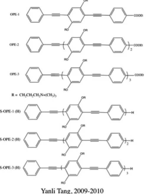
The synthesis of the OPE containing 1, 2 and 3 repeat units proved challenging, especially for the unsymmetrical OPE with sidearms shown in Figure 2. Nonetheless, we were able to obtain pure samples of the three OPE with ethyl ester substituents on one end and hydrogen on the opposite end (OPE-1, OPE-2 and OPE-3).12 It was a simpler and shorter synthetic path to “symmetrical OPE” (S-OPE-n(X)) with sidearms and the same substituents on each end.13 Concurrent with the synthesis of these two series of OPE with sidearms, a synthesis of a simpler series of OPE, absent of sidearms, with charged substituents on the ends was developed (Figure 2).14
Both the OPE with sidearms and these “end-only” OPE-1 (EO-OPE) derivatives have been found to have strong antimicrobial activity in both the dark and under light activation similar to that of the CPE described above.15,16 For the series of OPE with sidearms, the antimicrobial activity increases with the number of repeat units. The “end-only” OPEs have only a single repeat unit and are indicated by their limited solubility in water to be quite hydrophobic.14
In our first studies with the series of OPE, three unexpected results were observed as their photophysical properties were examined. The first was that while there is
Figure 2. Structures and names for representative OPEs discussed in this paper.
a substantial redshift in both absorption and fluorescence when we compared OPE-1 (or S-OPE-1(X)) with the corresponding OPE-2 derivatives, there was very little change when going from the two repeat unit to the three repeat unit OPEs.15 Previous to our studies with the charged OPEs, neutral, organic soluble OPEs had been studied both experimentally and computationally.17 In these studies it was suggested that the simplest OPE with one repeat unit should be planar, but with a very low barrier to rotation about the C−C bond between adjacent phenyl rings and acetylene units.17 We initiated a computational study of the series of OPE and these studies suggested that while an OPE with one repeat unit was planar, the OPEs with 2 and 3 repeat units were non-planar and that the highest occupied MO (molecular orbital) and lowest unoccupied MO did not extend over the entire conjugated system.15 Our computational results suggested that the π conjugated system may be composed of a series of segment chromophores that are equivalent in size to a zone slightly less than two repeat units.15 Support for this came from a study of similarly constituted CPE (same repeat unit structure) where the number of polymer repeat units (PRU) was controlled by an end-capping procedure. For these polymers where the average number of PRU varies from 7 to 49, the absorption and fluorescence maxima are at very nearly the same wavelengths, but the extinction coefficients and fluorescence intensity increase with the number of repeat units, n.18 This effect lends strong support to the idea that, for poly(phenylene ethynylene)s, there is no extended conjugation, but rather a series of segment chromophores where excitation may “delocalize” through hopping between segment chromophores in different parts of the polymer.
The second unanticipated result was the finding that for the first two series of OPE, the fluorescence of oligomers terminated with ethyl ester groups was quite strong in methanol and other organic solvents but very weak in water. This was found for both anionic and cationic oligomers. For all other oligomers studied thus far the fluorescence in water and in methanol is strong and similar in intensity and quantum efficiency.12,13,15
A third unanticipated result was that for cationic OPE, association with anionic scaffolds such as carboxymethyl amylose (CMA) and carboxymethyl cellulose (CMeC) leads to a redshift in absorption and in fluorescence; for the OPE terminated with ethyl ester groups, there is a big increase in fluorescence intensity upon association with CMA and CMeC.12,15 We had earlier seen similar changes with cationic cyanine dyes on association with CMA and CMeC and had attributed those changes to J-aggregation of the cyanines on these scaffolds.19 Initially we thought that similar aggregation phenomena might be occurring for the OPE. However an alternative possibility seemed to be that the non-planar OPEs might be undergoing planarization on association with these scaffolds. On the basis of our initial studies we could not offer a clear explanation of the latter two non-anticipated results.
detergent to oligomer is large, the same fluorescence and absorption of the oligomer as in water was observed. At these concentrations it can be deduced that the oligomer is a monomer. For the ethyl ester-terminated OPE, an onset of strong fluorescence is observed as soon as detergent is added and where the molecular ratio of detergent to oligomer is small. This is similar to the fluorescence observed on association of these derivatives with CMeC and CMA. An interesting aspect of the association of OPEs with detergents below the CMC is that it is essentially OPE-mediated since the detergent molecules do not associate with each other at these concentrations below the CMC. This can be seen impressively through molecular dynamics simulations of association of several molecules of detergent with a single OPE.20
Similar experiments were carried out with several “end-only” functionalized oligomeric phenylene ethynylenes (EO-OPEs) where there are no substituents on the central phenyl ring. Interestingly, different behavior is observed for the detergent complexes with oppositely charged EO-OPE. In the case where the molecular ratio of detergent to oligomer is small there is a blueshift in the absorption spectrum and a small shift and weakening of the fluorescence spectrum. As the molecular ratio of detergent to OPE is increased the absorption and fluorescence spectra “return” to those of the corresponding monomer in aqueous solution.20,21
We infer that an association of the detergent molecules with oligomer occurs for both the side armed OPE and the EO-OPE. We further infer that in both cases association of cationic OPEs with anionic detergent (and the reverse with anionic OPE and cationic detergent) enables the OPE to form dimers in the presence of the detergent due to the reduction of Columbic repulsion between the two like-charged oligomer molecules. In the case of the EO-OPE, it is likely that an H-dimer is formed, since face-to-face or end-to-face dimers can form giving maximum π overlap. This is supported by computational studies that suggest a dynamic complex involving several detergent molecules and a pair of EO-OPE in a complex that is driven by association of several detergents with two OPEs. For the side armed OPE the steric effect of the charged groups extending off the central phenyl ring likely prevents face-to-face or edge-face-to-face dimer formation but rather results in an offset dimeric structure resembling a J-dimer. The similar spectral shifts observed with these OPEs and CMeC or CMA suggest that the side armed OPE may form J-dimers on these scaffolds at that at least for the OPE-1 this is sufficient to shift the spectra of the already planar OPE. Returning to the second unanticipated result, the lack of strong fluorescence for ethyl ester terminated
derivatives of the side armed OPE, we reasoned that some process is reducing severely the singlet lifetimes of photoexcited OPE and thus preventing fluorescence when they are present as monomers in water. The finding that we see strong fluorescence on adding small amounts of oppositely charged detergent suggest that some interaction between closely associated water molecules and the OPE provides for rapid deactivation of the excited singlet. This idea was reinforced by the finding that comparing the weak fluorescence in H2O with that in D2O there was an increase in fluorescence in D2O by factors of 2.2, 2.3 and 1.6 for OPE-1, OPE-2 and OPE-3, respectively. Based on this deuterium isotope effect and computational studies which indicate hydrogen bonding between solvent water and the carbonyl oxygen of the ethyl ester group we suggest that there is strong association of a bound water proton with the carbonyl and that a partial proton transfer in the excited state results in deactivation that prevents strong fluorescence. The finding that oppositely charged detergent or oppositely charged scaffolds such as CMA, CMC and DNA associate with the OPE and allow strong fluorescence to occur suggests that the oppositely charged relatively hydrophobic or amphiphilic molecules disrupt the water solvation shell sufficiently to prevent the strong hydrogen bonding to the carbonyl oxygen and allow strong fluorescence to occur.22
3. Fluorescence Sensing of Enzyme Activity with Ethyl Ester terminated OPE and Exten-sions to Chemical Enzyme Inhibitor Sensing
The finding that ethyl ester terminated OPE undergo a large increase in fluorescence when the interfacial water shell of OPEs is interrupted by complex formation suggested several potential sensor applications.22 Notably, it seemed that OPE-mediated association with lipids could provide a new mode for fluorescence-based sensing. We reasoned that using a reactive detergent or lipid that could be degraded by an enzyme should provide a means for sensing of the enzyme. Previous examples of fluorescence-based sensors of enzyme activity have been developed in several laboratories.23-25
Figure 4. S-OPE-1 (n = 1) (cationic and anionic), (n = 2) (cationic) and lipid substrates DLPG and lauroyl choline chloride. Cationic OPE used with DLPG, anionic OPE used with lauroyl choline chloride. Reprinted with permission from Ref. 27. Copyright 2015 American Chemical Society.
ester linkage that is cleaved. In contrast, phospholipase C (PLC) cleaves the phospholipid at the phosphate, releasing diacyl glycerol as a product. Interestingly, Schanze and co-workers26 have examined sensing of the cleavage of a phospholipid by phospholipase C using a charged poly-phenylene ethynylenes-also as a fluorescence-based sensor.To develop a fluorescence-based sensing using the type of detergent-OPE complex described above we chose as a detergent-like lipid, dilauroyl phosphtidyl glycerol (DLPG).27 We selected the OPE series with cationic side arms and ethyl ester end groups as potential fluorescent sensing groups (Figure 4).
Our idea was that a fluorescent complex could be formed between the DLPG and cationic OPE and that
enzymatic cleavage removing a fatty acid chain should lead to breakup of the complex and quenching of the oligomer fluorescence. It was found that a sharp and rapid decrease in fluorescence was observed on addition of either PLA1 or PLA2 was added to aqueous solutions of the OPE-DLPG complex if we used a minimum amount of DLPG. If excess DLPG is present, addition of the enzyme results in first no change in fluorescence for a time dependent on (DLPG) and then a rapid drop. Presumably this occurs because the enzyme is more reactive towards free DLPG than the DLPG in the OPE-DLPG complex. Although similar behavior is observed with PLA1 and PLA2, the reaction with the lipid is more rapid with PLA2. The behavior is shown in Figure 5.27 The DLPG-OPE complex was also used in attempt to investigate PLC enzyme activity.27 Interestingly, there was no change in absorption or fluorescence when PLC was added to an aqueous solution of the complex. Neither intensity changed over time. This suggests that either PLC will not cleave the complexed phospholipid, or that the cleavage does not result in decomposition of the complex. It seems likely that the latter is most likely and that one or both of the cleavage products, diacyl glycerol and 1-lauroyl-sn-glycerol 3-phosphate are retained in the complex.
The next enzyme activity sensor investigated was that of acetylcholinesterase (AChE).27 Acetylcholinesterase (AChE) is an important enzyme that is responsible for terminating
Figure 5. (a) Cationic OPE-2 fluorescence vs. time for PLA1; (b) PLA2. Fluorescence measured at a temperature of 25 °C, and a pH of 7.5. Reprinted with permission from Ref. 27. Copyright 2015 American Chemical Society.
synaptic transmission by hydrolyzing the neurotransmitter acetylcholine. We decided to use lauroyl choline chloride (Figure 6) to form a complex with the anionic sidearmed S-OPE-1 (COOEt)2 since we felt that acetylcholine would not have a sufficiently long hydrocarbon tail to form a stable fluorescent complex with the OPE.
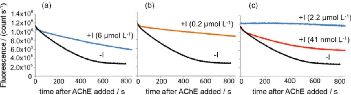
Figure 6 illustrates that the addition of AChE to a solution of the fluorescent complex between lauroyl choline and anionic S-OPE-1 (COOEt)2 does result in a decay of the OPE fluorescence. As expected the decay of fluorescence increases as the amount of AChE added is increased. Since AChE is responsible for the termination of nerve signals, many inhibitors of AChE are highly neurotoxic and are generally considered nerve agents and pesticides. To test whether the AChE sensor could be used to detect chemical agents or pesticides, three inhibitors of AChE were selected: itopride, meptazinol and TAE-1. While these compounds are less toxic and less volatile than the actual nerve agents of interest for detection, they are powerful inhibitors. They were added (individually) to samples of the sensor prior to addition of AChE. While addition of TAE-1 does produce some changes in the sensor absorption and fluorescence there was no change in sensor spectral properties when itopride or meptazinol was added.
As shown in Figure 7, addition of each inhibitor attenuates the loss of fluorescence seen in the black lines
where no inhibitor has been added. Most recently we have turned our attention to some unforeseen properties of the cationic and anionic OPE for both sensing and unexpected reactivity.
4. Induction of Spore Germination by Antimicrobial OPEs
Bacteria of the genus Bacillus are highly resistant to antimicrobials.28 Unlike other pathogenic bacteria, bacilli are capable of undergoing drastic morphology changes in the presence of stressful environmental conditions.28,29 This morphological change is referred to as sporulation, and coincides with ceased multiplication and division. Sporulation of bacilli confers a profound resistance to DNA-damaging UV light,30-33 temperature extremes,34 and various reactive oxygen species (ROS)-generating chemicals.35-37 Any effort to deactivate spores must be thorough and profound, as small-acid-soluble proteins (SASP) are in place to repair minor DNA damage upon germinating: the act of resuming active multiplication and division as a vegetative cell.32,33,38-40
Bacillus anthracis is particularly notorious, as it has been historically used as a biowarfare agent. Similarly to other bacilli, Bacillus anthracis spores are small (ca. 1 µm in size) and easily aerosolized as a result of their extremely
Figure 6. (a) Absorbance; (b) fluorescence of S-OPE-1− (COOEt) and LaCh at 0.2, 0.4 and 0.6 U of AChE. Reprinted with permission from Ref. 27.
Copyright 2015 American Chemical Society.
Figure 7. S-OPE-1 (COOEt)/LaCh complex (5 µmol L-1 OPE, 32 µmol L-1 LaCh) showing fluorescence change after addition of 0.6 U of AChE in the
Figure 8. Structure of EO-OPE (Th,C2).
Figure 9. Bacillus atrophaeus spores visualized via SEM. (a) Spores suspended in physiological saline solution for 5 h in the dark; (b) spores exposed to 20 µg mL-1 OPE for 5 h in the dark; (c) spores suspended in physiological saline solution for 5 h in UVA light; (d) and (e) spores exposed to 20 µg mL-1
OPE for 5 h in UVA light. Scale bars spanning 3 µm are included. Arrows indicate spore coat remnants. Reprinted with permission from Ref. 42. Copyright 2015 American Chemical Society.
low density. In addition, Bacillus anthracis is extremely pathogenic to mammals due to its two virulence plasmids-pOX1 and pOX2. For these reasons, Bacillus spores are frequently the focal point of decontamination regimes.41
We recently found that EO-OPE (Th,C2 Figure 8) is highly effective at deactivating B. atrophaeus and B. anthracis Sterne (-pOX2) in the presence of UVA light, and can facilitate germination-even in the absence of nutrients and UVA light.42 As can be seen in the scanning electron microscopy (SEM) image (Figure 9) untreated B. atrophaeus spores (used as a surrogate to B. anthracis and B. anthracis Sterne) are uniform in size, shape and surface roughness. However, the inclusion of 20 µg mL-1 EO-OPE (Th,C2) causes many, but not all, spores to germinate into viable vegetative cells. As can be seen, vegetative cells exhibit a higher aspect ratio than their dormant spore counterparts, and frequently exceed 3 µm in length. Spore coat remnants are also identified, which is yet another indicator of recent germination. The presence of UVA light, alone, does not damage spores, but a slight increase (ca. 5%) of germination is observed. Adding EO-OPE (Th,C2) to spores in UVA light also results in germination, yet the
resulting viability is drastically decreased. SEM imaging clearly depicts numerous instances of stunted germination, and in select cases, profound membrane damage.
B. anthracis Sterne was found to be equally vulnerable to EO-OPE (Th,C2). Using modified plating techniques, it is possible to gauge the extent of germination and cell death in a quantitative manner. As can be seen in Figure 10, germination in the dark is heavily dependent on spore concentration. Since volume is held constant, we believe EO-OPE (Th,C2) is damaging the B. anthracis Sterne spores to the extent that quorum-sensing chelate Ca2+-dipicolinic acid is released intercellularly, thereby facilitating the germination of neighboring spores. Despite the widespread germination into relatively vulnerable vegetative cells, cell death is minimal, as EO-OPE (Th,C2) is incapable of sensitizing the production of singlet oxygen and other ROS in the dark.
concentration is decreased, killing is greatly enhanced. The most likely explanation is that the OPE:spore ratio is so high that we are no longer depending on germination into a vulnerable vegetative cell to achieve cell death. Research is ongoing to determine whether other OPEs and PPEs are capable of this level of spore deactivation.
5. Fluorescent Detection of Amyloid Protein Aggregates
The fortuitous discovery of the ethyl ester-terminated OPEs led to another sensor application: their development, in collaboration with Eva Chi at UNM, as dyes for amyloid.43 Amyloid protein aggregates are fibrillar protein conformers with unusually high thermodynamic stability, mechanical integrity and resistance to proteolysis, formed of many protein monomers arranged with intermolecular beta-sheets perpendicular to the fibril axis.44,45 Understanding the biology and biochemistry of amyloid formation is critical to developing knowledge and therapies for Alzheimer’s disease,46 type II diabetes,47 transmissible spongiform encephalopathies and other diseases, as well as structural and chemical properties of bacterial extracellular matrix.48 Unique to amyloid is their ability to bind certain linear organic dyes with high specificity,49 which has led to the development of various small molecules as optical sensors for amyloid in solution and in tissue. The benzothiazinium salt thioflavin T and its derivatives are the most widely used amyloid dyes. Experimental and computational studies of amyloid dyes has identified a rigid, linear, highly conjugated backbone as a key shared element,50 which led us to hypothesize that OPEs, which have a rigid, linear,
π conjugated phenylene ethynylene backbone, might be
suitable amyloid dyes.
Using hen egg white lysozyme model amyloids formed in vitro,51,52 we found that many OPEs bind selectively and strongly to amyloid, with the COOEt-terminated OPEs showing an especially large increase in fluorescence signal upon amyloid binding (Figure 11).
Figure 11. (a) TEM (top) and AFM (bottom) images of hen egg white lysozyme amyloids incubated for 4 hours, showing short bunched fibrils. Scale bars 200 nm. Inset: isolated fibril showing double-braid morphology, scale bar 50 nm; (b) structure of example OPE investigated (-1C); (c) spectroscopic changes observed upon amyloid binding: left, excitation spectrum of OPE alone (black dashed line), OPE (500 nmol L-1) with monomeric HEWL (5 µmol L-1) (red
dotted line) and OPE with HEWL amyloid (5 µmol L-1 monomer per 250 µg mL-1) (blue solid line); middle, emission spectrum of OPE alone (black dashed
line), OPE with monomeric HEWL (red dotted line) and OPE with HEWL amyloid (blue solid line); right, circular dichroism of OPE (10 µmol L-1) with
HEWL monomer (500 µg mL-1) (black trace) and OPE with HEWL amyloid (500 µg mL-1) (red trace). OPE alone has no CD signal. Partially reprinted
with permission from Ref. 43. Copyright 2015 American Chemical Society.
Figure 12. Emission spectra at λex = 280 nm, showing decrease of HEWL intrinsic fluorescence due to solvent exposure of aromatic residues during refolding and aggregation process and appearance of HEWL OPE FRET signal in OPE/amyloid mixture. Partially reprinted with permission from Ref. 43. Copyright 2015 American Chemical Society.
Preliminary work shows that OPEs are also effective in dyeing intraneuronal tau amyloid aggregates in brain tissue from mouse models of tau diseases, using both one- and two-photon microscopy. We hope to see OPEs further exploited as tool compounds for detection of amyloids in many arenas. Crucial to the development of this application, like the others in this report, was the unexpected discovery of the photophysical properties imparted to OPEs by ethyl ester end groups.
6. Conclusions
We have presented a survey of several years’ research-focusing on surfactant complex based sensors for enzymes and chemical warfare agents, induced germination
and others, to pursue the new ideas suggested to them by their research, rather than merely “iterating and improving” upon the same old ones. Furthermore, making an artificial separation between “fundamental” and “applied” research, or between “science” and “engineering,” is dangerous and indeed fatal to creativity. Today’s researchers must, if possible, embrace the often-unnatural interfaces between ideas, between people, and between sciences.
Acknowledgements
We are grateful for generous support from the Defense Threat Reduction Agency for support of this research over the past seven years (Current Grant HDTRA1 08-1-0053). We also thank previous co-workers who have contributed to this project including Yanli Tang and Zhijun Zhou for most of the synthetic work, Ying Wang, Thomas Corbitt, and Liping Ding for their mechanistic work and Eric Hill for his work in mechanistic and computational studies and the development of sensing systems. We also thank our collaborators at UNM and UF for all of their assistance and suggestions. D. G. W. wishes to thank the CNPq for a Ciência sem Fronteiras Fellowship that sponsored extended stays at USP with Professors Frank H. Quina and Erick L. Bastos and for specific ideas for inclusion in this review.
Harry C. Pappas obtained his bachelor’s degree from the College o f E n g i n e e r i n g, U n i v e rs i t y o f Massachusetts Amherst in 2010. He is currently a PhD student under the supervision of Prof David G. Whitten at the University of New Mexico. His research is focused on evaluating the biocidal activity of phenylene ethynylene-based compounds, and in particular, the mechanisms by which they induce stress responses in bacteria.
Patrick L. Donabedian received his BSc in Liberal Arts from St. John’s College in 2012, and is currently a PhD student in Nanoscience Engineering in the labs of Prof David Whitten and Prof Eva Chi at the University of New Mexico, Albuquerque, New Mexico, USA.
Kirk Schanze earned his BSc in Chemistry from Florida State University in 1979 and his PhD in Chemistry from the University of North Carolina at Chapel Hill in 1983. He was appointed a Miller Postdoctoral Fellow at the University of California, Berkeley, from 1984-1986 and began his independent faculty career at the University of Florida in 1986. Schanze is currently University Distinguished Professor and Prominski Professor of Chemistry at the University of Florida. He has authored or co-authored more than 275 peer-reviewed articles on basic and applied research topics, with a primary focus on organic and organometallic materials chemistry.
David G. Whitten earned a PhD degree at Johns Hopkins University in 1963. After 2 years as an Army officer and a postdoctoral at Caltech, he joined the University of North Carolina in 1966 and rose to become Smith Professor. He joined the University of Rochester in 1983 as C. E. Kenneth Mees Professor of Chemistry. In 1997 he moved to Los Alamos and studied conjugated polyelectrolytes. In 2000 he co-founded QTL Biosystems, a company developing biosensing technology for drug discovery and defense. He moved to the University of New Mexico in 2005 where he is Professor of Chemical Engineering.
References
1. Whitesides, G. M.; Angew. Chem., Int. Ed. 2015, 54, 3196. 2. Lu, L.; Rininsland, F. H.; Wittenburg, S. K.; Achyuthan, K. E.;
McBranch, D. W.; Whitten, D. G.; Langmuir2005, 21, 10154. 3. Chen, L.; McBranch, D. W.; Wang, H.-L.; Helgeson, R.; Wudl, F.;
Whitten, D. G.; Proc. Natl. Acad. Sci. USA1999, 96, 12287. 4. Chen, L.; Xu, S.; McBranch, D.; Whitten, D. G.; J. Am. Chem.
Soc.2000, 122, 9302.
5. Jones, R. M.; Bergstedt, T. S.; McBranch, D. W.; Whitten, D. G.;
J. Am. Chem. Soc.2001,123, 6726.
6. Tan, C.; Pinto, M. R.; Schanze, K. S.; Chem. Commun.2002,
5, 446.
7. Chemburu, S.; Corbitt, T. S.; Ista, L. K.; Ji, E.; Fulghum, J.; Lopez, G. P.; Ogawa, K.; Schanze, K. S.; Whitten, D. G.;
Langmuir2008, 24,11053.
8. Corbitt, T. S.; Ding, L.; Ji, E.; Ista, L. K.; Ogawa, K.; Lopez, G. P.; Schanze, K. S.; Whitten, D. G.; Photochem. Photobiol. Sci.2009, 8,998.
10. Ding, L.; Chi, E.Y.; Ji, E.; Schanze, K. S.; Lopez, G. P.; Whitten, D. G.; Langmuir2010, 26, 5544.
11. Wang, Y.; Jones, E. M.; Tang, Y.; Ji, E.; Lopez, G. P.; Chi, E. Y.; Schanze, K. S.; Whitten, D. G.;Langmuir2011, 27, 10770. 12. Tang, Y.; Zhou, Z.; Ogawa, K.; Lopez, G. P.; Schanze, K. S.;
Whitten, D. G.; Langmuir 2009, 25, 21.
13. Tang, Y.; Zhou, Z.; Ogawa, K.; Lopez, G. P.; Schanze, K. S.; Whitten, D. G.; J. Photochem. Photobiol., A 2009,207, 4. 14. Zhou, Z.; Corbitt, T. S.; Parthasarathy, A.; Tang, Y.; Ista, L. K.;
Schanze, K. S.; Whitten, D. G.; J. Phys. Chem. Lett. 2010,1, 3207.
15. Tang, Y.; Hill, E. H.; Zhou, Z.; Evans, D. E.; Schanze, K. S.; Whitten, D. G.;Langmuir 2011, 27,4945.
16. Tang, Y.; Corbitt, T. S.; Parthasarathy, A.; Zhou, Z.; Schanze, K. S.; Whitten, D. G.; Langmuir 2011, 27,4956.
17. Sudeep, P. K.; James, P. V.; Thomas, K. G.; Kamat, P. V.; J. Phys. Chem. A 2006,110, 5642.
18. Ji, E.; Corbitt, T. S.; Schanze, K. S.; Whitten, D. G.; Langmuir
2011, 27,10763.
19. Lu, L.; Jones, R. M.; McBranch, D.; Whitten, D. G.; Langmuir
2002, 18, 7706.
20. Hill, E. H.; Sanchez, D.; Evans, D. G.; Whitten, D. G.; Langmuir
2013, 29, 15732.
21. Hill, E. H.; Evans, D. G.; Whitten, D. G.; Langmuir2013, 29, 9712.
22. Hill, E. H.; Evans, D. G.; Whitten, D. G.; J. Phys. Org. Chem.
2014, 27, 252.
23. Pinto, M. R.; Schanze, K. S.; Proc. Natl. Acad. Sci. USA2004,
101, 7505.
24. Feng, F.; Liu, L.; Wang, S.; Macromol. Rapid Commun.2010,
31, 1405.
25. Dai, N.; Kool, E. T.; Chem. Soc. Rev. 2011, 40, 5756. 26. Liu, Y.; Ogawa, K.; Schanze, K. S.; J. Photochem. Photobiol.,
C 2009,10, 173.
27. Hill, E. H.; Zhang, Y.; Evans, D. G.; Whitten, D. G.; ACS Appl. Mater. Interfaces2015,7, 5550.
28. Balaban, N. Q.; Merrin, J.; Chait, R.; Kowalik, L.; Leibler, S.;
Science 2004, 305, 1622.
29. Bakken, L. R. In Modern Soil Microbiology; van Elsas, J., ed.; CRC Press: New York, 1997, pp. 47-61.
30. Iannotti Jr., M. R. P.; Braz. J. Chem. Eng.2013, 30, 507. 31. Devine, D. A.; Keech, A. P.; Wood, D. J.; Killington, R. A.;
Boyes, H.; Doubleday, B.; Marsh, P. D.; J. Appl. Microbiol.
2001, 91, 786.
32. Nicholson, W.; Munakata, N.; Microbiol. Mol. Biol. Rev.2000,
64, 548.
33. Setlow, P.; J. Appl. Microbiol. 2006, 101, 514.
34. Gates, S. D.; McCartt, A. D.; Jeffries, J. B.; Hanson, R. K.; Hokama, L. A; Mortelmans, K. E.; J. Appl. Microbiol. 2011,
111, 925.
35. Khadre, M.; Yousef, A.; Int. J. Food Microbiol. 2001, 71, 131. 36. Labas, M. D.; Zalazar, C. S.; Brandi, R. J.; Cassano, A. E.;
Biochem. Eng. J. 2008, 38, 78.
37. Raffellini, S.; Schenk, M.; Guerrero, S.; Alzamora, S. M.; Food Control 2011, 22, 920.
38. Miller, M. C.; Resnick, J. B.; Smith, B. T.; Lovett, C. M.; J. Biol. Chem. 1996, 271, 33502.
39. Setlow, B.; Setlow, P.; J. Bacteriol. 1996, 178, 3486. 40. Setlow, P.; Annu. Rev. Microbiol. 1995, 49, 29.
41. Young, S. B.; Setlow, P.; J. Appl. Microbiol.2003, 95, 54. 42. Pappas, H. C.; Lovchik, J. A.; Whitten, D. G.; Langmuir 2015,
31, 4481.
43. Donabedian, P. L.; Pham, T. K.; Whitten, D. G.; Chi, E. Y.; ACS Chem. Neurosci. 2015, 6, 1526.
44. Rambaran, R. N.; Serpell, L. C.; Prion 2008, 2, 112.
45. Knowles, T. P. J.; Vendruscolo, M.; Dobson, C. M.; Nat. Rev. Mol. Cell Biol. 2014, 15, 384.
46. Ittner, L. M.; Götz, J.; Nat. Rev. Neurosci.2011, 12, 65. 47. Westermark, P.; Andersson, A.; Westermark, G. T.; Physiol.
Rev. 2011, 91, 795.
48. Barnhart, M. M.; Chapman, M. R.; Annu. Rev. Microbiol.2006,
60, 131.
49. Nilsson, K. P. R.; FEBS Lett. 2009, 583, 2593.
50. Skeby, K. K.; Sørensen, J.; Schiøtt, B.; J. Am. Chem. Soc.2013,
135, 15114.
51. Swaminathan, R.; Ravi, V. K.; Kumar, S.; Kumar, M. V. S.; Chandra, N.; Adv. Protein Chem. Struct. Biol.2011, 84, 63. 52. Hill, S. E.; Robinson, J.; Matthews, G.; Muschol, M.; Biophys. J.
2009, 96, 3781.
53. Würthner, F.; Kaiser, T. E.; Saha-Möller, C. R.; Angew. Chem.,
Int. Ed.2011, 50, 3376.
54. Piston, D. W.; Kremers, G. J.; Trends Biochem. Sci.2007, 32, 407.
Submitted: August 16, 2015