Rev Bras Cir Plást. 2013;28(2):260-3 260
Orsi VV et al.
Surgical correction of breast inferior pole hypoplasia
Correção da hipoplasia de polo inferior da mama
This study was performed at the Hospital Cristo Redentor – Grupo Hospitalar Conceição (Cristo Redentor Hospital – Conceição Hospital Group), Porto Alegre, RS, Brazil.
Submitted to SGP (Sistema de Gestão de Publicações/Manager Publications System) of RBCP (Revista Brasileira de Cirurgia Plástica/Brazilian Journal of Plastic Surgery).
Article received: March 18, 2013 Article accepted: May 10, 2013
Victor Vieira orsi1
GustaVo LeVacoV BerLim2
carLos eduardo ochoa
taGLiari3
dieGo iLha thomasi3
Pedro BiLiBio WestPhaLen3
João VaLter Pires Junior3
Franco T et al. Vendramin FS et al.
ORIGINAL ARTICLE
ABSTRACT
Background: Breasts with inferior pole hypoplasia have a disharmonious appearance owing to a high inframammary fold and the prevalence of glandular volume in the upper quadrants that results in a lower inclination of the nipple–areola complex. In these patients, the goal is to correct the disharmonious shape of their breasts and possible asymmetries while causing minimal or inconspicuous scars. The aim of this study was to present a sur-gical technique to correct breast inferior pole hypoplasia. Methods: The surgical technique
involved a rotated internal lap to reshape the breast and relocate the inframammary fold,
a result achieved using only the scores where the implants would be positioned as surgical access routes. Results: The procedure described here was used in four patients aged 19–27 years with breast inferior pole hypoplasia. The results demonstrate that the correction of breast deformities was properly achieved, and the inframammary fold was repositioned without any irregularities in the inferior breast contour. Conclusions: The technique pro-posed here is a suitable alternative for the treatment of selected cases of breast deformities with hypoplasia of the inferior pole.
Keywords: Breast/surgery. Mammaplasty. Plastic surgery/methods.
RESUMO
Introdução: As mamas com hipoplasia de polo inferior apresentam aspecto desarmôni-co, com o sulco inframamário alto, predominância de volume glandular nos quadrantes superiores, e, consequentemente, direcionamento inferior do complexo areolopapilar. É meta desejável, nesses casos, a correção da forma desarmônica das mamas, bem como de eventuais assimetrias, com cicatrizes mínimas ou inconspícuas. O objetivo deste trabalho é apresentar técnica cirúrgica para correção de hipoplasia de polo inferior da mama. Método:
A técnica cirúrgica utilizou retalho interno rodado para reconigurar a mama e reposicionar
o sulco inframamário, tendo como vias de acesso cirúrgico tão somente as cicatrizes por onde se colocam as próteses. Resultados: A técnica descrita foi utilizada em 4 pacientes com hipoplasia de polo inferior de mama, com idades variando entre 19 anos e 27 anos. Os resultados demonstram que a correção da deformidade mamária foi alcançada adequada-mente, bem como o reposicionamento do sulco inframamário, sem provocar irregularidades no contorno inferior da mama. Conclusões: A técnica proposta é uma alternativa adequada para o tratamento de casos selecionados de deformidade mamária com hipoplasia de polo inferior.
Descritores: Mama/cirurgia. Mamoplastia. Cirurgia plástica/métodos.
1. Plastic surgeon, full member of the Sociedade Brasileira de Cirurgia Plástica (Brazilian Society of Plastic Surgery - SBCP), Master in Medicine-Surgery by the Universidade Federal do Rio Grande do Sul (Federal University of Rio Grande do Su), preceptor of the Medical Residence of the Plastic Surgery Service of Hospital Cristo Redentor (Cristo Redentor Hospital), Porto Alegre, RS, Brazil.
2. Plastic surgeon, associated member of SBCP, staff of the Plastic Surgery Service of Hospital Cristo Redentor (Cristo Redentor Hospital), Porto Alegre, RS, Brazil.
Rev Bras Cir Plást. 2013;28(2):260-3 261
Surgical correction of breast inferior pole hypoplasia
INTRODUCTION
Breasts with inferior pole hypoplasia have a disharmo-nious appearance due to a high inframammary fold and the prevalence of glandular volume in the upper quadrants that results in a lower inclination of the nipple–areola complex (NAC)1-6.
Von Heimburg et al.2 classiied deformities of the base of
the breast into four different types:
• Type I – only the lower-medial quadrant of the breast is deicient;
• Type II – the presence of deiciencies in both inferior quadrants of the breast but no skin deiciency within the subareolar region;
• Type III – deiciencies in both inferior quadrants of the breast and one or more subareolar skin deicien
-cies; and
• Type IV – deiciencies within all four quadrants of
the breast (tuberous breast).
Breasts presenting with inferior pole hypoplasia,
regar-dless of von Heimburg classiication types I, II, or III, are not tuberous, although they share several treatment dificulties.
Moreover, patients with deformities of the base of the breast often exhibit mammary asymmetry as well, which renders the treatment even more complex.
The available methods for the treatment of breast inferior pole hypoplasia typically include mastopexia, which implies the use of implants and an extensive scar burden, and peria-reolar, associated with a lower vertical or “inverted T” type3.
In these cases, the goal is to correct the disharmonious shape of the breasts and the possible asymmetries using minimal or inconspicuous scars. Taking this into account, some patients
may beneit from techniques that use rotated internal laps to
reshape the breast and reposition the inframammary fold, a result achieved using only the scars where the implants would then be positioned as routes for surgical access as described in this case series.
METHODS
This study presents a series of four patients who underwent correction of breast inferior pole hypoplasia between 2010 and 2011. The patients underwent the following:
• Design and planning of the incisions – with the
patient standing, an ideal inframammary fold was de lineated, and sometimes, the fold was delineated to > 5 cm below the pre-existing inframammary
fold. During the procedure, the incision along the ideal inframammary fold had suficient extension.
In the cases reported in this study, we chose incisions
approximately 6 cm long;
• Dissection – beginning in the subglandular plane
and continuing until above the NAC projection. In
the upper portion of the mammary gland, a breast
tissue lap was prepared (Figure 1A) to be positioned
with the inferior base parallel to the skin and was
then rotated 180° inferiorly (Figure 1B);
• Fixation of the lap – the upper extremity of the lap to the upper edge of the surgical wound was sutured with 3-0 monoilament nylon threads. This promotes illing of the inferior breast pole and pu
-shing of the skin from the inside to the outside to cause disappearance of the pre-existing inframam-mary fold (Figure 1C). This step also ensures that, after surgery, the new inframammary fold would be
positioned adjacent to the surgical wound;
• Double plane dissection and implant insertion – the
plane below the pectoral muscle was dissec ted for breast implant insertion according to the dou ble plane procedure. Subglandular dissection was
verti-cally extensive owing to preparation of the lap, and
it extended above the NAC. For this rea son, it was considered necessary to re-suture the edge of the pectoral muscle to the breast using 3-0 monocryl
threads prior to implant insertion (Fi gu re 1D); and
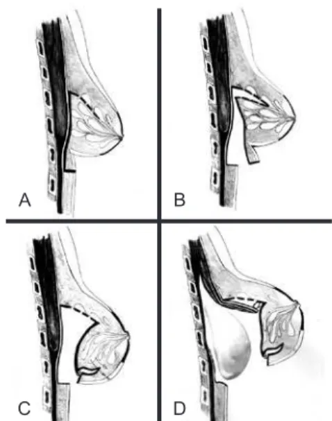
Figure 1 – Schematic drawing of the procedure used to correct breast inferior pole hypoplasia. In A, incisions of the inframammary fold and initial dissection of the subglandular plane.
In B, internal lap dissection. In C, rotation of the extremities
of the lap inferiorly and ixation next the edge of the surgical wound. This rotation promotes illing of the inferior breast
pole and pushing of the skin from the inside to the outside to cause the disappearance of the pre-existing inframammary fold. In D, dissection of the submuscular plane for the insertion of breast implants in the dual plane. Prior to the implant insertion,
the edge of the pectoral muscle was re-sutured to the breast using 3-0 monocryl thread.
A
C
B
Rev Bras Cir Plást. 2013;28(2):260-3 262
Orsi VV et al.
• Closing – suturing by planes using 3-0 monocryl
threads was performed, and the skin was closed using intradermal sutures.
RESULTS
The procedure described here was used in four patients aged 19–27 years with breast inferior pole hypoplasia. In
all cases, we used texture-coated, cohesive, and high-proile
silicone implants with volumes of 225–325 cc.
No complications were observed in the postoperative period. The longer followed up was of 2 years.
Figures 2 to 5 illustrate the results obtained in the four cases. The breast deformities were corrected, and the infra-mammary fold was appropriately repositioned without any irregularities in the inferior breast contour.
DISCUSSION
The procedure used in this case series closely resembled the technique described by Oroz e al.1 in 2005 in which in
-ter nal breast laps were used to reconstitute the volume of
the inferior breast pole and reposition the inframammary
fold. The laps were dissected starting from the same incision
through which the implants were inserted. This is the main (or only) difference between the procedure described by Oroz et al.1 and the technique used in this study.
The procedure described in the study by Oroz et al.1 used
a lower periareolar incision, whereas the route for surgical access in this series was through the inframammary fold.
A
C
B
D
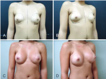
Figure 2 – Case 1. A 25-year-old patient. In A and B, frontal and left preoperative proile, respectively. In C and D, 1-year and 10-month frontal and left postoperative
proile, respectively. We used textured, cohesive gel and 250 cc high-proile silicone implants.
A
C
B
D
Figure 3 – Case 2. A 27-year-old patient.
In A and B, frontal and right preoperative proile, respectively,
showing mammary asymmetry. In the right proile view, observe the
detailed demarcation below the pre-existing inframammary fold. In C and D, 1 year and 6 month frontal and right postoperative
proile, respectively. We used textured, cohesive gel and 325 cc (right) and 300 cc (left) high-proile silicone implants.
A
C
B
D
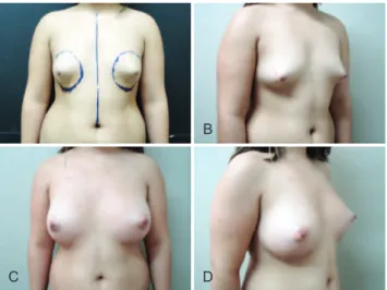
Figure 4 – Case 3. A 22-year-old patient.
In A and B, frontal and right preoperative proile, respectively. In C and D, 3-month frontal and right postoperative proile,
respectively. We used textured, cohesive gel and 225 cc high-proile silicone implants.
Otherwise, the approach within the breast and the dissection
of the lap are identical.
Rev Bras Cir Plást. 2013;28(2):260-3 263
Surgical correction of breast inferior pole hypoplasia
A
C
B
D
Figure 5 – Case 4. A 19-year-old patient.
In A and B, frontal and right preoperative proile, respectively. In the frontal view, observe the detailed demarcation below the pre-existing inframammary fold. In C and D, 1-week frontal and
right postoperative proile, respectively. We used textured,
cohesive gel and 275 cc3 high-proile silicone implants.
increase breast volume, and safely achieve a new and proper inframammary fold position without causing any irregula-rities in the inferior breast contour.
The cases presented here evolved positively. The patients
were satisied with the result, and thus far, no complications
have been observed in the postoperative period.
CONCLUSIONS
Despite the small sample size and limited follow-up
pe riod, the results obtained here suggest that the proposed procedure is a suitable alternative for the treatment of se -lected cases of breast deformities and inferior pole hypo-plasia.
REFERENCES
1. Oroz J, Pelay MJ, Escudero FJ. Reconstruction of the tuberous breast.
An Sist Sanit Navar. 2005;28 Suppl 2:101-8.
2. von Heimburg D, Exner K, Kruft S, Lemperle G. The tuberous breast deformity: classiication and treatment. Br J Plast Surg. 1996;49(6):
339-45.
3. Mandrekas AD, Zambacos GJ, Anastasopoulos A, Hapsas D, Lambri
-naki N, Ioannidou-Mouzaka L. Aesthetic reconstruction of the tuberous breast deformity. Plast Reconstr Surg. 2003;112(4):1099-108. 4. Costa SS, Blotta RM. Assimetrias da mama. Rio de Janeiro: Revinter;
2007.
5. Ribeiro L, Canzi W, Buss A Jr, Accorsi A Jr. Tuberous breast: a new approach. Plast Reconstr Surg.1998;101(1):42-50.
6. Ribeiro L, Accorsi A Jr, Buss A, Pessĵa MC. Short scar correction of the tuberous breast. Clin Plast Surg. 2002;29(3):423-31.
Correspondence to: Victor Vieira Orsi