Use of mid-forehead lap in nasal reconstruction
Uso do retalho médio-frontal na reconstrução do nariz
This study was performed at the
38a Enfermaria da Santa Casa da Misericórdia do Rio de Janeiro, Serviço do Professor Ivo Pitanguy
(38th Inirmary of the Santa Casa da Misericórdia of Rio de Janeiro, Professor Ivo Pitanguy’s Service), Rio de Janeiro, RJ, Brazil. Submitted to SGP (Sistema de Gestão de Publicações/Manager Publications System) of RBCP (Revista Brasileira de Cirurgia Plástica/Brazilian Journal of Plastic Surgery). Article received: December 12, 2012 Article accepted: March 29, 2013
Henrique P. L. Cintra1 aLi BouCHama2 tHiago HoLanda2 CarLos aLBerto JaimoviCH1 ivo Pitanguy3
ABSTRACT
Background: Six hundred years before Christ, the mid-forehead lap was described by the Indian Sushruta Samhita. Until today, this lap called ‘’Indian lap”, has a major role in the reconstruction of the nose. The aim of this work was to analyze the results of the 38th
Inirmary of the Santa Casa da Misericórdia of Rio de Janeiro, Professor Ivo Pitanguy’s Service, in nasal reconstruction with the mid-forehead lap. Methods: We did a retrospective study of 10 cases operated in the service for nasal reconstruction with Indian lap, during a 21 year period (1991-2012). Results: The number of nasal sub-units affected varied from 4 to 9, with an average of 6.5 subunits. In 70% of the patients, was performed a previous expansion of the mid-forehead lap and in 90% were used cartilage grafts and/or bones. Five patients had postoperative distortions, that were corrected with other surgeries. No cases of infection, necrosis of the lap or graft extrusion were recorded. Conclusions: This study allowed to demonstrate that the mid-forehead lap still have an important role in nasal reconstruction of major defects, showing satisfactory results due to its vascular safety, the amount of skin which is obtained, likeness of color, texture and skin thickness.
Keywords: Reconstructive surgical procedures. Nose/surgery. Surgical lap. Forehead/sur -gery. Plastic surgery/methods.
RESUMO
Introdução: Seiscentos anos antes de Cristo, foi descrito o retalho médio-frontal pelo indiano Sushruta Samhita. Até hoje, esse retalho, chamado ‘’retalho indiano”, é usado na reconstrução do nariz. O objetivo deste trabalho foi analisar os resultados da 38a Enfermaria
da Santa Casa da Misericórdia do Rio de Janeiro, Serviço do Professor Ivo Pitanguy, na reconstrução nasal com emprego de retalho médio-frontal. Método: Foi realizado estudo retrospectivo com 10 casos operados no serviço referido para reconstrução nasal com reta -lho indiano, no período de 21 anos (1991 a 2012). Resultados: O número de subunidades nasais atingidas variou de 4 a 9, com média de 6,5 subunidades. Em 70% dos pacientes foi realizada expansão prévia do retalho médio-frontal e em 90% foram utilizados enxertos cartilaginosos e/ou ósseos. Cinco pacientes apresentaram distorções pós-operatórias, que foram corrigidas por outras cirurgias. Nenhum caso de infecção pós-operatória, de necrose do retalho ou de extrusão de enxertos foi registrado. Conclusões: Este trabalho permitiu demonstrar que o retalho médio-frontal tem ainda importante papel na reconstrução nasal de grandes defeitos, com resultados satisfatórios, atribuídos a sua segurança vascular, à quantidade de pele que se obtém, e à semelhança de cor, textura e espessura cutâneas. Descritores: Procedimentos cirúrgicos reconstrutivos. Nariz/cirurgia. Retalhos cirúrgicos. Tes ta/cirurgia. Cirurgia plástica/métodos.
1. Plastic surgeon, full member of the Sociedade Brasileira de Cirurgia Plástica (Brazilian Society of Plastic Surgery - SBCP), assistant professor at Ivo Pitanguy Institute, Rio de Janeiro, RJ, Brazil.
2. General surgeon, postgraduate student at the Institute Ivo Pitanguy, Rio de Janeiro, RJ, Brazil.
INTRODUCTION
The history of nasal reconstruction is intertwined with the history of plastic surgery. Since the Vedic period (2000-500 BC) in ancient India, when adultery was punishable by amputation of the nose, has already been stories of plastic surgeries performed in the nasal region1. Sushruta Shamita
presented in Ayur-Veda (600 BC), a technique for total nasal reconstruction based on mid-forehead lap and advised the use of leaves of trees for the same marking. This technique is now called ‘’Indian method”2,3.
Some years before the birth of Jesus Christ, Aulus Cor -nelius Celsus (53 BC - 7 AD) marked the history of plastic sur gery with its work on grafts. In his book, ‘’De Re medica’’, he studied various nose, lips and ears defects and describes skin laps taken from the vicinity.
In the Renaissance, the neo-rinoplasties took a boost due to major injuries resulting from sequels of leprosy and syphilis. In this occasion, the nose reconstruction was per formed using a lap arm. Gaspare Tagliacozzi, in 1597, practices a lap of upper third of the inner side of the arm for a full or partial nasal reconstruction. This technique is now called “Italian method’’2-4.
For two centuries, nasal reconstruction goes through a period of bad reputation, as evidenced by the mockery of Amboise Parré who ridiculizes in 1575 the feasibility of this procedure2.
The reintroduction of “Indian lap” in the Occident was realized by Lucas in the London’s Gentlemen Magazine, in 1794, and by Carpue, in 1816, both in England2,3.
Von Graefe, in 1818, in Berlin and Delpech, in 1823, in Montpelier, started using the ‘’Indian Method’’ and the’’ Italian method”2.
In the immediate postoperative period, the results were considered good, but the vast subsequent retraction of the mid-forehead laps observed in the nineteenth century, made Dupuytren and Denonvilliers to criticize it2. The irst
breakthrough in the search for techniques to improve the results was the perception that the bloody surfaces of the lap were responsible of infection, ibrosis and retraction. Carpue, Von Graefe, Blandin and Dieffenbach started then folding the distal part of the laps to reduce the bloody surface2,3. The
fold of the lap also allows a more designed columella and alae. Volkmann, in 1874, and Thiersch, in 1879, advance the concept of making laps of the remaining nasal skin or partial skin grafts to cover the bloody areas.
At this time, Konig presents techniques of chondro-cu-taneous composite grafts that simplify the reconstruction of small to moderate defects2.
Gillies, in 1943, and Converse, in 1956, add chondro-cu-taneous composite graft of conchal cymba, chondro-mucosal graft of septum and naso-labial laps to replace the skin fold of mid-forehead lap, to improve the lining and support of alae2,3.
A deformity commonly seen after nasal reconstruction in the nineteenth century was the short columella with the nasal tip retracted caudally. Auvert draws the mid-forehead lap with an angle of 45 degrees, promoting stretching of the lap. Oblique laps started to be the favorite design in the end of nineteenth century2,4.
Gillies, in 1935, describes a “U-shaped” lap in the frontal region and Converse, in 1942, describes a lap of the scalp in cluding the skin of the frontal region. Both techniques increase the blood supply and the length of the lap2.
With laps of greater length is then possible the confec -tion of longer columellas, which in turn allow better nasal tip projection. Millard, in 1966, introduces a lap of labial mu cosa through a hole in the lip for lining the columella, dra matically reducing the postoperative retraction2,4.
Gillies advocates the anterior rotation of the remaining septum with inferior pedicle at the time of primary recons -truction, and Millard, in 1974, modiies this concept making a lap of the remaining septum with a superior pedicle through a ‘’L’’ shaped incision, also in the primary reconstruction. These authors also state that in the presence of a suitable length columella, addition of bone or cartilage grafts gives a “inal touch” in the nasal reconstruction2,3.
Burget & Menick5 introduce the concept of aesthetic units
of the nose, claiming that the incisions should be located at the limits of these aesthetic units. If it is necessary to remove more than one third of a unit, it must be completely removed and reconstructed.
This study aims to demonstrate the versatility of the lap in the mid-forehead lap in extensive nasal reconstructions, but it requires repeated surgical reinement process until we get adequate support and contour.
METHODS
A retrospective study of 10 cases of nasal reconstruc -tion with mid-forehead lap, between 1991 and 2012, was performed. The patients were operated at the 38ª Enfermaria da Santa Casa da Misericórdia do Rio de Janeiro, Serviço do Professor Ivo Pitanguy (38th Inirmary of the Santa Casa
da Misericórdia of Rio de Janeiro, Professor Ivo Pitanguy’s Service).
The following parameters were analyzed: the patient’s age, sex, cause of injury that led to the reconstruction, the number of sub-units hit in the nose, presence of associated injuries, the number of surgical steps that were necessary, the prior expansion of the lap, the type of graft used, the surgical methods used for reconstruction of the nasal li -ning, the number of surgeries per patient and postoperative complications.
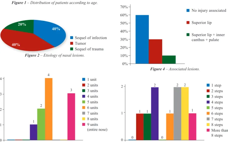
The most frequent etiologies that motivated the nasal reconstruction were sequelae of infection (40%) and tumor (40%) (Figure 2).
The number of affected nasal sub-units range from 4 to 9 (entire nose) with an average of 6.5 subunits affected (Figure 3).
In 4 (40%) patients, lesions of the nose were associated with other injuries. In 3 (30%) cases, lesions of the upper lip were associated, and in 1 (10%) case, various injuries were found (Figure 4).
The patient’s demand for medical consultation was mainly due to the aesthetic aspect, with complaint present
in 100% of them. Respiratory dificulty was observed in 4 (40%) patients.
All patients underwent nasal reconstruction by mid-fo-rehead lap with or without prior expansion. In all cases, the lap was based on the supra-trochlear artery.
The number of surgical steps that were required for each nasal reconstruction ranges from 2 to 32, with an average of 8.1 surgical steps per patient (Figure 5).
In 7 (70%) patients was performed a prior expansion of the mid-forehead lap and in 9 patients (90%) cartilaginous and/or bone grafts were used. Fifteen grafts were placed, the most used was the costal cartilage graft, which represents 46.7% of the grafts (Figure 6).
The Figure 7 shows the several surgical methods used to reconstruct nasal lining.
The irst surgical step corresponded to the placement of the expander in the frontal region when necessary. The second surgical step corresponded to the removal of the expander and rotation of the mid-forehead lap. The third step was cha racterized by the section of the pedicle. All surgeries beyond these three surgical steps were considered “reine -ment surgeries”: its objectives were to improve the aesthetic appearance of the nose and correct the distortions observed in the postoperative period.
40% 35% 30% 25% 20% 15% 10% 5% 0%
0 a 9 years 10 a 19 years 20 a 29 years 30 a 39 years 40 a 49 years 50 a 59 years 60 years or more
0% 0% 0%
30%
20%
40%
10%
Figure 1 – Distribution of patients according to age.
20%
40%
40%
Sequel of infection Tumor
Sequel of trauma
Figure 2 – Etiology of nasal lesions.
1 unit 2 units 3 units 4 units 5 units 6 units 7 units 8 units 9 units (entire nose)
4
3
2
1
0
Figure 3 – Number of nasal affected sub-units.
1 2
4
3
1 step 2 steps 3 steps 4 steps 5 steps 6 steps 7 steps 8 steps More than 8 steps 2
1
0
Figure 5 – Number of surgical steps per patient.
1 1 1 1
2 2
2
0 0
70% 60% 50% 40% 30% 20% 10% 0%
No injury associated
Superior lip
Superior lip + inner canthus + palate
RESULTS
Five patients had postoperative distortions which were corrected by other surgeries (Figure 9).
No cases of postoperative infection or necrosis of the lap or keloid was recorded.
Figures 10 to 12 illustrate some cases in this series. The number of “reinement surgeries” that were required
to achieve the inal result ranged from 0 to 26, with an average of 5.1 procedures per patient (Figure 8). Regarding patient who was operated 32 times (patient 3), were made 2 mid-forehead laps (ie 4 surgical steps) and were placed 2 expanders in the frontal region (ie 2 more additional surgical steps). In the end, 26 “reinement surgeries” were needed to achieve the inal result.
30% 25% 20% 15% 10% 5% 0%
Folded mid-forehead lap
Flap of the remaining nasal skin
Naso-labial lap Skin graft 28.6%
Figure 7 – Surgical procedures used for reconstruction of nasal lining (7 patients).
28.6% 28.6%
11.2% 50%
45% 40% 35% 30% 25% 20% 15% 10% 5% 0%
Opposite alar cartilage Conchal cartilage Costal cartilage Costal bone Iliac crest bone
6.7% 26.6%
6.7% 46.7%
23.3%
Figure 6 – Grafts used for nasal support.
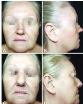
Figure 10 – Patient 1, 54 years old, had a tumor of the nose.
After resection, was performed a mid-forehead lap for skin
coverage. The bony-cartilaginous structures and nasal lining were intact. In A and B, preoperative appearance in frontal view and
right proile, respectively. In C and D, 8 months postoperative
appearance in frontal view and right proile, respectively.
A B
C D
Figure 8 – Number of “reinement surgeries” per patient.
0 surgery 1 surgery 2 surgeries 3 surgeries 4 surgeries 5 surgeries 6 surgeries More than 6 surgeries
3
2
1
0
1 2
1 1 1
1
0
3
50% 45% 40% 35% 30% 25% 20% 15% 10% 5% 0%
Figure 9 – Complications and distortions.
50%
10%10% 10%
0% 0% 0% 20%
No complication or distortion
Retraction of the alae Retraction of columella Retraction of columella + alae
Displacement of the osseous/cartilagenous graft
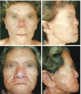
Figure 11 – Patient 2, 61 years old, presented a sequel
of trauma (dog bite). A mid-forehead lap was used for skin
coverage, conchal and costal cartilage grafts for the support’s
structures, and full skin graft for nasal lining. In A and B,
preoperative appearance in frontal view and right proile,
respectively. In C and D, 2 years postoperative appearance in
frontal view and right proile, respectively.
A B
C D
A B
C D
E F
Figure 12 – Patient 3, 24 years old had a sequel of
nasal infection due to leishmaniasis. A irst mid-forehead lap was made for skin coverage, iliac crest graft for support’s structures,
naso-labial laps for reconstruction of nasal lining.
In A and B, preoperative appearance showing a severe retraction
of the columella and ala, in frontal view and right proile,
respectively. In C and D, 7 years postoperative appearance showing severe retraction of the columella and alae, in frontal view
and right proile, respectively. In E and F, 2 years postoperative
appearance after surgical sequel of nasal reconstruction.
Distorted cartilage grafts were removed, old lap was used for reconstruction of the nasal tip lining and a new mid-forehead lap was done after expansion. This new lap was sutured on the dorsal lap desepithelized, and the support’s structure was done with costal
cartilage arranged in L. In total 32 surgeries were performed between 1991 and 2010.
DISCUSSION
The average age observed in this study was 43 years, ran ging from 20 to 61 years. The younger patients underwent surgery to treat sequel of nasal infection (leishmaniasis) and older to cure nasal tumors.
The most frequent complaint of these patients was the cosmetic deformity, present in 100% of cases. Dificulty breathing was reported by 40% of patients, due to the collapse and the impairment of nasal support structures, as well as soft tissue stenosis by scar retraction5,6.
The number of affected nasal subunits varied from 4 to 9 (entire nose) with an average of 6.5 subunits, which re presents more than half of the nose. In extensive lesions that affect more than half of the nose, the mid-forehead flap was the best local flap according to several authors7-10.
This flap proved to be most suitable for its safety, amount of skin obtained, similarity of color, texture and thickness of the skin.
Prior expansion of this flap allows overcoming its grea test limitation, the length, providing enough material for making long columella, alae and the coverage of large defects11,12. In this study, prior expansion of the frontal
region was used in 70% of cases. This facilitates primary closure of the donor area, improving scar due to lower ten -sion. Concerning characteristics of the flap, with the effect
of the skin expansion, it provides a thinner skin which facilitates the surgical modeling of the flap. However, it is difficult to predict the degree of secondary tissue retraction following the expansion, which may represent the shorte -ning of the nose in the immediate postoperative period or later. The expansion also can be used secondarily to improve the scar of the donor area3,13-15.
nasal coverage. However, it was used in 28.6% of cases for reconstruction of the lining. Other surgical modalities have been used to reconstruct the nasal lining: the mid-forehead lap folded in its distal part was used when the length of the lap was enough to make this fold (28.6% of cases), the hinge lap of remaining nasal skin was done when the remaining skin was suficient to cover the defect (14.2%), and the latter was the full-thickness skin graft (28.6% cases).
Patients underwent several surgeries (8.1 on average), due to the large deformity that often has to be rebuild. Some authors have suggested that cartilage or bone grafts, when necessary, were performed in a second surgical procedure, when the soft parts had already reached an adequate stabili -zation16. Other authors prefer to do the cartilaginous support
structure and skin coverage in a single surgical procedure for prevent soft tissue collapse and late contraction3,14,17. In
this study, the sequence of surgical steps depended on the pre ference of each surgeon.
If only bone is used, the patient often develops a rigidity of the nasal tip, whereas if only cartilage is used, this tends to fold up18. Cartilage remains the best material for recons
-titution of the dorsum, especially with bone support in the cranial two thirds of the nose, but it is limited in quantity and shape19. The costal cartilage is the best option if the
septal or auricular cartilage is not suficient19. In this study,
several types of grafts were used, combination of cartilage and bone grafts, for patients who lost supporting structures of the nose (30% of cases). The costal cartilage was the most used (46.7% of grafts) due to the large amount of cartilage that was needed, knowing that 90% of patients had a major loss of cartilaginous support.
There are many options toreine the primary reconstruc -tion of the nose10. The “reinement surgeries” are aimed at improving the aesthetic aspect of the mid-forehead lap and the correction of the distortions observed in the postopera -tive period. The techniques employed in the Professor Ivo Pitanguy Service are local laps for lining or external skin, grafts of skin, bone and cartilage, alar resection, correction of nostril position, degreasing laps, and z-plasties. On average, 5.1 “reinement surgeries” per patient were required, with ex tremes of 0 to 26. The patient who needed 26 “reinement surgeries” presented an important retraction of the columella and alae in the postoperative period. Several local laps were performed without favorable result, and so an expansion of the frontal region was realized allowing the use of a new mid-forehead lap with satisfactory results (patient 3).
Finally, 32 surgeries were performed: 2 mid-forehead laps (ie 4 surgical steps), 2 expanders in the frontal region (ie 2 more surgical steps) and 26 “reinement surgeries”.
CONCLUSIONS
The mid-forehead lap, with or without previous expan -sion, proved a suitable technique in nasal reconstruction, easy to use, but requires numerous surgical procedures of reinement.
REFERENCES
1. Melega JM. Cirurgia plástica - fundamentos e arte: cirurgia reparadora de cabeça e pescoço. Vol. II. Rio de Janeiro: Medsi; 2002.
2. Converse JM. Corrective and reconstructive surgery of the nose. In: Converse JM, ed. Reconstructive plastic surgery. Vol 2. 2nd ed. Phila-delphia: Saunders; 1977.
3. Rohrich RJ, Barton FE, Hollier L. Nasal reconstruction. In: Aston SJ, Beasley RW, Thorne CHM, eds. Grabb and Smith’s plastic surgery. 5th ed. Philadelphia: Lippincott-Raven; 1997. p. 513-29.
4. Talmant JC. Reconstruction du Nez. In: EMC. Techniques chirurgicales: chi rurgie plastique reconstructive et esthétique. Vol. 1. Paris: Elsevier; 2000. 5. Burget GC, Menick FJ. The subunit principle in nasal reconstruction.
Plast Reconstr Surg. 1985;76(2):239-47.
6. Cardoso AD. Reconstruction of cicatricial nasal retraction after leish
-maniosis. Plast Reconstr Surg (1946). 1951;7(4):309-15.
7. Pitanguy I, Franco T, Escobar R. Reconstrução de nariz. Trib Med. 1968;345:22-4.
8. Garcia-Velasco J. Half nose reconstruction. Br Plast Surg. 1973;26(4):412-3. 9. Millard DR Jr. Pitfalls and complications in reconstructive rhinoplasty.
In: Goldwyn RM, ed. The unfavorable result in plastic surgery. 2nd ed. Boston: Little Brown; 1984. p. 325-41.
10. Menick FJ. Aesthetic reinements in use of the forehead lap for nasal reconstruction: the paramedian forehead lap. Clin Plast Surg. 1990; 17(4):607-22.
11. Adamson JE. Nasal reconstruction with the expanded forehead lap. Plast Reconstr Surg. 1988;81(1):12-20.
12. Zucker RM, Capek L, Haas W. The expanded scalping lap: a new method of total nasal reconstruction. Plast Reconstr Surg.1997;98(1):155-9. 13. Jackson IT. Local laps in head and neck reconstruction. St. Louis:
Mosby; 1985.
14. Menick FJ. A 10-year experience in nasal reconstruction with the three- stage forehead lap. Plast Reconstr Surg. 2002;109(6):1839-55. 15. Rohrich RJ, Sheen JH, Burget GC. Rinoplastia y reconstrucción nasal.
Caracas: Actualidades Médico Odontológicas Latinoamérica; 2000. 16. Pitanguy I, Ramos H, Saraiva S. Reconstrução de nariz. Rev Bras Cir.
1972;62(7/8):287-91.
17. Burget GC, Menick FJ. Nasal support and lining: the marriage of beauty and blood supply. Plast Reconstr Surg. 1989;84(2):189-202.
18. Neu BR. Segmental bone and cartilage reconstruction of major nasal dorsal defects. Plast Reconstr Surg. 2000;106(1):160-70.
19. Sheen JH. The ideal dorsal graft: a continuing quest. Plast Reconstr Surg. 1998;102(7):2490-3.
Correspondence to: Ali Bouchama