Proliferation and osteogenic activity of
fi
broblasts
induced with
fi
bronectin
W.-H. Zhang, X.-L. Li, Y. Guo and Y. Zhang
Tianjin Hospital, Tianjin, China
Abstract
The aim of this study was to determine the proliferation and osteogenic activity offibroblasts induced withfibronectin and their possible dose-dependent relationship. Thefibroblasts obtained by tissue explants adherent method were induced withfibronectin at different concentrations of 0, 10, 20, 40, 60, and 80mg/mL for 14 days. The3H-thymidine and3H-proline incorporation test was used to evaluate the synthesis of DNA and collagen by fibroblasts, respectively. The mineralized nodules and osteocalcin secretion, as vital osteogenic indicators, were detected with tetracycline labeling and125I-labeled competitive immunoassay, respectively. Fibronectin significantly increased the synthesis of DNA and collagen byfibroblasts, especially at the concentra-tion of 40mg/mL (Po0.05). The increased secretion of osteocalcin in the supernatant was also statistically significant at the
concentration of 40mg/mL (Po0.05). The mineralized nodules with trabecula-like structure derived from inducedfibroblasts
were positive for tetracycline labeling. The granulation tissue-derivedfibroblasts induced withfibronectin exhibited increased proliferative, functional and osteogenic potential. Fibroblasts are considered a possiblein situstem cell in tissue engineering.
Key words: Fibroblast; Fibronectin; Proliferation; Osteogenesis
Introduction
The infectious soft tissue and bone defects induced by trauma is often followed by bone non-union and osteolysis and has been a critical problem to be solved by orthopedic surgeons. These patients experience poor limb function and decreased life quality. Most orthopedic surgeons make great efforts to treat infectious soft tissue and bone defects using Western medicine. The first step is to reconstruct the soft tissue coverage of the defect area withflaps rich in blood supply, aiming to control infection with the assis-tance of efficient antibiotics. Secondarily, bone graft and rigid internalfixation are used to treat bone nonunion (1,2). However, the results are not certain.
In thefield of traditional Chinese medicine, the medicinal herbal extract named ShengJi Ointment (Tianjin Hospital, Tianjin, China) is applied to the surface of infectious soft tissue and bone defect. With time, granulation tissue exhibits growth and re-epithelization, and gradually covers the soft tissue defect. More interestingly, the island-shaped mineral-containing granulation tissue can be seen in the bone defect area, which gradually fuses and links the fragments of the fracture. It simultaneously repairs the infectious soft tissue and the bone defect. In the procedure, the secretion from the wound area, derived from the interaction of the local tissue and ShengJi Ointment, is fester-like and thought to play an important part in the healing. What could be
responsible for the mineralization of the granulation tissue in the bone defect area? It is known that the main cell com-ponent of the granulation tissue is the fibroblast and the main biomolecule of the fester-like secretion isfibronectin (3). Therefore, our experiment was designed to determine whether the granulation tissue-derived fibroblasts show osteogenic activity when induced with fibronectin, and to determine their possible dose-dependent relationship in the proliferation offibroblasts and osteogenesis.
Material and Methods
Animal experiments andfibroblasts isolation
A circular full-thickness skin defect, with a diameter of 2 cm, was made on the back of two healthy adult rabbits (New Zealand rabbits, Institute of Laboratory Animal Sciences, CAMS & PUMC) under aseptic condition. After 3 to 5 days, granulation tissue appeared in the circular area of the skin defect. We acquired the granulation tissue and cut it into 11 mm pieces. The fragments of granula-tion tissue were washed three times with Hanks solugranula-tion (Invitrogen, USA), plated in DMEM (Invitrogen) supple-mented with 10% fetal bovine serum (Gibco, USA) and 1% mycillin (Invitrogen), in a 50-mL culture bottle, and incubated at 37°C with 5% CO2. Adherentfibroblasts were
Correspondence: Y. Zhang:<[email protected]>
harvested using 0.25% trypsin-EDTA (Invitrogen), and re-plated at 1105/mL in the medium described above.
All animal use protocols were reviewed and approved by the Animal Management Committee of Tianjin City (China).
Fibroblast culture
Passage 3fibroblasts were plated at 2104cells per well in a 24-well plate and incubated at 37°C with 5% CO2. After the cells adhered overnight, fibronectin (Sigma, USA) was added to the medium at the following con-centrations: 0 (control group), 10, 20, 40, 60, and 80mg/mL. Cells were incubated at 37°C for 14 days, with three wells for each group. The medium containing fibronectin was regarded as a conditioned culture. The medium of each group was replaced every 3 days.
Cell morphology
The changes in cell morphology, cell structure and mineralized nodule formation were visualized every day with hematoxylin and eosin (HE) staining using an inverted phase contrast microscope (Olympus, Japan).
Mineralized nodule labeling with tetracycline
Tetracycline staining was used to demonstrate miner-alization. After 14-day culture, PBS solution (Invitrogen) containing tetracycline (Sigma) at 50mg/mL was added to each well with incubation at 37°C for 1 h. The cells were incubated for another 1 h with the above medium, washed twice with Hanks solution andfixed with 80% cold alcohol (Invitrogen). Cell mineralization was visualized with a
fluorescence microscope (Olympus).
Labeling with3H-thymidine
The proliferative capacity offibroblasts was evaluated with the amount of DNA synthesis labeled with3H-thymidine (3H-TdR; China Institute of Atomic Energy, China), namely the 3H-TdR incorporation test. After fibroblasts were cultured for 7 and 14 days, 3H-TdR at 9.25 kBq/mL (1 Ci=3.71010Bq) was added to the medium and incubated at 37°C for 24 h. The cells were collected, washed, dried at 80°C and detected by a liquid scintillation counter (Perkin Elmer, USA) with the result reported as count per minute (cpm).
Labeling with3H-proline
The amount of collagen synthesis labeled with3H-proline (China Institute of Atomic Energy) was assessed by a liquid scintillation counter (Perkin Elmer) with the result reported as cpm. The method was the same as that performed for3H-TdR detection.
Osteocalcin assessment
Osteocalcin is regarded as an important indicator of osteogenesis. The supernatant was collected to assess the amount of secreted osteocalcin with125I-labeled competitive
immunoassay, afterfibroblasts were cultured for 4, 7, and 14 days. The procedures were performed according to the description of the osteocalcin kit (Invitrogen). The unit of measurement wasmg/L.
Statistical analysis
Data are reported as means±SD for each group. Com-parisons between groups were performed by one-way analysis of variance (ANOVA) with version 12.0 of Statistical Package of Social Sciences software (SPSS Inc., USA). Po0.05 was considered to be statistically significant.
Results
Cell morphology
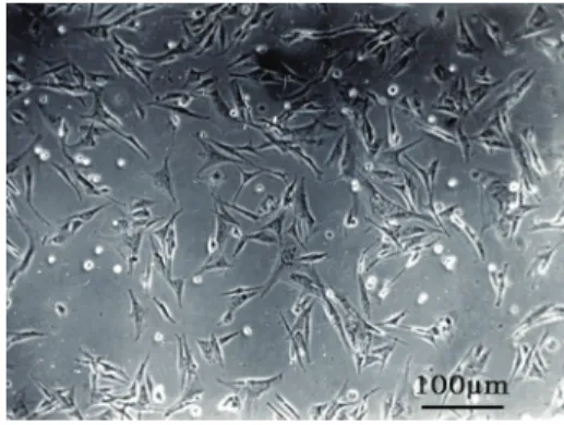
Passage 3fibroblasts began to adhere after seeding for 2 h. After 24 h, all fibroblasts adhered and stretched (Figure 1). The control group fibroblasts looked spindle-shaped, reached 100% fusion rate after 3 days and displayed a fingerprint-shaped order after 5 days of incubation (Figure 2). As days passed,fibroblasts proliferated slowly and dropped sporadically. When cultured for 14 days,
fibroblasts had little mineralized nodules.
Figure 1.After seeding for 24 h, passage 3fibroblasts adhered and stretched. Phase contrast microscope, 100.
Figure 2. The control group fibroblasts reached confluence and displayed afingerprint-shaped order at 5 days. HE staining,
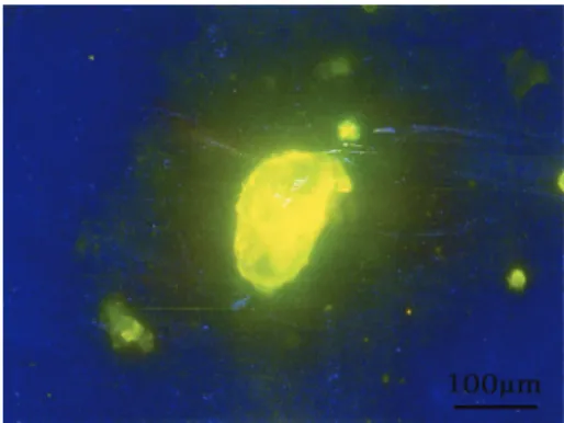
The fibroblasts in groups conditioned with fibronectin showed stronger proliferation compared with those of the control group. The fibroblasts looked triangle-shaped and exhibited obvious karyoschisis in the nucleolus after 3 days of culture. After culture for 5 days,fibroblasts had a decentered nucleus and distinct particles in cytoplasm (Figure 3). After 7 days, there were nebulous extracellular secretions cover-ing cells (Figure 4). As days passed, nebulous secretions gathered and formed dark nodules (Figure 5). The dark nodules gradually fused and formed a trabecula-like structure.
Mineralized nodule labeling with tetracycline
The visualized dark nodules were positive for the tetracycline staining, indicating mineralization. Compared with the control group, there were more and larger golden nodules containing a trabecula-like structure in the condi-tioned groups, especially in the group with afibronectin concentration of 40mg/mL (Figure 6).
Labeling with (3H-TdR)
The results of the3H-TdR incorporation test indicated the amount of the fibroblast DNA synthesis. Fibronectin
regulated the DNA synthesis offibroblasts. The group with afibronectin concentration of 60mg/mL at 7 days and the group with a fibronectin concentration of 40 mg/mL at 14 days had significantly higher DNA synthesis (Po0.05) compared with the other groups (Figure 7).
Labeling with3H-proline
Fibronectin up-regulated the collagen synthesis of
fibroblasts. The group with fibronectin concentration of 40mg/mL peaked at 7 and 14 days (Po0.05), the highest among all the groups (Figure 8).
Osteocalcin assessment
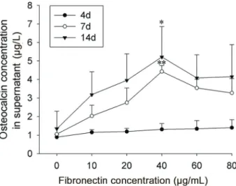
The increasing osteocalcin amount showed the osteogenesis capacity offibroblasts induced byfi bronec-tin. There was no statistical significance among groups at day 4 (P40.05). The group of 40 mg/mLfibronectin was statistically significant at day 7 (Po0.01) and day 14 (Po0.05), compared with the other groups (Figure 9).
Discussion
In this study,fibroblasts were obtained from the granula-tion tissue by tissue explants adherent method. Fibronectin
Figure 3. Fibroblasts induced with 40 mg/mL fibronectin had decentered nucleus and distinct particles in cytoplasm at 5 days. HE staining, 200.
Figure 4. Nebulous extracellular secretions coveredfibroblasts induced with 40 mg/mL fibronectin at 7 days. Phase contrast microscope, 100.
Figure 5.Dark tight nodules formed at 14 days infibroblasts induced with 40mg/mLfibronectin. Phase contrast microscope, 100.
in different concentrations was used to inducefibroblasts, and the ability of proliferation and osteogenesis was eval-uated. The3H-TdR and3H-proline incorporation test exhibited the high proliferative and functional capacity offibroblasts, respectively. Fibronectin at 40 mg/mL dramatically up-regulated the proliferation and collagen synthesis of
fibroblasts. The mineralized nodules and osteocalcin levels were vital osteogenic indicators. With the induction offibronectin,fibroblasts looked triangle-shaped, prolifer-ated, gathered with nebulous extracellular secretions and formed dark nodules ultimately. The nodules were posi-tive for tetracycline-labeling and identified as mineralized nodules. Combined with the significantly increased
osteocalcin levels, fibroblasts showed a definite osteo-genic capacity when induced with fibronectin, especially at the concentration of 40mg/mL. In summary,fibroblasts induced with fibronectin showed a high potential of pro-liferation, function and osteogenesis.
Some reviews have reported that fibroblasts derived from the adventitia (4), cardiac (5,6), burn scar (7) and normal skin (8) could be induced to myofibroblasts, which express a-smooth muscle actin. Periodontal fibroblasts were considered to have the potential of osteoblastic differentiation with an osteogenic medium containing 50 mg/mL of ascorbic acid-2-phosphate and 10 mM of b-glycerophosphate (9). Periodontal fibroblasts strongly expressed osteogenic genes such as ALP, Runx2, osterix and osteocalcin and formed mineralized nodules (10). The human dermalfibroblasts cultured on macroporous gelatin microcarriers encapsulated in platelet-rich plasma into three-dimensional constructs were successfully differen-tiated towards chondrogenic and osteogenic phenotypes using specific induction media (11). In 2005, Rieske et al. (12) reported thatfibroblasts had some of the characteristics of stem cells, and could be induced to become neuroecto-dermal cells and perhaps even mature neurons. Moreover, the human vascular adventitialfibroblasts from pulmonary arteries, were cultured in appropriate media and showed osteogenic, adipogenic and myogenic differentiation (13).
Based on the above literature and our study onfibroblasts, we present the theory thatfibroblasts have a potential of
in situ pleuripotent stem cell. Under specific conditions,
fibroblasts could exhibit pleuripotent differentiation in accordance with inductive factors. Mesenchymal stem cells (MSCs) are considered to be a promising source of stem cells in regenerative medicine. There has been evidence that MSCs could be induced to differentiate into
Figure 7. 3H-TdR incorporation test indicating thatfibronectin regulated the DNA synthesis offibroblasts. The 60mg/mLfibronectin concentration at 7 days and the 40mg/mLfibronectin at 14 days had significantly higher DNA synthesis compared to the other groups (*Po0.05, ANOVA). Data are reported as means±SD.
cpm: counts per minute.
Figure 8. 3H-proline incorporation test showing that
fibronectin up-regulated the collagen synthesis offibroblasts. Collagen synthe-sis in the 40mg/mLfibronectin group was significantly higher at 7 and 14 days than the other groups (*Po0.05, ANOVA). Data
are reported as means±SD. cpm: counts per minute.
Figure 9.Osteocalcin concentration increased with culture time in all groups. The 40mg/mLfibronectin was statistically significant at 7 (**Po0.01) and 14 days (*Po0.05) compared to the other
fibroblasts with conditioned induction media (14). Perhaps
fibroblasts are a kind of intermediate cell in the differentia-tion system from mesenchymal stem/progenitor cells to terminally differentiated cells, not strictly confined to dif-ferentiatedfibrocytes.
The induced pluripotent stem (iPS) cells have also been extensively studied. Fully reprogrammed iPS cells display numerous properties similar to those of embryonic stem cells and exhibit the capacity of self-renewal and differentiation into three germ layers (15). Up to now, there are few documents that suggest the use of MSCs or iPS cells to treat infectious soft tissue and bone defects. A series of undergoing experiments at the molecular and cellular levels are rooted in clinical observations on the subject. Our experiment demonstrated the fibroblast capa-city of proliferation and osteogenesis with the induction of
fibronectins. More importantly, the results supported the clinical observations that the granulation tissue, whose main cellular component is the fibroblast, could treat infectious soft tissue and bone defects.
Why can fibroblasts survive in an infectious environ-mentfilled with virulent bacteria, such asstaphylococcus aureus or/and pseudomonas aeruginosa, and display strong proliferation and osteogenesis? Although not tested in this study, a key factor could be the employment of the ShengJi Ointment, a Chinese herbal preparation.
Our preliminary work reported that the ShengJi Ointment and its fester-like secretions effectively enhanced capil-lary permeability of the local soft tissue and activated a monocyte-macrophage system that could efficiently secret various bioactive molecules, including fibronectins, not aiming at killing bacteria (16–18). Thus, we regarded ShengJi as an efficient biological agent, due to its immune modulation function. Althoughfibronectins play a vital role in adhesion, migration, chemotaxis and opsonization (19), many factors contribute to the local environment in a synergistic way. The employment of the Chinese herbal preparation as a promising biological agent and inductive factor is a novel strategy to induce the pluripotent stem cells in tissue engineering.
In conclusion, our experiment demonstrated that granula-tion tissue-derived fibroblasts induced with fibronectin exhibited increased proliferative, functional and osteo-genic potential. Fibroblasts are considered a possible
in situstem cell in tissue engineering.
Acknowledgments
This work was supported by the Natural Science Funds of Tianjin (Grant No.11JCYBJC10200) and the Science and Technology Supporting Plan Key Project of Tianjin (Grant No.08ZCGYSF01300).
References
1. Tiemann AH, Braunschweig R, Hofmann GO. Bone infec-tions.Unfallchirurg2012; 115: 480–488, doi: 10.1007/s00113-012-2187-y.
2. Hart B. Osteomyelitis (refractory) with literature review supplement.Undersea Hyperb Med2012; 39: 753–775. 3. Li XL, Han H, Shi YJ, Zhao FY, Xu EZ. A motive study of the
capillary permeability during wound healing–First session of research program on ‘‘leaning on the pus to promote regeneration’’.China J Orthop Trauma1994; 7: 5–7. 4. Shen W L, Gao PJ, Che ZQ, Ji KD, Yin M, Yan C, et al. NAD
(P)H oxidase-derived reactive oxygen species regulate angiotensin-II induced adventitial fibroblast phenotypic differentiation.Biochem Biophys Res Commun2006; 339: 337–343, doi: 10.1016/j.bbrc.2005.10.207.
5. Yuko AN, Ping D, Yoshinori H, Jiang Y, Hamaoka K, Takamatsu T. Cx43 contributes to TGF-b signaling to regulate differentiation of cardiacfibroblasts into myofi bro-blasts.Exper Cell Res2009; 315: 1190–1199, doi: 10.1016/ j.yexcr.2008.12.021.
6. Yano T, Miura T, Ikeda Y, Matsuda E, Saito K, Miki T, et al. Intracardiacfibroblasts, but not bone marrow derived cells, are the origin of myofibroblasts in myocardial infarct repair. Cardiovasc Pathol2005; 14: 241–246, doi: 10.1016/j.carpath. 2005.05.004.
7. Junker JP, Kratz C, Tollbäck A, Kratz G. Mechanical tension stimulates the transdifferentiation offibroblasts into myofi -broblasts in human burn scars.Burns2008; 34: 942–946, doi: 10.1016/j.burns.2008.01.010.
8. Huang CY, Fu X L, Liu J, Qi Y, Li S, Wang H. The involvement of integrin b1 signaling in the migration and myofibroblastic differentiation of skin fibroblasts on aniso-tropic collagen-containing nanofibers. Biomaterials 2012; 33: 1791–1800, doi: 10.1016/j.biomaterials.2011.11.025. 9. Chen YC, Ninomiya T, Hosoya A, Hiraga T, Miyazawa H,
Nakamura H. 1a,25-Dihydroxyvitamin D3 inhibits osteoblas-tic differentiation of mouse periodontalfibroblasts.Arch Oral Biol 2012; 57: 453–459, doi: 10.1016/j.archoralbio.2011. 10.005.
10. Hiraga T, Ninomiya T, Hosoya A, Takahashi M, Nakamura H. Formation of bone-like mineralized matrix by periodontal ligament cellsin vivo: a morphological study in rats.J Bone Miner Metab2009; 27: 149–157, doi: 10.1007/s00774-009-0039-9.
11. Sommar P, Pettersson S, Ness C, Johnson H, Kratz G, Junker JP. Engineering three-dimensional cartilage- and bone-like tissues using human dermalfibroblasts and macro-porous gelatine microcarriers.J Plast Reconstr Aesthet Surg 2010; 63: 1036–1046, doi: 10.1016/j.bjps.2009.02.072. 12. Rieske P, Krynska B, Azizi SA. Humanfibroblast-derived cell
lines have characteristics of embryonic stem cells and cells of neuro-ectodermal origin.Differentiation 2005; 73: 474– 483, doi: 10.1111/j.1432-0436.2005.00050.x.
14. Han YF, Chai JK, Sun TJ, Li D, Tao R. Differentiation of human umbilical cord mesenchymal stem cells into dermal fibroblastsin vitro. Biochem Biophys Res Commun2011; 413: 561–565, doi: 10.1016/j.bbrc.2011.09.001.
15. Takahashi K, Yamanaka S. Induction of pluripotent stem cells from mouse embryonic and adult fibroblast cultures by defined factors.Cell 2006;126: 663–676, doi: 10.1016/ j.cell.2006.07.024.
16. Li XL, Xu EZ, Shi YJ, Zhao FY. Phagocytic function and heterogeinicitic motive studies of surface wound healing– Studies on the mechanism of Wei Nong Zhang Rou (2). China J Orthop Trauma1995; 8: 8–11.
17. Li XL, Ji GY, Zhao FY, Shi YJ. Influence of immuno-activity of cellular oxidation metabolic function of the external used Chinese herb during wound healing –Study on the mechanism of Wei Nong Zhang Rou (3).China J Orthop Trauma1995; 8: 9–11.
18. Li XL, Shi YJ, Xu EZ, Zhao FY. Motive study offibrin binding protein in wound healing–Mechanism of Wei Nong Zhang Rou (IV) .China J Orthop Trauma1995; 8: 10–12. 19. Wu D, Hong Q, Chen XM, Bo F, Aipingi L, Hang L, et al.