Induction of chagasic-like arrhythmias
in the isolated beating hearts of
healthy rats perfused with
Trypanosoma
cruzi
-conditioned medium
H. Rodrı´guez-Angulo
1, J. Toro-Mendoza
2, J. Marques
3,
R. Bonfante-Cabarcas
4and A. Mijares
11Centro de Biofı´sica y Bioquı´mica, Instituto Venezolano de Investigaciones Cientı´ficas, Caracas, Venezuela 2Centro de Estudios Interdisciplinarios de la Fı´sica, Instituto Venezolano de Investigaciones Cientı´ficas,
Caracas, Venezuela 3Servicio de Cardiologı´a, Instituto de Medicina Tropical, Universidad Central de Venezuela, Caracas, Venezuela 4Unidad de Investigacio´n en Bioquı´mica, Decanato de Ciencias de la Salud,
Universidad Centroccidental ‘‘Lisandro Alvarado’’, Barquisimeto, Venezuela
Abstract
Chagas’ myocardiopathy, caused by the intracellular protozoan Trypanosoma cruzi, is characterized by microvascular alterations, heart failure and arrhythmias. Ischemia and arrythmogenesis have been attributed to proteins shed by the parasite, although this has not been fully demonstrated. The aim of the present investigation was to study the effect of substances shed byT. cruzion ischemia/reperfusion-induced arrhythmias. We performed a triple ischemia-reperfusion (I/R) protocol whereby the isolated beating rat hearts were perfused with either Vero-control or VeroT. cruzi-infected conditioned medium during the different stages of ischemia and subsequently reperfused with Tyrode’s solution. ECG and heart rate were recorded during the entire experiment. We observed that triple I/R-induced bradycardia was associated with the generation of auricular-ventricular blockade during ischemia and non-sustained nodal and ventricular tachycardia during reperfusion. Interestingly, perfusion with Vero-infected medium produced a delay in the reperfusion-induced recovery of heart rate, increased the frequency of tachycardic events and induced ventricular fibrillation. These results suggest that the presence of parasite-shed substances in conditioned media enhances the arrhythmogenic effects that occur during the I/R protocol.
Key words: Chagas’ disease; Arrhythmias; Ischemia
Introduction
Chagas’ disease is a tropical disease, initially confined to Central and South America, caused by the intracellular protozoan Trypanosoma cruzi. This illness is charac-terized by an acute phase, which is generally asympto-matic or oligosymptoasympto-matic; an indeterminate phase, which may persist for several years, and a chronic phase, when dilated cardiomyopathy and arrhythmias are primarily observed. Chagasic cardiomyopathy has been attributed to an imbalance between adrenergic or cholinergic innervations, cellular and humoral autoimmunity, parasitic effects, or microischemic disturbances (1).
The phenomenon of microvascular alterations and the generation of microischemic foci have been studied extensively during the acute and chronic phases of
Chagas’ disease. Several researchers have described changes indicative of ischemia in the hearts of chagasic patients, mainly associated with platelet aggregation (2), thinning of the capillary basement membranes (3), and areas of focal vascular constriction, microaneurysms, and dilatation of vascular beds in murine hearts during the acute phase of this infection (4). These disturbances could generate or enhance arrhythmias that are observed during chronic Chagas’ disease.
Cardiac arrhythmias are one of the most important disorders occurring during Chagas’ disease. Traditionally, arrhythmias have been considered to be a consequence of cardiac structural changes that occur during the chronic phase. In this respect, the main alterations reported are
Correspondence: J. Toro-Mendoza, Centro de Estudios Interdisciplinarios de la Fı´sica, Instituto Venezolano de Investigaciones Cientı´ficas, Caracas, 1020A, Venezuela. E-mail: [email protected] and/or [email protected]
atrial and ventricular extrasystoles, intraventricular and/or AV conduction disturbances, and primary ST-T wave changes (5). However, ST and T abnormalities, ventri-cular and supraventriventri-cular arrhythmias and low-voltage QRS have been reported in a recent acute oral outbreak characterized by high parasitemia (6), which suggest that the parasite may have a role in arrhythmia generation. The mechanisms associated with non-structural-related arrhythmias in Chagas’ disease are poorly understood.
Soluble factors that are generated during ischemia have been linked to cardiac arrhythmias, which are observed in isolated beating chagasic hearts (7). Moreover, it is known thatT. cruzi can release several substances into the extracellular milieu that produce alterations in microvascular (8) and intracellular calcium homeostasis (9,10), thereby contributing to arrhythmo-genesis. Nevertheless, the association between proteins that are secreted and/or shed by T. cruzi and the disturbances in heart conduction system and arrhythmias has not been fully elucidated. The aim of the present study was to determine whether perfusion of non-chagasic beating hearts with substances released by
T. cruzi can potentiate arrhythmias under ischemic conditions. Therefore, we performed a triple ischemia/ reperfusion (I/R) protocol to induce the ischemic and pro-arrhythmic conditions that are present in chronic chagasic hearts. To obtain quantitative information, the effects on heart rate (HR) were analyzed by fitting against a double-logistic model, and the results were compared to the changes observed in the ECG recordings during the different protocol stages.
Material and Methods
Cell culture and conditioned medium
Vero cells were plated in a 75-cm2 Easy Flask in 20-mL complete minimum essential medium (MEM) supplemented with 10% (v/v) fetal bovine serum (FBS), and were infected with 26105trypomastigotes/mL of the
EP strain at a ratio of 2 parasites per cell. Noninvasive parasites were removed after 24 h and the initial medium was changed at this time. The conditioned media were collected on the 5th or 6th day post-infection. The criteria for medium collection were that a minimum of 75% Vero cells should have adhered and that at least 2.56 106
trypomastigotes/mL should be present in the supernatant. The medium was centrifuged at 1500 g for 10 min to separate the parasites, and the supernatant was subse-quently filtered using a 0.2-mM membrane filter
(MilliporeH, USA) and stored at -20
˚
C until use.Preparation of isolated Langendorff hearts
For the isolated beating heart experiments, hearts were removed from adult female Sprague Dawley rats weighing 300-400 g anesthetizedipwith 40 mg/kg pento-barbital. The isolated hearts were placed in cold Tyrode’s
solution (25 mM sodium bicarbonate, 10 mM glucose, 116 mM sodium chloride, 3.3 mM potassium chloride, 2.5 mM calcium chloride, and 1 mM magnesium sulfate), cannulated through the aorta, and perfused in a retro-grade manner with warm Tyrode’s solution (37
˚
C) for 30 min by using the isolated beating heart system (AD Instruments, Australia). The presence of sinus rhythm, HR greater than 180 bpm, perfusion pressure higher than 30 mmHg, and a flow rate of 8-10 mL/min were considered to be stable values for all experiments. All procedures were approved by the Institutional Committee of the Venezuelan Institute for Scientific Research.After stabilization was achieved, a protocol consisting of three consecutive I/R cycles (10 and 20 min, respec-tively) was initiated. During each ischemic cycle, deoxygen-ated complete MEM (1.8 mM calcium chloride, 0.81 mM magnesium sulfate, 5.33 mM potassium chloride, 117.24 mM sodium chloride, 1.01 mM sodium phosphate monobasic, 5.56 mM D-glucose, plus L-glutamine, phenol red and essential amino acids) supplemented with 24 mM sodium bicarbonate was perfused at a low flow rate (2 mL/ min) for 10 min, followed by reperfusion with oxygenated Tyrode’s solution for 20 min (flow of 8 to 10 mL/min). During the ischemic periods, the hearts were perfused with medium that was conditioned with either control Vero cells control medium, n = 12) or infected Vero cells (Vero-infected medium, n = 14). Both of these media contained 10% FBS (v/v).
Measurement of enzyme activity
For biochemical analysis of the I/R process, we determined aspartate aminotransferase (AST) activity by sampling the effusate to indicate cardiac damage. The samples were collected every 2 min for a period of 30 min. The samples were immediately frozen and stored at -20
˚
C until further processing. The enzyme activity was measured with a commercially available ELISA kit (InvelabTM,Venezuela) according to manufacturer specifications.
Curve fitting
In this study, we used a double-logistic model (11)
f(t)~P1z P2
1zeP3(P4{t)z P2
1zeP5(P6{t) (1)
where fis the time (t)-dependent HR, P1represents the
minimum HR value,P2is the difference between the initial
and final HR values in the ischemic stage, andP3andP5
represent the HR slope values during the final ischemic and the initial reperfusion stages, respectively.P4andP6
are the ascending and descending inflection times, respectively.
Statistical analysis
with P , 0.05 were considered to be statistically significant. The d2 test was used to compare the ventricular fibrillation rate between the groups. Data analysis was performed using the Prism5H software (GraphPad Software, Inc., USA).
Results
Validation of the ischemia-reperfusion model
To evaluate cardiac damage during the I/R cycles, we collected the media after heart reperfusion and measured AST activity (Figure 1). A rapid increase in enzymatic activity was observed during the three ischemic phases (0-10, 30-40 and 60-70 min; Figure 1, thick black lines), which returned to basal levels during reperfusion. The enzymatic activity was not found to be different between the different ischemic cycles; therefore, these results suggest that our protocol produced a transient damage to the isolated beating hearts. Similarly, no significant differences were observed between the hearts that were perfused with Vero-control or Vero-infected medium (Figure 1).
Effect of conditioned medium on ECG values and recordings during I/R
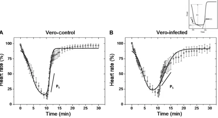
Figure 2 demonstrates the fitting of Equation 1 to HR kinetics during the I/R protocol for perfusion with Vero-control (A) or Vero-infected conditioned (B) medium (r2= 0.94 and 0.92, respectively). The plot shows that HR recovery (P5) was significantly slower in hearts
perfused with infected medium compared to Vero-control medium (0.53 ± 0.1422vs2.87 ± 0.36555, P = 0.0006). Interestingly, these results were associated with bradyarrhythmias, particularly sinus arrest during the
Figure 1. Average evolution of enzymatic activity (AST) in ischemia-reperfusion cycles. The graph shows the values of enzymatic activity measured every 2 min during the three ischemia-reperfusion cycles. Thick horizontal lines (top) indicate the different ischemia stages and are identified as I1 (ischemia 1), I2 (ischemia 2), and I3 (ischemia 3) and match the increased AST activity. The graph averages the data of Vero-control (n = 12) and Vero-infected hearts (n = 14).
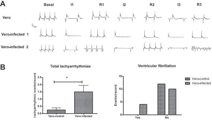
reperfusion period (middle portion of Figure 3), and were not observed in hearts perfused with Vero-control (top portion of Figure 3) that showed AV blockade during the final phase of ischemia.
In addition, tachyarrhythmias and the low-voltage QRS complex generated during perfusion of Vero-infected medium are displayed in Figure 3A (Vero-infected ‘‘2’’, bottom). Ventricular tachycardia was observed during the first reperfusion phase, whereas ventricular fibrillation was noted during the last reperfu-sion phase. Hearts perfused with Vero-infected medium demonstrated atrial flutter and nodal and ventricular tachycardia during the I/R stages, whereas in hearts perfused with Vero-control medium, nodal and ventricular tachycardia was observed in the reperfusion stage only. Overall, 1.5 ± 0.43 tachyarrhythmic events/record were recorded in hearts perfused with Vero-infected medium, whereas 0.25 ± 0.13 tachyarrhythmic events/record were recorded in hearts perfused with Vero-control medium (P = 0.0156). Ventricular fibrillation was detected in 4 of 14 hearts perfused with Vero-infected medium compared to 0 of 12 hearts perfused with Vero-control medium (P = 0.0441).
Discussion
Ischemia-reperfusion can lead to arrhythmias and sudden death. In particular, ischemia can lead to ventricular tachycardia and, finally, ventricular fibrillation (12). These events correlate with a concomitant increase in the intracellular H+, Na+, and Ca2+ levels (12), affecting the
cardiac action potential and thereby generating arrhyth-mias. Furthermore, microvascular alterations observed in Chagas’ disease could produce transient ischemia (2), and this is probably due to parasite-shed proteins (13). However, their participation in arrythmogenesis, which is observed in Chagas’ disease, are still unclear.
The fitting of a double-logistic equation (Equation 1) to model the I/R process is suitable to quantify and compare the different situations explained above. Despite the high complexity of the heart dynamics, the tendency shown allows us to use this tool to have an idea of the kinetics of the process with a reasonable degree of precision. However, a full mathematical description of the heart is far from being obtained and is out of the scope of the present study.
During our experiments, we observed an increase in the frequency of tachyarrhythmias during perfusion with
Vero-infected medium; therefore, we suggest that pro-teins present in the conditioned medium have the ability to enhance arrhythmias, probably by increasing the intra-cellular calcium levels. During parasite-host cell interac-tion, several secreted or released proteins can bind to the host cell receptors and modify cellular signaling. The different mucins present on the surface of trypomasti-gotes have been associated with the process of invasion. The protein gp82, a surface mucin ofT. cruzi, causes an increase in intracellular calcium in Hela cells infected with metacyclic trypomastigotes, an effect mediated by IP3, phospholipase C and protein tyrosine kinase (9,14). Gp90 and gp35/50 proteins are involved in the modulation of calcium increase in the host cell and the invasiveness of the strain of the parasite in which they are present (15). On the other hand, it has been reported that protein members of the transialidase and mucin family attach to the host cells and increase the intracellular calcium levels (13). Similarly, parasite cysteine proteases can cleave extracellular precursors, such as kininogens, which in turn bind to the host cell BK2R receptors and increase the
intracellular calcium concentration (16). These phenomena facilitate the entry of the parasite into its respective host cell. Interestingly, the existence of calcium overload in the ventricular myocytes of chagasic patients was recently reported (17). It is well known that increases in the intracellular calcium levels are responsible for the exten-sion of the plateau phase of the cardiac action potential. This phenomenon contributes to the generation of re-entry circuits and therefore the appearance of malignant ventricular arrhythmias (18). It is possible that the calcium overload that occurs during parasitic invasion of the cardiac cells can potentiate the proarrhythmogenic status produced by the ischemia due to microvascular altera-tions, which is observed in Chagas’ disease.
Ventricular arrhythmias observed in the isolated beat-ing chagasic hearts have been attributed to the activation of adenosine and adrenergic systems, both of which are present during ischemia (7). To our knowledge, this is the first report that demonstrates a causal link between trypomastigote-conditioned medium and arrythmogenesis in Chagas’ disease.
Other ionic currents may be associated with electrical alterations produced by T. cruzi-conditioned medium. Pacioretty et al. (19) reported a reduction of transient outward potassium current in epicardial myocytes during the acute phase of Chagas’ disease. This reduction was associated with the parasitemia peak, suggesting that a direct action of the parasite may be involved in this effect. Also, sera from chagasic patients with beta-adrenergic activity shortened the QT interval and the action potential duration and increased slow inward potassium current (IKs) and inward calcium current (Ica) (20).
On the other hand, parasite-cell interaction relies on specific signaling pathways in the host cells that facilitate the entry of the parasite, which could contribute to the
physiopathology of the disease. Andrade et al. (21) reported that exogenous endothelin-1 (ET-1) potentiated
T. cruziuptakein vitroand was associated with interstitial edema elicited by extracellular trypomastigotes. ET-1 is a potent vasoconstrictor and is present in high levels in the serum of chagasic patients. ET-1 induces vasospasm in
T. cruzi-infected mice, consequently contributing to the development of myocardial ischemia and myonecrosis (16). Additionally, other non-protein soluble mediators are secreted by the parasite and may be related to ischemic and electrical disturbances in chagasic hearts. T. cruzi -derived thromboxane A2 (TXA2) has been detected in the circulation of infected mice (22). Interestingly, TXA2 has been associated with reperfusion arrhythmias (23). Therefore, it could be involved in the arrythmogenesis observed in Chagas’ disease.
Perfusion with conditioned media produced a delay in HR recovery during the early reperfusion phase (see Figures 2 and 3). This is particularly relevant because it has been widely reported that Chagas’ disease is characterized by an alteration in the heart conduction pathways, which leads to AV block and sinus arrest among other complications (24). In addition, it has been reported that sera obtained from chronic chagasic patients who suffer from complex cardiac arrhythmias show depressed electrogenesis and conduction when used in different isolated beating heart preparations (25). It has been suggested that dampening of electrogenesis and bradyarrhythmias is due to the presence of auto-antibodies against the second extracellular loop of cardiac M2 acetylcholine receptor and b2 adrenoceptor (26,27). Interestingly, some investigators have reported a decrease in the expression of connexin 43, a gap junction protein, in the infected cardiac myocytes (28). This implies a causal relationship between parasitic infection and cardiac conduction disorders. However, to our knowledge, this is the first report that demonstrates the association of cardiac bradyarrhythmias with the perfusion of T. cruzi -conditioned medium.
We developed an experimental protocol to reproduce the ischemic and pro-arrhythmic conditions that exist in chronic chagasic hearts. The application of a double-logistic model allows the comparison of HR in the different media perfused. Our results suggest that substances present in the conditioned media have the ability to enhance I/R-induced arrhythmic and conduction dis-orders, which are similar to those observed in chagasic cardiomyopathy.
Acknowledgments
References
1. Higuchi Mde L, Benvenuti LA, Martins Reis M, Metzger M. Pathophysiology of the heart in Chagas’ disease: current status and new developments.Cardiovasc Res2003; 60: 96-107, doi: 10.1016/S0008-6363(03)00361-4.
2. Rossi MA, Carobrez SG. ExperimentalTrypanosoma cruzi cardiomyopathy in BALB/c mice: histochemical evidence of hypoxic changes in the myocardium.Br J Exp Pathol1985; 66: 155-160.
3. Andrade ZA, Andrade SG, Sadigursky M, Wenthold RJ Jr, Hilbert SL, Ferrans VJ. The indeterminate phase of Chagas’ disease: ultrastructural characterization of cardiac changes in the canine model.Am J Trop Med Hyg1997; 57: 328-336.
4. Factor SM, Cho S, Wittner M, Tanowitz H. Abnormalities of the coronary microcirculation in acute murine Chagas’ disease.Am J Trop Med Hyg1985; 34: 246-253.
5. Elizari MV, Chiale PA. Cardiac arrhythmias in Chagas’ heart disease.J Cardiovasc Electrophysiol1993; 4: 596-608, doi: 10.1111/j.1540-8167.1993.tb01247.x.
6. Alarcon de Noya B, Diaz-Bello Z, Colmenares C, Ruiz-Guevara R, Mauriello L, Zavala-Jaspe R, et al. Large urban outbreak of orally acquired acute Chagas disease at a school in Caracas, Venezuela.J Infect Dis2010; 201: 1308-1315, doi: 10.1086/651608.
7. Alvarado-Tapias E, Rivas-Coppola M, Alvarado A, Bello M, Briceno M, Rodriguez-Bonfante C, et al. [Adenosine induces ventricular arrythmias in hearts with chronic chagas cardiomyopathy].Rev Esp Cardiol2010; 63: 478-482, doi: 10.1016/S0300-8932(10)70069-5.
8. Libby P, Alroy J, Pereira ME. A neuraminidase from Trypanosoma cruziremoves sialic acid from the surface of mammalian myocardial and endothelial cells.J Clin Invest 1986; 77: 127-135, doi: 10.1172/JCI112266.
9. Dorta ML, Ferreira AT, Oshiro ME, Yoshida N. Ca2+signal induced byTrypanosoma cruzi metacyclic trypomastigote surface molecules implicated in mammalian cell invasion. Mol Biochem Parasitol 1995; 73: 285-289, doi: 10.1016/ 0166-6851(94)00123-5.
10. Manque PM, Neira I, Atayde VD, Cordero E, Ferreira AT, da Silveira JF, et al. Cell adhesion and Ca2+signaling activity in stably transfected Trypanosoma cruzi epimastigotes expressing the metacyclic stage-specific surface molecule gp82. Infect Immun 2003; 71: 1561-1565, doi: 10.1128/ IAI.71.3.1561-1565.2003.
11. Head GA, Lukoshkova EV, Mayorov DN, van den Buuse M. Non-symmetrical double-logistic analysis of 24-h blood pressure recordings in normotensive and hypertensive rats. J Hypertens2004; 22: 2075-2085, doi: 10.1097/00004872-200411000-00008.
12. Cascio WE. Myocardial ischemia: what factors determine arrhythmogenesis? J Cardiovasc Electrophysiol2001; 12: 726-729, doi: 10.1046/j.1540-8167.2001.00726.x.
13. Yoshida N, Cortez M. Trypanosoma cruzi: parasite and host cell signaling during the invasion process. Subcell Biochem 2008; 47: 82-91, doi: 10.1007/978-0-387-78267-6_6.
14. Yoshida N, Favoreto S Jr, Ferreira AT, Manque PM. Signal transduction induced in Trypanosoma cruzi metacyclic trypomastigotes during the invasion of mammalian cells.
Braz J Med Biol Res 2000; 33: 269-278, doi: 10.1590/ S0100-879X2000000300003.
15. Ruiz RC, Favoreto S Jr, Dorta ML, Oshiro ME, Ferreira AT, Manque PM, et al. Infectivity ofTrypanosoma cruzistrains is associated with differential expression of surface glycopro-teins with differential Ca2+ signalling activity. Biochem J 1998; 330 (Part 1): 505-511.
16. Scharfstein J, Andrade D. Infection-associated vasculopa-thy in experimental chagas disease pathogenic roles of endothelin and kinin pathways.Adv Parasitol2011; 76: 101-127, doi: 10.1016/B978-0-12-385895-5.00005-0.
17. Lopez JR, Espinosa R, Landazuru P, Linares N, Allen P, Mijares A. [Dysfunction of diastolic [Ca(2)(+)] in cardio-myocytes isolated from chagasic patients].Rev Esp Cardiol 2011; 64: 456-462, doi: 10.1016/j.recesp.2011.01.008. 18. Sipido KR, Bito V, Antoons G, Volders PG, Vos MA. Na/Ca
exchange and cardiac ventricular arrhythmias. Ann N Y Acad Sci 2007; 1099: 339-348, doi: 10.1196/ annals.1387.066.
19. Pacioretty LM, Barr SC, Han WP, Gilmour RF Jr. Reduction of the transient outward potassium current in a canine model of Chagas’ disease.Am J Physiol1995; 268: H1258-H1264.
20. Medei EH, Nascimento JH, Pedrosa RC, Barcellos L, Masuda MO, Sicouri S, et al. Antibodies with beta-adrenergic activity from chronic chagasic patients modulate the QT interval and M cell action potential duration.Europace2008; 10: 868-876, doi: 10.1093/europace/eun138.
21. Andrade D, Serra R, Svensjo E, Lima AP, Ramos ES Jr, Fortes FS, et al. Trypanosoma cruzi invades host cells through the activation of endothelin and bradykinin recep-tors: a converging pathway leading to chagasic vasculo-pathy.Br J Pharmacol2012; 165: 1333-1347, doi: 10.1111/ j.1476-5381.2011.01609.x.
22. Ashton AW, Mukherjee S, Nagajyothi FN, Huang H, Braunstein VL, Desruisseaux MS, et al. Thromboxane A2 is a key regulator of pathogenesis during Trypanosoma cruziinfection.J Exp Med2007; 204: 929-940, doi: 10.1084/ jem.20062432.
23. Parratt JR, Coker SJ, Wainwright CL. Eicosanoids and susceptibility to ventricular arrhythmias during myocardial ischaemia and reperfusion. J Mol Cell Cardiol 1987; 19 (Suppl 5): 55-66, doi: 10.1016/S0022-2828(87)80610-7. 24. Garzon SA, Lorga AM, Nicolau JC. Electrocardiography in
Chagas’ heart disease.Sa˜o Paulo Med J1995; 113: 802-813, doi: 10.1590/S1516-31801995000200011.
25. Costa PC, Fortes FS, Machado AB, Almeida NA, Olivares EL, Cabral PR, et al. Sera from chronic chagasic patients depress cardiac electrogenesis and conduction. Braz J Med Biol Res 2000; 33: 439-446, doi: 10.1590/S0100-879X2000000400010.
26. Escobar AL, Fernandez-Gomez R, Peter JC, Mobini R, Hoebeke J, Mijares A. IgGs and Mabs against the beta2-adrenoreceptor block A-V conduction in mouse hearts: A possible role in the pathogenesis of ventricular arrhythmias. J Mol Cell Cardiol 2006; 40: 829-837, doi: 10.1016/ j.yjmcc.2006.03.430.
recognize the second extracellular loop of the beta 1-adrenergic and M2-muscarinic receptors and regulate cal-cium channels in isolated cardiomyocytes.Mol Cell Biochem 1996; 163-164: 107-112, doi: 10.1007/BF00408646.