Longitudinal study of lung function in pregnant women:
Influence of parity and smoking
Luciana Duzolina Manfre´ Pastro,I,* Miriam Lemos,IIFrederico Leon Arrabal Fernandes,IIISilvia Regina Dias Me´dici Saldiva,IVSandra Elisabete Vieira,VBeatriz Mangueira Saraiva Romanholo,VI,VIIPaulo Hila´rio Nascimento Saldiva,IIRossana Pulcineli Vieira FranciscoI
IDepartamento de Ginecologia e Obstetricia, Faculdade de Medicina FMUSP, Universidade de Sao Paulo, Sao Paulo, SP, BR.IIDepartamento de Patologia,
Faculdade de Medicina FMUSP, Universidade de Sao Paulo, Sao Paulo, SP, BR.IIILaboratorio de Funcao Pulmonar, Disciplina de Pulmonologia, Instituto do
Coracao (InCor), Hospital das Clinicas HCFMUSP, Faculdade de Medicina, Universidade de Sao Paulo, Sao Paulo, SP, BR.IVInstituto de Saude, Secretaria de
Estado da Saude, Sao Paulo, SP, BR.VDepartamento de Pediatria, Faculdade de Medicina FMUSP, Universidade de Sao Paulo, Sao Paulo, SP, BR. VI
Departamento de Medicina (LIM 20), Faculdade de Medicina, Universidade de Sao Paulo, Sao Paulo, SP, BR.VIIUniversidade Cidade de Sao Paulo (UNICID), Sao Paulo, SP, BR.
OBJECTIVES: To evaluate pulmonary function in the first and third trimesters of pregnancy and analyze the influence of parity and smoking on spirometry parameters.
METHODS:This longitudinal prospective study included a cohort of 120 pregnant women. The inclusion criteria were as follows: singleton pregnancy, gestational age less than 13.86 weeks, and no preexisting maternal diseases. The exclusion criteria were as follows: change of address, abortion, and inadequate spirometry testing. ClinicalTrials.gov: NCT02807038.
RESULTS:A decrease in values of forced vital capacity and forced expiratory volume were noted in the first second from the first to third trimester. In the first and third trimesters, multiparous women demonstrated lower absolute forced vital capacity and forced expiratory volume values in the first second compared with nulliparous women (po0.0001 andp=0.001, respectively). Multiparous women demonstrated reduced forced expiratory flow in 25% to 75% of the maneuver compared with nulliparous women in the first (p=0.005) and third (p=0.031) trimesters. The absolute values of forced expiratory flow in 25% to 75%, forced expiratory volume in the first second and predicted peak expiratory flow values in the third trimester were higher in smokers compared with nonsmokers (p=0.042,p=0.039,p=0.024, andp=0.021, respectively).
CONCLUSION:There was a significant reduction in forced vital capacity and forced expiratory volume values in the first second during pregnancy. Parity and smoking significantly influence spirometric variables.
KEYWORDS: Spirometry; Pregnancy; Pulmonary Function Test; Parity; Smoking.
Pastro LD, Lemos M, Fernandes FL, Saldiva SR, Vieira SE, Romanholo BM, et al. Longitudinal study of lung function in pregnant women: Influence of parity and smoking. Clinics. 2017;72(10):595-599
Received for publication onFebruary 17, 2017;First review completed onApril 11, 2017;Accepted for publication onMay 10, 2017 *Corresponding author. E-mail: [email protected]
’ INTRODUCTION
Pregnancy is a period during which the female body under-goes functional and anatomical alterations, including changes in lung function. These findings can be assessed by spirometry, a simple, inexpensive, and effective method (1,2).
There is some controversy regarding the results of spiro-metry in pregnancy, particularly in terms of tobacco use and parity. Several studies measured lung function during pregnancy and after delivery. Mixed results have been reported for forced
vital capacity (FVC), forced expiratory volume in the first second (FEV1), and the FEV1/FVC ratio with respect to whether they
remain the same, increase, or decrease slightly (1,3,4). The predictors for decreasing lung function during preg-nancy remain unknown. Multiparity and smoking status may be associated with decreased lung function.
Objectives
This study aimed to use spirometry to evaluate changes in pulmonary function in pregnant women between the first (T1) and third trimester (T3) of pregnancy and to evaluate the influence of smoking and parity during this period.
’ METHODS
This longitudinal prospective study was conducted from August 2011 to May 2013 at the University of São Paulo Medical School (FMUSP), Brazil. This study was approved
DOI:10.6061/clinics/2017(10)02
Copyright&2017CLINICS–This is an Open Access article distributed under the terms of the Creative Commons License (http://creativecommons.org/licenses/by/ 4.0/) which permits unrestricted use, distribution, and reproduction in any medium or format, provided the original work is properly cited.
by the Ethics Committee for Research Project Analysis at the Hospital of the Faculty of Medicine under number 193/11, and written informed consent was obtained from the sub-jects. ClinicalTrials.gov: NCT02807038.
Study population
We included 220 pregnant women who were consecutively recruited from the three primary care units located in the west of the city of São Paulo. The women were part of a larger cohort of 400 patients who were recruited to study the effects of environmental air pollution on pregnancy.
The following inclusion criteria were applied: receipt of prior informed consent, singleton pregnancy, gestational age of less than 13.86 weeks at the first evaluation, and absence of preexisting maternal diseases. The exclusion criteria included withdrawal from the project, abortion, and inadequate spirom-etry testing performed at T1 and T3.
Study design
After the confirmation of pregnancy, community health workers made the first contact with the pregnant women. During this first meeting, the research project was explained to the pregnant women, who were invited to participate in the study and provide informed consent. If the women agreed to participate, they received a card with the first assess-ment date and information about spirometry.
The measurement techniques and spirometry parameters were selected in accordance with the Guidelines for Pul-monary Function Test of the Brazilian Thoracic Association (5). All spirometric values were adjusted according to sex, age, height, and weight at the time of the exam to estimate the predicted values (6).
At the first assessment, the patients were asked about previous pregnancies and tobacco use during pregnancy according to the questionnaire in the guideline cited above.
Statistical analysis
For the characterization and summary of the study vari-ables, the mean and standard deviation and median, mini-mum and maximini-mum values were used for quantitative variables, and absolute and relative frequency were used for qualitative variables.
A paired t-test was used for comparison of spirometric variables in the first and third trimesters. The t-test was applied to compare the groups of nulliparous and multi-parous women in relation to spirometric variables in each trimesters, and the Mann-Whitney-test was used for compar-ing smokers and nonsmokers.
’ RESULTS
The initial sample included 220 patients. In total, 11.4% of the patients were excluded due to abortion, 12.7% were excluded due to loss to follow-up, and 22% were excluded due to unsatisfactory spirometric test results. The final sample consisted of 120 pregnant women with 240 valid spirometric exams. The maternal characteristics are presented in Table 1. We analyzed the mean BMI in the first and third trimesters in the two groups, with no significant difference found (first trimester: nulliparous=25.65±5.02; multiparous=26.75±5.81
p=0.273); (third trimester: nulliparous=28.70±4.73, multiparous=
29.55±5.84p=0.390). There also was no significant difference
in the percentage of obese patients in the two groups studied (nulliparous=11/54 (20.4%), multiparous=13/66 (19.7%)p=0.927).
Table 2 presents the spirometric values obtained in T1 and T3. The absolute and relative values of FVC and absolute and % predicted values of FEV1were significantly reduced in
T3 compared with T1.
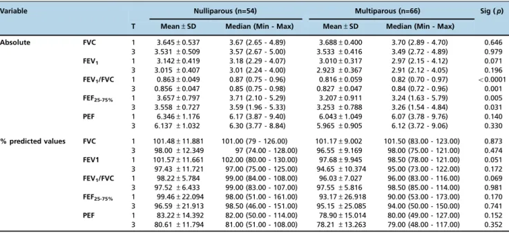
In terms of parity, as shown in Table 3, 54 patients (45%) were nulliparous, and the other 66 patients (55%) were multiparous. In T1, the multiparous women (0.816±0.059)
had smaller FEV1/FVC ratios compared with the nulliparous
women (0.863±0.049) (po0.0001) and smaller forced
expira-tory flow in 25% to 75% (FEF25-75%), atp=0.005 (multiparous
women (3.207±0.911) and nulliparous women (3.657±0.797)).
In T3, the multiparous women exhibited reductions in the same parameters compared with the nulliparous women: FEV1/FVC (multiparous women (30.827±0.047) and
nulli-parous women (0.856±0.047)) and FEF25-75% (multiparous
women (3.253±0.788) and nulliparous women (3.558±
0.727)) (p=0.00.1 andp=0.031, respectively). Maternal age
differed among nulliparous (23.00±5.57) and multiparous
(29.17±5.96) women atpp0.001. To eliminate the
confound-ing effect of age on absolute spirometry results, patients were categorized according to age: less or equal to 25 and more than 25 years age (Table 4).
Regarding smoking, as shown in Table 5, 17 of the 120 (14.17%) women smoked one to ten cigarettes a day throughout their pregnancies. Some parameters were significantly higher in nonsmokers compared with smokers.
Smokers had lower absolute values of FVC in T1 (3.48±
0.37) when compared to nonsmokers (3.70±0.47), p=0.042
lower absolute values of FEV1in T3 (2.78±0.29) when
com-pared to nonsmokers (2.99±0.39), p=0.039 lower absolute
values of FEF25-75%in T3 (3.00±0.69) when compared to
non-smokers (3.46±0.76), p=0.024 and lower values of predicted
peak expiratory flow (PEF) in T3 (smokers (71.71±11.39) and
nonsmokers (80.36±12.53),p=0.021.
’ DISCUSSION
In this study, it was possible to evaluate the spirometry results of 120 pregnant women during the first and third trimesters of pregnancy. In addition, the results were com-pared according to parity and smoking. Spirometry is the most widely used pulmonary function test, although it is estimated that approximately 15% of spirometry tests are inadequate (7,8). Because we required two complete exam-inations (i.e., in T1 and T3), some patients were excluded (22%). However, in one study of the long-term effect of air pollution on respiratory health in adult Swiss woman (9), a 28% loss to follow-up (related to inadequate spirometry tests) was reported, which was greater than the loss during follow-up in the current study.
All spirometric values in the pregnant subjects were within normal ranges. The FVC and FEV1values decreased
signifi-cantly in the third trimester. Redivo (10) described no changes in pulmonary function in pregnancy (10); however, Grindheim et al. (7) demonstrated differences, particularly in FVC and PEF (7). The results of recent studies agree with our find-ings (1,11). These reduced values may be due to decreased negative intrapleural pressure due to an upward tilt of the diaphragm caused by the enlarging uterus. Another expla-nation is the reduction in the alveolar partial pressure of carbon gas, which is caused by hyperventilation during pregnancy (12).
and multiparous women. Compared with the nulliparous women, the multiparous pregnant women had significantly reduced absolute values of FEV1/FVC and FEF25-75%. This
reduction was maintained among the younger multiparous
women (p25 years age) when compared to the younger
nulliparous women, suggesting that the decrease found is due to parity. In relation to multiparous women who are more than 25 years old, only the FEV1/FVC ratio in T1 maintained a significant effect. It is important to consider
that for this analysis, there was a significant decrease in the number of patients in each group, which can directly influence the calculation of the pvalue. It can be observed
that the average of these spirometric variables for different age groups remained similar to the general analysis.
The % predicted value of FEV1/FVC and FEF25-75%., which
took into account individual characteristics (sex, age, and body mass index), was not significantly different between the two groups. Grindheim et al. (7) found similar results in
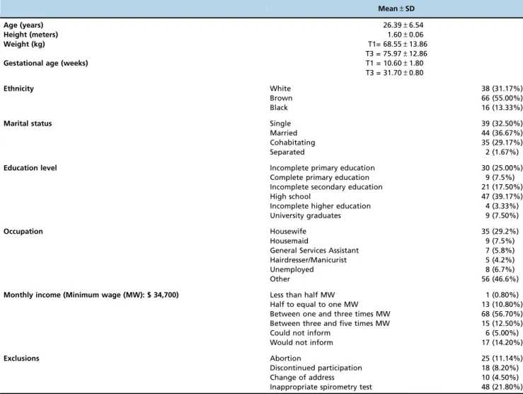
Table 1-Maternal characteristics.
Mean±SD
Age (years) 26.39±6.54
Height (meters) 1.60±0.06
Weight (kg) T1= 68.55±13.86
T3 = 75.97±12.86 Gestational age (weeks) T1 = 10.60±1.80
T3 = 31.70±0.80
Ethnicity White 38 (31.17%)
Brown 66 (55.00%)
Black 16 (13.33%)
Marital status Single 39 (32.50%)
Married 44 (36.67%)
Cohabitating 35 (29.17%)
Separated 2 (1.67%)
Education level Incomplete primary education 30 (25.00%)
Complete primary education 9 (7.5%)
Incomplete secondary education 21 (17.50%)
High school 47 (39.17%)
Incomplete higher education 4 (3.33%)
University graduates 9 (7.50%)
Occupation Housewife 35 (29.2%)
Housemaid 9 (7.5%)
General Services Assistant 7 (5.8%)
Hairdresser/Manicurist 5 (4.2%)
Unemployed 8 (6.7%)
Other 56 (46.6%)
Monthly income (Minimum wage (MW): $ 34,700) Less than half MW 1 (0.80%)
Half to equal to one MW 13 (10.80%)
Between one and three times MW 68 (56.70%)
Between three and five times MW 15 (12.50%)
Could not inform 6 (5.00%)
Would not inform 17 (14.20%)
Exclusions Abortion 25 (11.14%)
Discontinued participation 18 (8.20%)
Change of address 10 (4.50%)
Inappropriate spirometry test 48 (21.80%)
Table 2-Comparison of spirometric variables between the first and third trimesters of pregnancy.
Variable T1 T3 Sig (p)
Mean±SD Mean±SD
Absolute values FVC 3.670±0.467 3.532±0.459 o0.001
FEV1 3.070±0.397 2.964±0.387 o0.001
FEV1/FVC 0.837±0.059 0.840±0.049 0.486
FEF25-75% 3.409±0.887 3.390±0.773 0.667
PEF 6.179±1.113 6.042±0.964 0.044
% predicted values FVC 101.31±10.352 97.20±10.694 o0.001
FEV1 99.43±10.880 95.90±11.040 o0.001
FEV1/FVC 97.02±6.563 97.53±6.075 0.212
FEF25-75% 96.00±24.961 95.80±23.624 0.881
PEF 80.63±14.918 79.23±12.800 0.142
their study, which analyzed a cohort of 87 healthy pregnant women. The authors concluded that the predicted FVC values in multiparous pregnant women decreased by 4.4%
compared with those in nulliparous women (p=0.039).
No statistically significant differences were observed in the absolute and predicted PEF values. Although obesity was related to changes in spirometry and also to multiparity, there was no difference between the groups in relation to this maternal characteristic.
The absolute values of FVC, FEF25-75%, and FEV1 were
significantly decreased in smokers compared with nonsmokers. Vergara et al. (13) studied lung function in 136 girls (45.5%) in
relation to smoking (active and passive smokers) and investi-gated repercussions on spirometric parameters. A significant association was found between smoking and spirometric para-meters (ANOVA), which was lower in smokers: FVC (p=0.001),
FEV1 (p=0.0001), FEV1/FVC (p=0.004), PEF (p=0.0001), and
FEF25-75% (p=0.0001).
Prasad et al. (14) reported a study involving over 100 female smokers and 100 female non-smokers in the age group of 30-40 years in which three lung function tests, FEVI, FVC and PEFR, were employed, and it was observed that all parameters of the three lung function tests were significantly reduced among the smokers compared to the non-smokers.
Table 3-Comparison of spirometric variables in the first and third trimesters between nulliparous and multiparous women.
Variable Nulliparous (n=54) Multiparous (n=66) Sig (p)
T Mean±SD Median (Min - Max) Mean±SD Median (Min - Max)
Absolute FVC 1 3.645±0.537 3.67 (2.65 - 4.89) 3.688±0.400 3.70 (2.89 - 4.70) 0.646 3 3.531±0.509 3.57 (2.67 - 5.00) 3.533±0.416 3.49 (2.72 - 4.89) 0.979
FEV1 1 3.142±0.419 3.18 (2.29 - 4.07) 3.010±0.317 2.97 (2.15 - 4.12) 0.071
3 3.015±0.407 3.01 (2.24 - 4.00) 2.923±0.367 2.91 (2.12 - 4.05) 0.196 FEV1/FVC 1 0.863±0.049 0.87 (0.75 - 0.96) 0.816±0.059 0.82 (0.70 - 0.97) o0.0001
3 0.856±0.047 0.85 (0.75 - 0.98) 0.827±0.047 0.84 (0.72 - 0.96) 0.001 FEF25-75% 1 3.657±0.797 3.71 (2.10 - 5.29) 3.207±0.911 3.24 (1.63 - 5.79) 0.005
3 3.558±0.727 3.59 (1.96 - 5.33) 3.253±0.788 3.26 (1.54 - 4.84) 0.031 PEF 1 6.346±1.176 6.17 (3.87 - 9.40) 6.043±1.049 6.07 (3.78 - 9.76) 0.140 3 6.137±1.032 6.30 (3.77 - 8.84) 5.965±0.905 6.12 (3.72 - 9.06) 0.330 % predicted values FVC 1 101.48±11.881 101.00 (79 - 126.00) 101.17±9.002 101.50 (83.00 - 123.00) 0.873
3 98.00±12.349 97 (74.00 - 128.00) 96.55±9.169 98.00 (75.00 - 121.00) 0.474 FEV1 1 101.57±11.661 102.00 (80.00 - 130.00) 97.68±9.945 98.50 (78.00 - 121.00) 0.051
3 97.43±11.721 97.00 (75.00 - 125.00) 94.65±10.374 95.00 (73.00 - 122.00) 0.172 FEV1/FVC 1 98.22±5.784 99.00 (84.00 - 108.00) 96.03±7.027 96.00 (83.00 - 116.00) 0.069
3 97.52±6.433 99.00 (83.00 - 107.00) 97.55±5.816 98.50 (85.00 - 114.00) 0.981 FEF25-75% 1 99.46±22.094 98.00 (51.00 - 161.00) 93.17±26.918 90.00 (53.00 - 173.00) 0.170
3 96.59±21.913 98.50 (46.00 - 151.00) 95.15±25.085 94.00 (50.00 - 150.00) 0.741 PEF 1 83.22±14.392 82.00 (50.00 - 114.00) 78.90±15.014 80.00 (49.00 - 127.00) 0.152 3 80.61±11.794 81.00 (51.00 - 108.00) 78.21±13.263 79.00 (48.00 - 117.00) 0.352
Table 4-Comparison of spirometric variables in the first and third trimesters between nulliparous and multiparous women atp25 and425 years old.
Variable T Womenp25 years sig (p) Women425 years sig (p)
Nulliparous (n=40) Multiparous (n=20) Nulliparous (n=14) Multiparous (n=46) Mean±SD Mean±SD Mean±SD Mean±SD
Absolute FVC 1 3.626±0.532 3.670±0.315 0.691 3.712±0.569 3.696±0.436 0.910 3 3.515±0.493 3.516±0.318 0.993 3.579±0.573 3.542±0.456 0.801
FEV1 1 3.143±0.419 3.038±0.265 0.310 3.141±0.438 2.999±0.412 0.269
3 3.016±0.411 2.911±0.269 0.302 3.014±0.412 2.928±0.409 0.499 FEV1/FVC 1 0.870±0.053 0.829±0.054 0.007 0.847±0.032 0.812±0.062 0.043
3 0.860±0.050 0.830±0.045 0.026 0.845±0.036 0.826±0.049 0.185 FEF25-75% 1 3.721±0.825 3.230±0.673 0.025 3.474±0.707 3.197±1.004 0.342
3 3.564±0.755 3.220±0.650 0.088 3.542±0.667 3.268±0.849 0.273
PEF 1 6.292±0.987 6.169±1.125 0.666 6.502±1.639 5.989±1.023 0.284
3 6.121±1.088 5.762±0.911 0.210 6.185±0.892 6.052±0.899 0.630 % predicted values FVC 1 101.250±10.970 99.800±7.031 0.538 101.143±14.623 101.761±9.746 0.928
3 97.450±11.031 94.450±7.577 0.223 99.571±15.907 97.457±9.715 0.644 FEV01 1 101.050±10.651 96.050±7.715 0.065 103.071±14.715 98.391±10.771 0.198
3 96.625±10.860 91.300±7.349 0.053 99.571±14.095 96.109±11.202 0.325 FEV1/FVC 1 97.375±6.270 94.350±5.613 0.074 100.643±3.153 96.761±7.499 0.007
3 96.475±6.752 95.300±5.516 0.504 100.500±11.202 98.522±5.726 0.239 FEF25-75% 1 96.950±22.368 83.150±18.082 0.020 106.643±20.353 97.522±29.061 0.279
One limitation is the fact that smoking research was per-formed only through a structured questionnaire with the possibility of one response (yes or no). Currently, the use of monoximetry is suggested to more accurately discriminate smokers who, for any reason, will declare themselves to be non-smokers. Nonetheless, we must point out that the great majority of studies in the literature that use spirometry for lung function evaluation do not advocate the performance of monoximetry to exclude smoking. In this study, we concluded that the absolute values of FVC, FEF25-75%, and
FEV1were significantly decreased in smokers compared with
nonsmokers, with significant reductions in absolute and in % predicted FVC and FEV1 values during pregnancy (T1 to T3). Parity and smoking significantly influenced spirometric para-meters, and these are important factors that must be considered when analyzing spirometry results during pregnancy.
FVC and FEV1values decreased significantly in the third
trimester. The reductions in these parameters may be explained by the upward tilt of the diaphragm caused by the enlarging uterus, causing a decreased negative intrapleural pressure, and because of a reduction in the alveolar partial pressure of carbon gas, which is caused by hyperventilation during pregnancy. Decreased lung function at both the beginning and end of the gestational period is more evident in multiparous women and those who smoke.
’ ACKNOWLEDGMENTS
Financial support: FAPESP, process number 2009/17315-9 and CNPq, process number 140260/2011-0.
’ AUTHOR CONTRIBUTIONS
Pastro LD performed spirometry and data analyses and was responsible for thefinal preparation of the manuscript. Lemos M assisted in the design of the study and spirometry. Fernandes FL provided training regarding spirometry and analyzed spirometric tests. Saldiva SR assisted in the pre-paration of the project and the recruitment of pregnant women. Vieira SE assisted in the preparation of the project and the recruitment of pregnant women. Romanholo BM performed spirometry. Saldiva PH assisted in the
preparation of the project and thefinal data analysis. Francisco RP assisted
in the preparation of the project, manuscript writing andfinal data analysis.
’ REFERENCES
1. Gupta L, Dixit R. A linear study of pulmonary function tests in normal pregnant and non-pregnant women. J Indian Med Assoc. 2013;111(10): 666-9.
2. Tan EK, Tan EL. Alterations in physiology and anatomy during preg-nancy. Best Pract Res Clin Obstet Gynaecol. 2013;27(6):791-802, http://dx. doi.org/10.1016/j.bpobgyn.2013.08.001.
3. Hegewald MJ, Crapo RO. Respiratory physiology in pregnancy. Clin Chest Med. 2011;32(1):1-13, http://dx.doi.org/10.1016/j.ccm.2010.11.001. 4. Kolarzyk E, Szot WM, Lyszczarz J. Lung function and breathing regula-tion parameters during pregnancy. Arch Gynecol Obstet. 2005;272(1):53-8, http://dx.doi.org/10.1007/s00404-004-0691-1.
5. Pereira C. Diretrizes para o teste de func¸ão pulmonar da Sociedade Bra-sileira de pneumologia e tisiologia–Espirometria. J Pneumol. 2002;1-82. 6. American Thoracic Society. Standardization of Spirometry, 1994 Update.
Am J Respir Crit Care Med. 1995;152(3):1107-36, http://dx.doi.org/ 10.1164/ajrccm.152.3.7663792.
7. Grindheim G, Toska K, Estensen ME, Rosseland LA. Changes in pul-monary function during pregnancy: a longitudinal cohort study. BJOG. 2012;119(1):94-101, http://dx.doi.org/10.1111/j.1471-0528.2011.03158.x. 8. Mbatchou Ngahane BH, Afane Ze E, Chebu C, Mapoure NY, Temfack E,
Nganda M, et al. Effects of cooking fuel smoke on respiratory symptoms and lung function in semi-rural women in Cameroon. Int J Occup Environ Health. 2015;21(1):61-5, http://dx.doi.org/10.1179/2049396714Y.00000 00090.
9. Schikowski T, Schaffner E, Meier F, Phuleria HC, Vierkötter A, Schindler C, et al. Improved air quality and attenuated lung function decline: modification by obesity in the SAPALDIA cohort. Environ Health Perspect. 2013;121(9):1034-9, http://dx.doi.org/10.1289/ehp.1206145. 10. Redivo MB. Avaliac¸ão da func¸ão pulmonar em gestantes no período
gestacional entre 28 a 36 semanas. Universidade do Sul de Santa Catarina. 2007.
11. Neeraj, Sodhi C, Pramod J, Singh J, Kaur V. Effect of advanced uncom-plicated pregnancy on pulmonary function parameters of North Indian subjects. Indian J Physiol Pharmacol. 2010;54(1):69-72.
12. Ogbodo S, Nwagha U, Okaka A, Okeke A, Chukwurah F, Ezeonu P. Low levels of some nutritional parameters of pregnant women in a rural community of South East Nigeria: implications for the attainment of the millennium developmental goal. Ann Med Health Sci Res. 2012;2(1): 49-55, http://dx.doi.org/10.4103/2141-9248.96939
13. Suárez López de Vergara RG, Fernández C, Oliva Hernández C, Domé-nech Martínez E, Dorta Delgado JM, Dorta Suárez M. Lung function and exposure to tobacco smoke among adolescents. An Pediatr. 2007;67(6): 559-66, http://dx.doi.org/10.1016/S1695-4033(07)70804-4
14. Prasad BK, Sahay AP, Singh AK. Smoking women and their lung function tests. Kathmandu Univ Med J (KUMJ). 2004;2(2):142-4.
Table 5-Comparison of spirometric variables in the first and third trimesters between pregnant smokers and nonsmokers.
Variable Nonsmokers (n=103) Smokers (n=17) Sig (p)
T Mean±SD Median (Min - Max) Mean±SD Median (Min - Max)
Absolute FVC 1 3.70±0.47 3.71 (2.71 - 4.89) 3.48±0.37 3.45 (2.65 - 4.39) 0.042 3 3.56±0.47 3.53 (2.67 - 5.00) 3.36±0.29 3.32 (2.92 - 3.94) 0.074
FEV1 1 3.10±0.40 3.11 (2.15 - 4.12) 2.91±0.32 2.96 (2.29 - 3.38) 0.115
3 2.99±0.39 2.95 (2.12 - 4.05) 2.78±0.29 2.84 (2.12 - 4.05) 0.039 FEV1/FVC 1 0.84±0.06 0.84 (0.70 - 0.97) 0.84±0.05 0.84 (0.71 - 0.96) 0.842
3 0.84±0.04 0.84 (0.73 - 0.98) 0.83±0.05 0.83 (0.72 - 0.95) 0.154 FEF25-75% 1 3.44±0.90 3.43 (1.63 - 5.59) 3.22±0.75 3.26 (1.63 - 4.21) 0.447
3 3.46±0.76 3.42 (1.55 - 5.33) 3.00±0.69 3.07 (1.54 - 4.09) 0.024 PEF 1 6.25±1.14 6.22 (3.78 - 9.76) 5.73±0.81 5.92 (4.02 - 7.24) 0.055 3 6.11±0.96 6.20 (3.72 - 9.06) 5.65±0.85 5.58 (3.89 - 6.83) 0.078 % predicted values FVC 1 101.95±10.67 102 (79 - 126) 97.41±7.15 96 (87 - 110) 0.064
3 97.78±11.00 98 (78 - 128) 93.71±7.92 94 (78 - 111) 0.131
FEV1 1 100.07±11.11 100 (78 - 130) 95.59±7.63 99 (80 - 108) 0.144
3 96.70±11.10 96 (73 - 125) 91.06±9.53 95 (75 - 106) 0.081
FEV1/FVC 1 97.02±6.62 97 (83 - 116) 97.00±6.39 99 (85 - 105) 0.734
3 97.79±6.09 99 (83 - 114) 96.00±5.88 97 (85 - 103) 0.252
FEF25-75% 1 96.86±25.15 94 (51 - 173) 90.76±23.79 93 (53 - 156) 0.416
3 97.43±23.54 98 (50 - 151) 85.94±22.30 93 (46 - 135) 0.076
PEF 1 81.63±15.02 82 (49 - 127) 84.57±12.47 78 (50 - 97) 0.092
3 80.36±12.53 80 (48 - 117) 71.71±11.39 70 (51 - 88) 0.021