99mTc-thymine scintigraphy may be a promising
method in the diagnosis of breast cancer
Monica Pires Ribeiro, Sergio Augusto Lopes de Souza, Flavia Paiva Proenc¸a Lobo Lopes, Paulo Henrique Rosado-de-Castro, Lea Mirian Barbosa da Fonseca, Bianca Gutfilen
Universidade Federal do Rio de Janeiro, Faculdade de Medicina, Hospital Universita´rio Clementino Fraga Filho, Departamento de Radiologia, Rio de Janeiro/RJ, Brazil.
OBJECTIVE:Mammography has been established as the gold standard for the detection of breast cancer, and imaging techniques such as ultrasonography, magnetic resonance imaging, scintigraphy and positron emission tomography may be useful to improve its sensitivity and specificity. The objective of this study with breast scintigraphy was to evaluate the uptake of 99mTc-thymine in mammary lesions.
METHODS:A total of 45 patients were included in this study. Thirty-three patients (73%) were subjected to surgery or percutaneous biopsy, providing histopathological data. The other 12 patients who remained under surveillance received clinical examinations and biannual mammography with a normal follow-up of at least three years, the data from which were used for comparison with the scintimammography results.
RESULTS:The majority of patients (64.4%) had clinically impalpable lesions with a mammogram diagnosis of microcalcifications, impalpable nodules, or focal asymmetry. Of the studied lesions, 87% were smaller or equal to 20 mm in diameter, and 22% had malignant histopathological findings. Scintigraphy with 99mTc-thymine had a sensitivity of 70%, a specificity of 85.7%, positive and negative predictive values of 58.3% and 90.9%, respectively, and an accuracy of 82.2%.
CONCLUSIONS:The results of this study are consistent with those previously reported by other authors. The good specificity and high negative predictive value of this technique and the absence of uptake in the heart indicate that it may be a promising complementary method in clinical practice and that it may contribute to reducing unnecessary benign biopsies.
KEYWORDS: Breast Scintigraphy; 99mTc-thymine; Breast Cancer.
Ribeiro MP, Souza SA, Lopes FP, Castro PH, Fonseca LM, Gutfilen B. 99mTc-thymine scintigraphy may be a promising method in the diagnosis of breast cancer. Clinics. 2013;68(3):283-289.
Received for publication onSeptember 25, 2012;First review completed onOctober 3, 2012;Accepted for publication onOctober 19, 2012 E-mail: [email protected]
Tel.: 55 21 2562-6274 / 55 21 2562-6273
& INTRODUCTION
Breast cancer is a major public health problem, the incidence and mortality of which are continuously increas-ing. Breast cancer represents the most common malignancy in women, affecting more than one million individuals every year worldwide (1,2).
An early diagnosis of breast cancer may result in a better prognosis. Thus, regular screenings are recommended by health systems around the world. Mammography has been established as the gold standard method for the detection of breast cancer (3-5). The efficacy of cancer screening using mammography has been demonstrated in both randomized
trials and observational studies (3-5). Mammographic screening reduces the mortality from breast cancer in women between 40 and 69 years of age by 20 to 35% (4,6). However, not all malignancies are detected using this screening method. Major factors that lead to false-negative findings using mammography include breast density, implants, severe dysplastic disease, and significant archi-tectural distortion following breast surgery or radiation therapy (7-10).
Approximately 90 to 95% of women with abnormal mammogram findings do not have breast cancer. The relative inefficiency reflected by this statistic highlights the necessity of new diagnostic methods that can improve the sensitivity and specificity of mammography (10,11). The development of improved non-invasive diagnostic methods for the screening of breast lesions is needed, which would reduce the requirement for invasive procedures such as breast biopsy for a precise diagnosis (10,11). Conventional biopsies, in addition to the potential for the involvement of hospitalization, anesthesia, and clinical complications, are unnecessary in 80% of cases and may induce changes in the
Copyrightß2013CLINICS– This is an Open Access article distributed under the terms of the Creative Commons Attribution Non-Commercial License (http:// creativecommons.org/licenses/by-nc/3.0/) which permits unrestricted non-commercial use, distribution, and reproduction in any medium, provided the original work is properly cited.
No potential conflict of interest was reported.
breast parenchyma that hinder subsequent mammographic readings (4,10). In addition, procedures such as percuta-neous or surgical biopsy can cause anxiety, inconvenience, and discomfort to the patient and can add medical costs to both the patient and the health care system (10-12).
In the last decade, progress in pharmacology combined with evolving technology has enabled the establishment of scintimammography as a non-invasive technique for the localization and staging of malignant tumors of the breast using molecules such as Sestamibi and Tetrofosmin labeled with technetium-99m (99mTc) (13-15). Other molecules such as 99mTc(V) dimercaptosuccinic acid (DMSA) and 99mTc-labeled alkyl triphenyl phosphonium (99mTc-Mito10
-MAG3) are also being studied to improve detection (16,17). The quality of scintimammography is not compro-mised by breast density, which limits the effectiveness of mammography particularly in young women. Breast scinti-graphy has a major indication for select groups of patients with dense breasts; patients using hormone therapy; patients suspected of having recurrent disease resulting in architectural distortions secondary to previous surgery; patients undergoing chemotherapy or radiotherapy or who have breast implants; and patients at high risk for breast cancer (15,18-21). Scintimammography is also useful for the study of breast cancer multicentricity (13,15).
A recent meta-analysis of 42 studies that used ultrasound, computed tomography (CT), magnetic resonance imaging (MRI), scintimammography, and positron emission tomogra-phy (PET) determined that MRI seemed to be a more useful complement to existing surveillance methods to assess patients with suspected recurrent and/or metastatic breast cancer (17). Although MRI can be applied in these patient populations, its usage is limited by higher costs and a higher rate of false-positive results compared with mammography (4).
In previous reports, we presented the use of 99mTc-thymine in the evaluation of nodules and the thickening of breast tissue. The pattern of 99mTc-thymine uptake could differentiate malignant lesions of the breast from benign lesions and breast densities in which there is uptake of the radiopharmaceutical (22). We also compared the uptake of 99mTc-thymine and 99mTc-sestamibi in malignant breast lesions and found that 99mTc-thymine exhibited higher sensitivity, specificity, accuracy, and positive predictive values compared to 99mTc-sestamibi (19).
Here, we evaluated the use of 99mTc-thymine for the study of malignant microcalcifications and initial palpable and impalpable malignant lesions of the breast.
& METHODS AND MATERIALS
Study subjects consisted of 45 patients of the gynecology and mastology services of two public hospitals. Inclusion criteria were: (I) patients who detected changes in their breast self-examination, (II) patients with microcalcifications detected using mammography consisting of asymmetry or focal nodules smaller or equal to 25 mm in diameter, and (III) patients with BI-RADSH categories 3, 4, or 5. We excluded patients based on the following criteria: (I) breast lesions greater than 25 mm in diameter, (II) prior chemo or hormone therapy, (III) breast surgery or prior homolateral radiotherapy, and (IV) pregnancy or lactation.
The BI-RADS system includes categories that are used to standardize interpretation of mammograms among radiolo-gists (23). BI-RADS assessment categories can be summarized
as: Category 0 - need additional imaging evaluation; Category 1 - negative; Category 2 - benign finding; Category 3 - probably benign finding - follow-up in a short time frame is suggested; Category 4 - suspicious abnorm-ality, biopsy considered; Category 5 - highly suggestive of malignancy, appropriate action needed; Category 6 - known biopsy - proven malignancy.
The average age of the patients studied was 49.9 years and ranged from 26 to 80 years. All patients were informed about the procedure and, after acceptance for inclusion in the study, signed a written informed consent. The research protocol was approved by the research ethics committee of the institution.
To evaluate breast lesions, 370 MBq (10 mCi) of 99mTc-thymine was injected intravenously into the forearm, contralateral to the breast with the lesion. Patients were placed in a prone position on a foam mattress, and the studied breast was positioned to make it accessible. The other breast remained compressed to the mattress. The homolateral arm of the affected breast was placed above the patient’s head in a position as comfortable as possible. The surface of the collimator (a device of the gamma camera that is capable of collimating radiation to produce the patient’s images) touched the side of the body with the studied breast.
Counts were acquired in a 15% window centered at 140 keV. The acquisition of images was initiated 15 minutes after the administration of 99mTc-thymine, and each acquisition lasted for 10 minutes. Both breasts were evaluated. The first lateral image obtained was of the breast without lesions, followed by the breast with the suspected tumor. Following image acquisition, the patient was placed in the supine position with the hands behind the head.
Planar images were analyzed separately by two nuclear medicine physicians and, when the opinions were discor-dant, the results were determined by consensus. Using a qualitative analysis of images, scintigraphy was considered positive when there was a high uptake of the radio-pharmaceutical in a segment of the breast. Scintigraphy was considered negative in cases where there was a homogeneous distribution of the radiopharmaceutical in both breasts.
The data were evaluated by the analysis of the sensitivity, specificity, positive and negative predictive values, and measures of accuracy (confidence interval of 95%; 95% CI) for scintigraphy in relation to the standard for histology (positive and negative).
The histopathological samples were obtained from study patients with breast mammography results with BI-RADSH
classifications of category 4 or 5, which included 33 patients. Twelve patients with BI-RADSHclassification of category 3 had been recommended for monitoring with biannual mammography. We used the follow-up period of three years and the radiological gold standard for comparison with the outcome of breast scintigraphy in these patients.
The degree of agreement was evaluated using the Kappa coefficient. The criterion for determination of significance was 5%.
& RESULTS
biannual mammography with a normal follow-up of at least three years, the data from which were used for comparison with the scintimammography results.
A majority of patients (29/45; 64.4%) had clinically impalpable lesions with a mammogram diagnosis of microcalcifications, impalpable nodules, or focal asymme-try. Sixteen patients (35.6%) had palpable lesions. Clustered microcalcifications, which were or were not associated with a nodule, were present in 24/45 patients (53%). Among the studied lesions, 87% were smaller or equal to 20 mm in diameter (41/47). Two patients had two lesions.
Ten (22%) of the 45 patients studied had malignant histopathological findings (11 lesions - eight cases of infiltrating ductal carcinoma, one case of in situ ductal carcinoma and invasive lobular carcinoma, and one case of cribriform type intraductal carcinoma. All patients with confirmed breast cancer were between 42 and 75 years old. In comparison with breast mammography, 7/10 of the cancer cases were correctly identified by breast scintigraphy with 99mTc-thymine. In three patients (3/10), breast scintigraphy showed false-negative results (a palpable nodule with normal mammogram, an irregular nodule suspected for malignancy, and clustered microcalcifications on mammography).
Thirty patients with negative breast scintigraphy results had histopathology and/or radiological follow-up for three years. Four patients who had positive breast scintigraphy had benign histopathological biopsies. The false-positive results were: two cystic nodules, one fibroadenoma, and an atypical hyperplasia. In one patient (BI-RADSH 3) with a positive breast scintigraphy, histopathological examination was not performed by decision of the mastology service, which followed the patient for three years. Table 1 shows the patients’ characteristics, Table 2 lists the measures of accuracy of the patients studied, and Table 3 shows the patients sorted by BI-RADS.
We observed a significant concordance (Kappa = 0.520, standard error = 0.147,p= 0.0002) between the diagnosis by
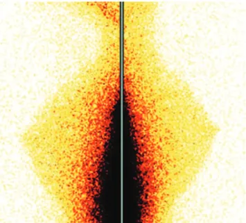
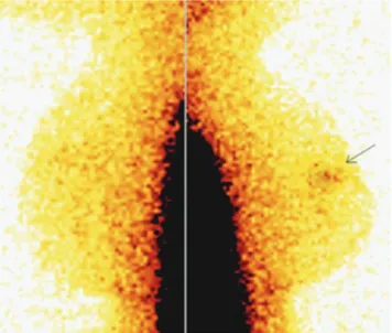
scintigraphy and the pattern of histology, but it was of a moderate degree. Figure 1 illustrates a normal distribution of 99mTc-thymine scintigraphy in the anterior view with a high liver uptake. We can observe that there is no heart uptake. Figure 2 is an example of a negative scintigraphy image obtained with 99mTc-thymine. A homogenous distribution of the radiopharmaceutical can be observed in both breasts. Figure 3 shows an example of a positive scintigraphy image obtained with 99mTc-thymine in which an increased uptake of the radiopharmaceutical is observed in the region of the right breast nodule.
& DISCUSSION
Although the efficacy of mammography has been demonstrated, perfect sensitivity or specificity is not achieved in women undergoing screening. As such, the issue of adverse consequences for women who do or do not have breast cancer has been a source of growing attention (15,24,25).
If breast cancer screening is to be successful, the majority of cancers among screened women must be detected when tumors are small and before the occurrence of distant or nodal metastases. To achieve this, a combination of imaging modalities may be superior to any single screening technique (9). 99mTc-sestamibi and 99mTc-tetrofosmin
were first described as myocardial perfusion tracers due to their property of accumulating in cells with higher mitochondrial activity and demonstrating perfused myo-cardium in contrast with ischemic and fibrotic regions. Afterwards, they were applied to detect tumors based on the principle that tumors such as breast cancer generally have higher mitochondrial activity than the surrounding cells. Despite promising initial results, breast scintigraphy with 99mTc-sestamibi is not yet included in the routine diagnosis of breast cancer. The sensitivity of the method was reported to range from 84 to 96% in initial studies, and the specificity ranged from 86 to 100% (13,26,27). However, in more recent publications, particularly in multicenter studies in which a large number of palpable and impalpable lesions were studied, sensitivity and specificity were both reduced (28-30).
Tiling et al. (31) reported that tumor size appears to be crucial in limiting the capacity of scintigraphy to detect breast cancer, and a number of studies have also identified a correlation between the sensitivity of breast scintigraphy and the diameter of the tumor (29,31).
In a modified meta-analysis of the use of 99mTc-sestamibi for the diagnosis of initial breast cancer in 2,727 lesions, Khalkhali and Itti (32) stated that the overall sensitivity and specificity of breast scintigraphy were 83.3% and 81.3%, respectively. The overall sensitivity reported included palpable and impalpable lesions, which likely explains the difference in reported results in this and other reviews (32). In a multicenter study to assess the effectiveness of breast scintigraphy with 99mTc-sestamibi in 1243 women with 33% palpable lesions, Sampalis et al. (33) concluded that breast scintigraphy was highly effective and potentially useful as a complement to mammography for the early detection of breast cancer. These authors reported a sensitivity and specificity of 93% and 87%, respectively, with a negative predictive value of 98% and a positive predictive value of 58% (33).
Lesion Localization (ROLL) and Sentinel Node and Occult Lesion Localization (SNOLL) techniques.
In nearly all reports of breast scintigraphy, sestamibi or tetrofosmin was used. The 99mTc-thymine results in this study are consistent with those
reported by other authors (30,38), even though the majority of patients in this study had impalpable lesions.
To be an acceptable complementary method to mammo-graphy, breast scintigraphy must provide high specificity. We obtained a high level of specificity and a high negative Table 1 -Patient characteristics.
Patient Age Location Side
Micro-calcifications BI-RADSH
Lesion size on mammography
(mm) Scintigraphy Biopsy Histopathology
1 80 UOQ R Yes 4 NEG NEG Fibrocystic disease
2 58 Retroareolar R 4 POS POS IDC (15 mm)
3 52 UOQ L Yes 4 NEG NEG Fibrocystic disease
4 42 LIQ L Yes 5 9.2 POS POS In situ ductal
carcinoma and invasive lobular carcinoma (20 mm)
5 52 UOQ L 3 25 NEG Follow-up
-6 50 UOQ L Yes 4 POS NEG Fibrocystic disease
7 55 UIQ L Yes 4 NEG NEG Usual ductal
hyperplasia
8 46 LIQ L Yes 3 18 and 7 NEG Follow-up
-9 43 UOQ L 4 18 POS POS IDC (18613 mm)
10 67 Retroareolar L Yes 3 NEG Follow-up
-11 50 Asymmetry on
UOQ, UIQ
R 3 NEG Follow-up
-12 54 UIQ and UOQ R 4 17 NEG NEG Fibrocystic disease
13 61 UOQ R 3 12 NEG Follow-up
-14 68 UOQ L 4 15 NEG NEG Fibrocystic disease
15 56 UIQ L 4 25 NEG NEG
-16 30 Retroareolar L 3 25 NEG Follow-up
-17 61 UOQ L Yes 4 NEG NEG Fibrocystic disease
18 56 UOQ L Yes 3 NEG Follow-up
-19 55 Asymmetry on LIQ
and UIQ
L 3 NEG Follow-up
-20 65 UOQ R Yes 4 NEG Follow-up
-21* 36 LIQ R+L Yes (L) 4 20 (R) POS (R) NEG Fibroadenoma
22 67 LOQ L Yes 4 NEG NEG Fibrocystic disease
23 58 UOQ R 4 13 NEG NEG Fibrocystic disease
24 39 UIQ R 4 15 POS NEG AEH
25 75 UOQ L 5 POS POS IDC (10 mm)
26 70 LIQ L Yes 4 NEG NEG
-27 45 LIQ R Yes 4 NEG NEG
-28 68 UOQ L 5 25 NEG POS IDC (25 mm)
29 54 UOQ L Yes 4 NEG Follow-up
-30 61 UOQ L Yes 4 NEG NEG
-31 55 UOQ R Yes 4 NEG POS IDC (10 mm)
32 61 Subareolar L Yes 4 POS POS Cribriform IDC
(20 mm)
33 26 UOQ R 4 20 NEG NEG Fibroadenoma
34 67 Retroareolar R Yes 4 NEG NEG Fibrocystic disease,
adenosis
35 63 Asymmetry, LIQ L 4 NEG NEG
-36 62 UOQ and LOQ L Yes 4 NEG NEG Fibrocystic disease,
adenosis, usual ductal hyperplasia
37 50 UOQ L Yes 4 POS NEG Fibrocystic disease,
adenosis
38 58 UOQ L Yes 4 NEG NEG Adenosis, usual ductal
hyperplasia
39 58 Asymmetry R 4 POS POS IDC (20 mm)
40 44 Asymmetry L 4 NEG POS IDC (25 mm)
41 48 UOQ L 4 POS POS IDC (23 mm)
42 49 UOQ R 4 20 NEG NEG Fibrocystic disease
43 48 UIQ and LIQ R Yes 4 NEG NEG
44 36 UIQ and LIQ L Yes 4 NEG Follow-up
-45 50 LIQ L 4 8 POS Follow-up
-Abbreviations: UOQ, upper outer quadrant; UIQ, upper inner quadrant; LOQ, lower outer quadrant; LIQ, lower inner quadrant; AEH, atypical epithelial hyperplasia; IDC, invasive ductal carcinoma; NEG, negative; POS, positive; R, right; L, left.
predictive value in this breast scintigraphy study. These make this test attractive because of the potential to reduce unnecessary biopsies. Other studies have reported a better sensitivity but a lower specificity than those reported in this study (39). Although the early detection of breast cancer needs to be improved with the development of equipment that can discover smaller lesions, the employment of 99mTc-thymine seems to be more advantageous than other radio-pharmaceuticals because it is a precursor to a DNA nitrogenous base that plays an important role in tumoral growth. Some of the main characteristics of the metabolism of tumor cells are the increase of glycolysis and mitochondrial activity, the synthesis of nucleic acids, and the production of proteins previous to mitosis. Unlike methods that simply demonstrate an increase in mitochondrial activity, which may occur in tissues that are not under cell division, 99mTc-thymine has the potential to demonstrate the synthesis of nucleic acids related to tumor growth. This effect increases the sensitivity and specificity of tumor detection.
Recently, Sun et al. (40) imaged DNA synthesis with PET using 18F- 1-(2’-deoxy-2’-fluoro-beta-D-arabinofuranosyl) thymine (FMAU), a pyrimidine analogue that is phosphory-lated by thymidine kinase and incorporated into DNA. Their results demonstrated that 18F-FMAU was selectively retained in the DNA of proliferating cells and was resistant to degradation (40). We have similarly shown that 99mTc-thymine is incorporated into cellular DNA (22). Unlike PET and PET-CT, which require expensive equipment with limited availability worldwide, conventional gamma cam-eras are able to perform SPECT imaging and are much more readily available. Therefore, the use of 99mTc-thymine as a radiopharmaceutical for SPECT imaging has the potential for broader use.
It is important to note that our study has limitations; for instance, our study included a great number (87%) of lesions smaller or equal to 20 mm and few patients with malignant lesions. These factors could explain the lower sensitivity that we report for scintigraphy. Further studies are needed to provide a greater understanding of the application of this technique in these two groups. Nonetheless, the high level of specificity and the high negative predictive value of this technique indicate that the use of breast scintigraphy with 99mTc-thymine may be a promising complementary method,
particularly in selected patients. It may be a useful adjunct method in high-risk patients such as those with prior breast cancer, a very strong family history of breast cancer, lobular cancer in situ, or Paget disease. Scintimammography may also be helpful for the study of borderline lesions of BI-RADS categories 3 and 4 or patients under evaluation for recurrence and adjuvant chemotherapy. Therefore, scintimammography Table 2 -Measures of accuracy of scintigraphy using
99mTc-thymine for the diagnosis of breast cancer.
Measures % LL 95% UL 95%
Sensitivity 70.0 41.6 98.4
Specificity 85.7 74.1 97.3
Positive Predictive Value 58.3 30.4 86.2 Negative Predictive Value 90.9 81.1 100
Accuracy 82.2 71.1 93.4
LL 95%: Lower limit of 95%; UL: Upper limit of 95%.
Table 3 -Patients sorted by BI-RADS category.
Number of patients
Age (mean¡SD)
Micro-calcifications
(%)
Lesion size on mammography in
mm (mean¡SD)
BI-RADS 3 8 52.1¡11.1 38% 18.5¡8.0
BI-RADS 4 34 54.3¡11.1 44% 17.1¡4.7 BI-RADS 5 3 61.7¡17.4 33% 17.1¡11.2
Figure 1 - Scintigraphy shows a normal distribution of 99mTc-thymine in anterior view with high liver uptake and no heart uptake.
may be a valuable tool in clinical practice that can reduce the number of unnecessary benign biopsies.
& ACKNOWLEDGMENTS
This study was supported by grants from Fundac¸a˜o Carlos Chagas Filho de Amparo a` Pesquisa do Estado do Rio de Janeiro (FAPERJ) and Conselho Nacional de Desenvolvimento Cientı´fico e Tecnolo´gico (CNPq).
& AUTHOR CONTRIBUTIONS
Ribeiro MP contributed to the screening of the patients, design and writing of the paper. Souza SA, Fonseca LM, and Gutfilen B contributed to the execution of scintigraphy, discussion of the results, design and writing of the paper. Lopes FP contributed to the discussion of the results, design and writing of the paper. Castro PH contributed to the design and writing of the paper.
& REFERENCES
1. Ferlay A, Shin HR, Bray F, Forman D, Mathers C, Parkin DM. GLOBOCAN 2008, Cancer Incidence and Mortality Worldwide: IARC CancerBase No. 10 [Internet]. Lyon, France: International Agency for Research on Cancer; 2010.
2. Instituto_Nacional_de_Caˆncer. Estimativa da incideˆncia de caˆncer no Brasil. Brasil: Ministe´rio da Sau´de; 2012; Available from: http://www1. inca.gov.br/estimativa/2012.
3. Gotzsche PC, Nielsen M. Screening for breast cancer with mammogra-phy. Cochrane Database Syst Rev. 2009(4):CD001877.
4. Smith RA, Saslow D, Sawyer KA, Burke W, Costanza ME, Evans WP, 3rd, et al. American Cancer Society guidelines for breast cancer screening: update 2003. CA Cancer J Clin. 2003;53(3):141-69, http://dx. doi.org/10.3322/canjclin.53.3.141.
5. Smith RA, Cokkinides V, Brawley OW. Cancer screening in the United States, 2012: A review of current American Cancer Society guidelines and current issues in cancer screening. CA Cancer J Clin. 2012 Jan 19. doi: 10.3322/caac.20143. [Epub ahead of print]
6. Alexander FE, Anderson TJ, Brown HK, Forrest AP, Hepburn W, Kirkpatrick AE, et al. 14 years of follow-up from the Edinburgh randomised trial of breast-cancer screening. Lancet. 1999;353(9168):1903-8, http://dx.doi.org/10.1016/S0140-6736(98)07413-3.
7. Nothacker M, Duda V, Hahn M, Warm M, Degenhardt F, Madjar H, et al. Early detection of breast cancer: benefits and risks of supplemental breast ultrasound in asymptomatic women with mammographically
dense breast tissue. A systematic review. BMC Cancer. 2009;9:335, http://dx.doi.org/10.1186/1471-2407-9-335.
8. Kriege M, Brekelmans CT, Boetes C, Besnard PE, Zonderland HM, Obdeijn IM, et al. Efficacy of MRI and mammography for breast-cancer screening in women with a familial or genetic predisposition. N Engl J Med. 2004;351(5):427-37.
9. Warner E, Plewes DB, Shumak RS, Catzavelos GC, Di Prospero LS, Yaffe MJ, et al. Comparison of breast magnetic resonance imaging, mammo-graphy, and ultrasound for surveillance of women at high risk for hereditary breast cancer. J Clin Oncol. 2001;19(15):3524-31.
10. Elmore JG, Barton MB, Moceri VM, Polk S, Arena PJ, Fletcher SW. Ten-year risk of false positive screening mammograms and clinical breast examinations. N Engl J Med. 1998;338(16):1089-96.
11. Kerlikowske K, Smith-Bindman R, Ljung BM, Grady D. Evaluation of abnormal mammography results and palpable breast abnormalities. Ann Intern Med. 2003;139(4):274-84.
12. Liang W, Lawrence WF, Burnett CB, Hwang YT, Freedman M, Trock BJ, et al. Acceptability of diagnostic tests for breast cancer. Breast Cancer Res Treat. 2003;79(2):199-206, http://dx.doi.org/10.1023/A:1023914612152. 13. Lumachi F, Zucchetta P, Marzola MC, Ferretti G, Povolato M, Paris MK,
et al. Positive predictive value of 99mTc sestamibi scintimammography in patients with non-palpable, mammographically detected, suspicious, breast lesions. Nucl Med Commun. 2002;23(11):1073-8, http://dx.doi. org/10.1097/00006231-200211000-00006.
14. Taillefer R. The role of 99mTc-sestamibi and other conventional radio-pharmaceuticals in breast cancer diagnosis. Semin Nucl Med. 1999; 29(1):16-40.
15. Taillefer R. Clinical applications of 99mTc-sestamibi scintimammogra-phy. Semin Nucl Med. 2005;35(2):100-15.
16. Gholamrezanezhad A, Mirpour S, Ardekani JM, Bagheri M, Alimoghadam K, Yarmand S, et al. Cytotoxicity of 111In-oxine on mesenchymal stem cells: a time-dependent adverse effect. Nucl Med Commun. 2009;30(3):210-6, http://dx.doi.org/10.1097/MNM.0b013e328 318b328.
17. Pan L, Han Y, Sun X, Liu J, Gang H. FDG-PET and other imaging modalities for the evaluation of breast cancer recurrence and metastases: a meta-analysis. J Cancer Res Clin Oncol. 2010;136(7):1007-22, http://dx. doi.org/10.1007/s00432-009-0746-6.
18. Ollivier L, Balu-Maestro C, Leclere J. Imaging in evaluation of response to neoadjuvant breast cancer treatment. Cancer Imaging. 2005;5(1):27-31, http://dx.doi.org/10.1102/1470-7330.2005.0009.
19. Gutfilen B, Fonseca LM. Comparison of Tc-99m THY and Tc-99m MIBI scans for diagnosis of breast lesions. J Exp Clin Cancer Res. 2001;20(3):385-91.
20. Xu HB, Li L, Xu Q. Tc-99m sestamibi scintimammography for the diagnosis of breast cancer: meta-analysis and meta-regression. Nucl Med Commun. 2011;32(11):980-8, http://dx.doi.org/10.1097/MNM.0b013e 32834b43a9.
21. Planche K, Vinnicombe S. Breast imaging in the new era. Cancer Imaging. 2004;4(2):39-50, http://dx.doi.org/10.1102/1470-7330.2003. 0033.
22. Gutfilen B, Rodrigues E, Soraggi R, Barbosa Da Fonseca LH. Preliminary observation of 99mTc-thymine imaging in breast neoplasms. Nucl Med Commun. 2001;22(10):1133-7, http://dx.doi.org/10.1097/00006231-200 110000-00013.
23. American_College_of_Radiology. Breast imaging report and data system (BI-RADS). 4th ed. Reston, VA: American College of Radiology; 2003. 24. Ferlay J, Shin HR, Bray F, Forman D, Mathers C, Parkin DM. Estimates of
worldwide burden of cancer in 2008: GLOBOCAN 2008. Int J Cancer. 2010;127(12):2893-917.
25. Green BB, Taplin SH. Breast cancer screening controversies. J Am Board Fam Pract. 2003;16(3):233-41, http://dx.doi.org/10.3122/jabfm.16.3.233. 26. Scopinaro F, Schillaci O, Ussof W, Nordling K, Capoferro R, De Vincentis
G, et al. A three center study on the diagnostic accuracy of 99mTc-MIBI scintimammography. Anticancer Res. 1997;17(3B):1631-4.
27. Kao CH, Wang SJ, Liu TJ. The use of technetium-99m methoxyisobuty-lisonitrile breast scintigraphy to evaluate palpable breast masses. Eur J Nucl Med. 1994;21(5):432-6.
28. Khalkhali I, Villanueva-Meyer J, Edell SL, Connolly JL, Schnitt SJ, Baum JK, et al. Diagnostic accuracy of 99mTc-sestamibi breast imaging: multicenter trial results. J Nucl Med. 2000;41(12):1973-9.
29. Palmedo H, Biersack HJ, Lastoria S, Maublant J, Prats E, Stegner HE, et al. Scintimammography with technetium-99m methoxyisobutylisonitrile: results of a prospective European multicentre trial. Eur J Nucl Med. 1998;25(4):375-85.
30. Liberman M, Sampalis F, Mulder DS, Sampalis JS. Breast cancer diagnosis by scintimammography: a meta-analysis and review of the literature. Breast Cancer Res Treat. 2003;80(1):115-26, http://dx.doi.org/ 10.1023/A:1024417331304.
31. Tiling R, Stephan K, Sommer H, Shabani N, Linke R, Hahn K. Tissue-specific effects on uptake of 99mTc-sestamibi by breast lesions: a targeted analysis of false scintigraphic diagnoses. J Nucl Med. 2004;45(11):1822-8. Figure 3 -A 58-year-old patient with a nodule (15 mm) in the
32. Khalkhali I, Itti E. Functional breast imaging using the single photon technique. Nucl Med Commun. 2002;23(7):609-11, http://dx.doi.org/10. 1097/00006231-200207000-00003.
33. Sampalis FS, Denis R, Picard D, Fleiszer D, Martin G, Nassif E, et al. International prospective evaluation of scintimammography with (99m)technetium sestamibi. Am J Surg. 2003;185(6):544-9.
34. Mekhmandarov S, Sandbank J, Cohen M, Lelcuk S, Lubin E. Technetium-99m-MIBI scintimammography in palpable and nonpalpable breast lesions. J Nucl Med. 1998;39(1):86-91.
35. Tolmos J, Cutrone JA, Wang B, Vargas HI, Stuntz M, Mishkin FS, et al. Scintimammographic analysis of nonpalpable breast lesions previously identified by conventional mammography. J Natl Cancer Inst. 1998;90(11):846-9. 36. Fondrinier E, Muratet JP, Anglade E, Fauvet R, Berger V, Lorimier G, et al. Clinical experience with 99mTc-MIBI scintimammography in patients with breast microcalcifications. Breast. 2004;13(4):316-20, http:// dx.doi.org/10.1016/j.breast.2003.11.007.
37. Gommans GM, van der Zant FM, van Dongen A, Boer RO, Teule GJ, de Waard JW. (99M)Technetium-sestamibi scintimammography in non-palpable breast lesions found on screening X-ray mammography. Eur J Surg Oncol. 2007;33(1):23-7.
38. Itti E, Ahdoot H, Khalkhali I. Scintimammography for the Diagnosis of Breast Cancer. . Journal of Women’s Imaging. 2002;4(2):66-72, http://dx. doi.org/10.1097/00130747-200205000-00005.
39. Prats E, Banzo J, Merono E, Herranz R, Carril JM. 99mTc-MIBI scintimammography as a complement of the mammography in patients with suspected breast cancer. A multicentre experience. Breast. 2001;10(2):109-16.