Increased P-wave dispersion in patients with newly
diagnosed lichen planus
Musa Sahin,ISerap Gunes Bilgili,IIHakki Simsek,ISerkan Akdag,IIIAytac Akyol,IIIHasan Ali Gumrukcuoglu,I Mehmet Yaman,IYasemin Bayram,IVAyse Serap KaradagV
IYuzunci Yil University, Faculty of Medicine, Cardiology Department, Van, Turkey.IIYuzunci Yil University, Faculty of Medicine, Dermatology Department,
Van, Turkey. IIIVan High Education and Research Hospital, Cardiology Department, Van, Turkey. IVYuzunci Yil University, Faculty of Medicine, Microbiology Department, Van, Turkey.VIstanbul Medeniyet University, Department of Dermatology, Istanbul, Turkey.
OBJECTIVE:Lichen planus is a chronic inflammatory autoimmune mucocutaneous disease. Recent research has emphasized the strong association between inflammation and both P-wave dispersion and dyslipidemia. The difference between the maximum and minimum wave durations on an electrocardiogram is defined as P-wave dispersion. The prolongation of P-P-wave dispersion has been demonstrated to be an independent risk factor for developing atrial fibrillation. The aim of this study was to investigate P-wave dispersion in patients with lichen planus.
METHODS: Fifty-eight patients with lichen planus and 37 age- and gender-matched healthy controls were included in this study. We obtained electrocardiographic recordings from all participants and used them to calculate the P-wave variables. We also assessed the levels of highly sensitive C-reactive protein, which is an inflammatory marker, and the lipid levels for each group. The results were reported as the means¡standard deviations and percentages.
RESULTS:The P-wave dispersion was significantly higher in lichen planus patients than in the control group. Additionally, highly sensitive C-reactive protein, LDL cholesterol, and triglyceride levels were significantly higher in lichen planus patients compared to the controls. There was a significant positive correlation between highly sensitive C-reactive protein and P-wave dispersion (r = 0.549, p,0.001) in lichen planus patients. CONCLUSIONS:P-wave dispersion increased on the surface electrocardiographic measurements of lichen planus patients. This result may be important in the early detection of subclinical cardiac involvement. Increased P-wave dispersion, in terms of the tendency for atrial fibrillation, should be considered in these patients.
KEYWORDS: Atrial Fibrillation; Lichen Planus; P-Wave Dispersion.
Sahin M, Bilgili SG, Simsek H, Akdag S, Akyol A, Gumrukcuoglu HA, et al. Increased P-wave dispersion in patients with newly diagnosed lichen planus. Clinics. 2013;68(6):846-850.
Received for publication onFebruary 4, 2013;First review completed onFebruary 21, 2013;Accepted for publication onMarch 27, 2013
E-mail: [email protected]
Tel.: 90505 452 24 69
& INTRODUCTION
Lichen planus (LP) is a chronic inflammatory disease that affects the skin, genitalia, mucous membranes, and appen-dages (1). Although its etiology and pathogenesis are not fully understood, it is believed that LP represents a T-cell-mediated inflammatory disorder. Inflammation produces lipid metabolism disturbances, such as serum increases in triglycerides and low-density lipoprotein cholesterol (LDL-C) and decreases in high-density lipoprotein cholesterol (HDL-C) (2).
Chronic inflammation, lipid disturbances, and oxidative stress are also thought to be responsible for the increased prevalence of cardiovascular diseases (2,3). Psoriasis, which is a chronic inflammatory skin disease similar to LP, is associated with a high frequency of cardiovascular events (4). An increased risk for cardiovascular risk factors, such as hypertension, obesity, diabetes mellitus, dyslipidemia, and metabolic syndrome, has been reported in patients with psoriasis (4,5). LP has also been associated with dyslipide-mia(2,6), diabetes mellitus(7), and increased oxidative stress (8).
P-wave dispersion (PWD) can be measured on an electrocardiogram (ECG), and it is regarded as an electro-cardiographic marker of prolonged intra-atrial and inter-atrial conduction time in addition to the heterogeneous and discontinuous propagation of sinus impulses (9). The propagation of electrical activity in the atrial myocardium causes a different conduction rate and atrial re-entry because of the shortening of the refractory period. Atrial Copyrightß2013CLINICS– This is an Open Access article distributed under
the terms of the Creative Commons Attribution Non-Commercial License (http:// creativecommons.org/licenses/by-nc/3.0/) which permits unrestricted non-commercial use, distribution, and reproduction in any medium, provided the original work is properly cited.
No potential conflict of interest was reported.
re-entry is an electrophysiological mechanism that plays an important role in atrial fibrillation development.
Prolongation in PWD is related to an increased risk of atrial fibrillation development in patients with various heart diseases (10,11) and sleeping disorders, such as obstructive sleep apnea (12). PWD has been reported to be influenced by autonomic tone, which induces changes in atrial size and the velocity of impulse propagation (13). Increased PWD has been demonstrated to be an independent risk factor for developing atrial fibrillation (AF, the most common sustained arrhythmia in the general population); it also increases cardiovascular morbidity and mortality and decreases quality of life (14). A PWD value of 40 ms has been used in previous research to separate patients with paroxysmal AF from control subjects; studies using this method have reported that this PWD value has a sensitivity of 83% and a specificity of 85% (15).
In 2012, Ahlehoff et al. (16) investigated the relationship between psoriasis and atrial fibrillation; they reported that atrial fibrillation was increased in psoriasis patients. Bacaksiz et al. (3) found that P-wave dispersion was increased in psoriasis patients. Both studies demonstrated that a chronic inflammatory disorder, such as psoriasis, might increase the risk of developing atrial fibrillation. In the present study, we aimed to investigate the risk of developing atrial arrhythmia in LP, which is recognized as a chronic disease that displays a similar physiopathology as psoriasis. However, we have not found any studies in the literature that indicated a relationship between LP and atrial fibrillation. A large number of serial studies must be performed to determine the incidence of atrial fibrillation in LP. The aim of the current study was to evaluate P-wave dispersion in LP patients.
& MATERIALS AND METHODS
Patients
Fifty-eight LP patients and 37 age-, body mass index (BMI)-, and gender-matched healthy controls were included in this clinical study. All subjects underwent clinical examinations and provided detailed medical histories, including the presenting complaints, illness history, pre-vious drug usage (including gold salts, beta blockers, thiazide diuretics, furosemide, spironolactone, antimalar-ials, penicillamine, antihypertension, nonsteroidal anti-inflammatory drugs, and tetracycline), dentistry treatment, smoking, alcohol intake, and other lifestyle habits and concomitant diseases. None of the patients were on systemic treatments at the time of investigation. LP was diagnosed according to clinical assessment, histological study, and immunofluorescence examination. All subjects provided informed consent, and the study was approved by the local ethics committee.
Clinical and Biochemical Parameters
The participants’ weights and heights were measured, and their BMIs (kg/m2) were calculated. Systolic and diastolic blood pressures were measured after a 5-minute rest and again 10 minutes later; the mean value of both measurements was used. Serum triglycerides, LDL-C, glucose levels, and highly sensitive C-reactive protein (hs-CRP) levels were determined in samples collected between 8 and 9 AM after a 12-hour fasting period.
Echocardiographic examination
The echocardiographic examination was performed at rest, with the patient in a left, lateral, decubitus position, using a commercially available echocardiographic device (Vivid 3, General Electric) with a 3 MHz transducer; this test was performed by 2 experienced echocardiographers who were blinded to the clinical data. Long-axis measurements were obtained at the level distal to the mitral valve leaflets using M-mode echocardiography according to current recommendations (17). The left ventricular ejection fraction was calculated via a modified biplane Simpson’s method from apical four- and two-chamber views. The pulsed Doppler sampling volume was placed between the tips of the mitral valve leaflets to obtain maximum filling velo-cities. The early diastolic flow (E), atrial contraction signal (A), and E deceleration time (DT) were measured. The isovolumetric relaxation time (IVRT) was defined as the interval between the end of the aortic outflow and the beginning of the mitral inflow signal.
Twelve-lead electrocardiogram and P-wave duration analysis
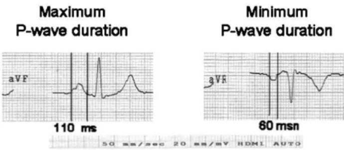
Twelve-lead ECGs were obtained after a 10-minute rest with 20-mm/mV amplitude and 50-mm/sec rates with standard lead positions in a supine position using commer-cially available machinery (Marquette Case, Hellige Medical System, Cardiosmart Hellige Instrument Company, Freiburg, Germany). The beginning of the P-wave was defined as the point at which the first atrial deflection crossed the isoelectric line; the end of the P-wave was defined as the point at which the atrial deflection returned to the isoelectric line. The P-wave durations (Pmax and Pmin) were calculated in all 12 ECG leads (Figure 1). The difference between Pmax and Pmin was defined as PWD (18).
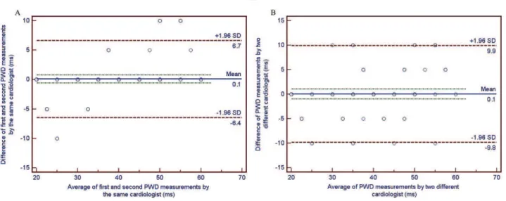
All measurements were performed by 2 experienced cardiologists who were blinded to the patient’s clinical status. The measurements were obtained manually with the help of calipers and a magnifying glass to define the electrocardiographic deflection. After completing the man-ual measurements, PWD was tested to identify the intra-observer and inter-intra-observer variability in 30 randomly-selected patients using the Bland-Altman method (19). The 95% limits of agreement for PWD were acceptable (26.4 to 6.7 ms and 29.8 to 9.9 ms; respectively) (Figures 2A and 2B).
Figure 1 -The P-wave durations (Pmax and Pmin) were calculated
Statistical analysis
The results were expressed as the mean¡SD. The data
between the groups were compared using SPSS (ver. 13). Student’s t-test was used for continuous variables, the chi-squared test was used for categorical variables, and the Mann-Whitney U-test was used for variables without normal distributions. Pearson’s correlation analysis was performed to assess the correlation between variables. In addition, a multivariate analysis of variance (MANOVA) was performed to compare groups of whole continuous variables in the tables. A two-tailed p-value of, 0.05 was
considered significant.
& RESULTS
Fifty-eight LP patients (30 females [51.7%] and 28 males [48.3%]) were included in the study. The patient ages ranged from 17 to 70 years (mean age, 43.42¡14.58 years).
They were age and gender matched with 37 healthy controls (mean age, 39.32¡11.67 years). The disease duration ranged
from 4 months to 6 years (mean, 29.62¡17.39 months).
Cutaneous lesions were present in all 58 cases. Mucosal lesions were present in 31 patients (53.4%) in the form of white, reticular streaks on the buccal mucosa. None of the
patients had erosive oral LP. Nail involvement was also present in 38 patients (65.5%) in the form of ridging, grooving, and longitudinal striations. Thirty-one patients (53.4%) presented with classic LP, 17 patients (29.3%) had the actinic type, and 10 patients (17.2%) had the hyper-trophic type. The descriptive, demographical characteristics of the groups are shown in Table 1.
According to the MANOVA results, there was a statisti-cally significant difference between the continuous variables in the two groups (Wilks’ lambda distribution = 0.007,
p= 0.001). However, when the univariate analysis results
were considered, there were no statistically significant differences between the groups in terms of age, gender, cigarette smoking, height, weight, or BMI. The mean systolic and diastolic blood pressures and heart rates were also similar between the groups. Echocardiographic variables, including the LV ejection fraction, LA dimension, and diastolic Doppler indices, were not significantly different between the groups (Table 2). The biochemical data, including serum glucose levels, were not significantly different between the groups, but the hs-CRP (3.5¡2.6
versus 1.7¡1.1, respectively, p,0.001), LDL cholesterol
Figure 2 -Bland-Altman plots demonstrating the 95% limits of agreement between (A) the repeated measurements of PWD by the
same observer and (B) between the manual measurement of PWD by two different observers in 30 randomly selected patients.
Table 1 -Demographic characteristics of the study population.
LP patients (n = 58)
Control group
(n = 37) p-value
Age (years) 43.4¡14.5 39.3¡11.6 NS
Male (n, %) 28 (48.3%) 19 (51%) NS
Smoking (n, %) 22 (37.9%) 10 (27%) NS
BMI (kg/m2) 26.7¡3.7 25.2¡2.8 NS Fasting glucose (mg/dL) 85.9¡7.74 86.3¡8.8 NS LDL cholesterol (mg/dL) 129.6¡31.7 96.8¡33.6 ,0.001 Triglycerides (mg/dL) 169.2¡93.6 110.5¡52.7 0.001 hs-CRP (mg/L) 3.5¡2.6 1.7¡1.1 ,0.001 Systolic BP (mmHg) 128¡8.5 123¡12.9 NS Diastolic BP (mmHg) 76.5¡11.5 78¡7.8 NS
BMI: body mass index, BP: blood pressure, HR: heart rate, hs-CRP: highly sensitivity C-reactive protein, LDL: low-density lipoprotein, HDL: high-density lipoprotein, NS: statistically non-significant.
Table 2 -Echocardiographic measurements of study population.
LP patients (n = 58)
Control group
(n = 37) p-value
2D echocardiography
LVEDD (mm) 45.9¡4.3 47.0¡3.6 NS
LVESD (mm) 26.3¡4.9 28.3¡4.1 NS
LV ejection fraction (%) 67¡5.1 68¡3.0 NS LA diameter (mm) 33.8¡4.4 33.9¡3.8 NS Doppler echocardiography
E (cm/sn) 79.0¡17.5 86.0¡9.1 NS
A (cm/sn) 65.1¡10.0 67.5¡9.5 NS
EDT (ms) 218.1¡46.1 208.2¡34.9 NS
IVRT (ms) 88.6¡22.6 79.7¡13.9 NS
(129.6¡31.7 versus 96.8¡33.6, respectively, p,0.001), and
triglyceride (169.2¡93.6 versus 110.5¡52.7, respectively, p= 0.001) levels were significantly higher in the LP patients
compared to the controls (Table 1).
Pmax and PWD were significantly higher in LP patients than in the control groups (78.44¡14.14versus68.64¡11.88, respectively, p= 0.001; 39.9¡12.9versus 32.4¡11.8,
respec-tively, p= 0.005). Pmin was not significantly different
between the groups (Table 3). In addition, there was a significant, positive correlation between hs-CRP and PWD (r = 0.549,p,0.001) in the LP patients.
& DISCUSSION
The major findings of this study are as follows: (i) LP patients had a significantly higher PWD compared to healthy subjects; (ii) the lipid and hs-CRP levels were higher in LP patients than in healthy subjects; and (iii) PWD was significantly correlated with hs-CRP.
Lichen planus is a chronic autoimmune mucocutaneous disease that affects the oral mucosa, skin, genital mucosa, scalp, and nails (20). The disease occurs in 0.4-1.9 percent of the population, mostly middle-aged patients and particu-larly women (7,21). The exact pathogenesis of the disease remains unclear, but both antibodies and T-cell mediation have been implicated. Activated T-cells release cytokines, leading to the attraction of inflammatory cells and the destruction of keratinocytes through cell-mediated cytotoxi-city (22). It has also been suggested that increased reactive oxygen species and lipid peroxides may affect the patho-genesis of LP (23). Studies have shown that LP is associated with extreme cardiovascular risks, including dyslipidemia (2,6), diabetes mellitus (7), and increased oxidative stress (8). Most cardiovascular disorders (including atherosclerosis, hypertension, insulin-resistance, dyslipidemia, and arrhyth-mias) share similar pathogenetic mechanisms, such as chronic inflammation, endothelial dysfunction, and increased oxidative stress (24).
LP has been reported to be associated with dyslipidemia in some studies (2,6,25). Some drugs used for dyslipidemia have been associated with lichen planus-like eruption, and many drugs used to treat lichen planus (e.g., systemic corticosteroids, retinoic acid, and methotrexate) have also been associated with dyslipidemia. Psoriasis, which is another chronic inflammatory skin disease, has been recently related to dyslipidemia (26). The frequent presence of chronic inflammation parameters in patients with psoriasis has been used to suggest a relationship with cardiovascular disease. Several cytokines, such as TNF-a, IL-2, and IL-6, have been implicated in the increased lipid levels in these patients (2). Otherwise, LP is an immune-mediated disease; antigens are processed by Langerhans cells and then presented to T-lymphocytes. This stimulated
lymphocytic infiltrate is epidermotropic and attacks kerati-nocytes, resulting in the generation of reactive oxygen species. During this lymphocytotoxic process, keratinocytes release more cytokines that, in turn, attract more lympho-cytes. The cytokines involved in LP pathogenesis (such as TNF-a, IL-6, IL-10, and IL-4) could explain the association with dyslipidemia (2).
Some studies have hypothesized that inflammatory markers, such as hs-CRP, may provide an adjunctive method to globally assess cardiovascular risk (27,28). The ongoing inflammation might affect the myocardium, and increased inflammatory activity is almost always associated with poor cardiac prognosis. Increased hs-CRP levels demonstrate the activity of this chronic disease and are an independent risk factor for arrhythmias (29). Recent studies have found a close association between atrial fibrillation and inflammation; atrial fibrillation was found to be more common in cases with high hs-CRP levels following coronary bypass (30) and acute pericarditis (31).The estimation of PWD, which is a reliable, non-invasive, and feasible variable with good reproducibility of intra- and inter-atrial heterogeneity, could be a useful method of identifying patients who are prone to suffer from AF. Several studies have shown that PWD has a predictive value for atrial fibrillation in patients with various conditions (18,32). Increased hs-CRP levels and the correlation between PWD and hs-CRP in our study indicated that inflammation might be a possible mechanism for increased PWD in LP. Many inflammatory diseases, such as rheumatoid arthritis (33), psoriasis (3), and systematic lupus erythematosus, have increased PWD, which may indicate that prolonged PWD depends on these diseases rather than itself to promote inflammation.
We demonstrated prolonged PWD using surface ECG in LP patients without other conditions that could increase PWD, such as coronary artery disease, systolic heart failure (ejection fraction ,45%), hypertension, arrhythmia, anti-arrhythmic treatment, obesity (BMI.35 kg/m2), valve
dis-ease, pulmonary hypertension, sleep apnea syndrome, chronic obstructive lung disease, and neurological disease. To the best of our knowledge, this study is the first to demonstrate prolonged PWD and atrial conduction delay in LP patients without additional systemic disease. Although LP is a disease characterized by increased inflammatory activity, no clinical data exist regarding whether atrial inflammation and the risk of AF are increased in these patients. Furthermore, we do not have any knowledge regarding the relationship between the incidence of AF and inflammation in this special patient group. In future studies, it will be important to investigate the prevalence of AF and the relationship between atrial fibrillation and the degree of inflammation in LP patients.
Limitations
The small number of LP patients and lack of long-term clinical follow-up are major limitations to our study. Automated ECG measurements were not available, and manual calculations of P-wave measurements may be subject to error. Whether increased PWD in patients with LP predicts poorer clinical outcomes or mandates any special treatment warrants further study.
In conclusion, the present study showed that PWD increased on surface ECG measurements in LP patients. This result may be an early detection of subclinical cardiac
Table 3 -Comparison of P-wave values of the groups.
LP patients (n = 58)
Control group
(n = 37) p-value
Pmax 78.44¡14.14 68.64¡11.88 0.001
Pmin 38.62¡5.44 36.48¡6.95 0.098
PWD 39.9¡12.9 32.4¡11.8 0.005
involvement. Increased PWD should be considered, in terms of tendency to AF, in these patients. We suggest that long-term LP patients be considered for referral for cardiovascular evaluation. Larger studies with longer follow-up are necessary to evaluate any clinical implications of our findings.
& AUTHOR CONTRIBUTIONS
Sahin M contributed to the study design, data collection and interpreta-tion, statistical analysis, manuscript preparainterpreta-tion, and literature search. Bilgili SG contributed to the study design, data collection and interpreta-tion, and literature search. Simsek H contributed to the data interpretainterpreta-tion, manuscript preparation, and literature search. Akdag S, Akyol A, Gumrukcuoglu HA, Yaman M, Bayram Y, and Karadag AS contributed to the data collection and interpretation and statistical analysis.
& REFERENCES
1. Arias-Santiago S, Buendia-Eisman A, Aneiros-Fernandez J, Giron-Prieto MS, Gutierrez-Salmeron MT, Garcia-Mellado V, et al. Lipid levels in patients with lichen planus: a case-control study. Journal of the European Academy of Dermatology and Venereology : JEADV. 2011;25(12):1398-401, http://dx.doi.org/10.1111/j.1468-3083.2011.03983.x.
2. Arias-Santiago S, Buendia-Eisman A, Aneiros-Fernandez J, Giron-Prieto MS, Gutierrez-Salmeron MT, Mellado VG, et al. Cardiovascular risk factors in patients with lichen planus. The American journal of medicine. 2011;124(6):543-8, http://dx.doi.org/10.1016/j.amjmed.2010.12.025. 3. Bacaksiz A, Erdogan E, Tasal A, Vatankulu MA, Kul S, Sevgili E, et al.
Electrocardiographic P-wave characteristics in patients with psoriasis vulgaris. Upsala journal of medical sciences. 2012.
4. Rocha-Pereira P, Santos-Silva A, Rebelo I, Figueiredo A, Quintanilha A, Teixeira F. Dislipidemia and oxidative stress in mild and in severe psoriasis as a risk for cardiovascular disease. Clinica chimica acta; international journal of clinical chemistry. 2001;303(1-2):33-9, http://dx. doi.org/10.1016/S0009-8981(00)00358-2.
5. Neimann AL, Shin DB, Wang X, Margolis DJ, Troxel AB, Gelfand JM. Prevalence of cardiovascular risk factors in patients with psoriasis. Journal of the American Academy of Dermatology. 2006;55(5):829-35, http://dx.doi.org/10.1016/j.jaad.2006.08.040.
6. Dreiher J, Shapiro J, Cohen AD. Lichen planus and dyslipidaemia: a case-control study. The British journal of dermatology. 2009;161(3):626-9, http://dx.doi.org/10.1111/j.1365-2133.2009.09235.x.
7. Seyhan M, Ozcan H, Sahin I, Bayram N, Karincaoglu Y. High prevalence of glucose metabolism disturbance in patients with lichen planus. Diabetes research and clinical practice. 2007;77(2):198-202, http://dx.doi. org/10.1016/j.diabres.2006.12.016.
8. Aly DG, Shahin RS. Oxidative stress in lichen planus. Acta dermatove-nerologica Alpina, Panonica, et Adriatica. 2010;19(1):3-11.
9. Centurion OA. Clinical implications of the P wave duration and dispersion: relationship between atrial conduction defects and abnor-mally prolonged and fractionated atrial endocardial electrograms. International journal of cardiology. 2009;134(1):6-8, http://dx.doi.org/ 10.1016/j.ijcard.2008.12.072.
10. Kosar F, Aksoy Y, Ari F, Keskin L, Sahin I. P-wave duration and dispersion in obese subjects. Annals of noninvasive electrocardiology : the official journal of the International Society for Holter and Noninvasive Electrocardiology, Inc. 2008;13(1):3-7, http://dx.doi.org/ 10.1111/j.1542-474X.2007.00194.x.
11. Ozer N, Aytemir K, Atalar E, Sade E, Aksoyek S, Ovunc K, et al. P wave dispersion in hypertensive patients with paroxysmal atrial fibrillation. Pacing and clinical electrophysiology : PACE. 2000;23(11 Pt 2):1859-62. 12. Can I, Aytemir K, Demir AU, Deniz A, Ciftci O, Tokgozoglu L, et al.
P-wave duration and dispersion in patients with obstructive sleep apnea. International journal of cardiology. 2009;133(3):e85-9, http://dx.doi.org/ 10.1016/j.ijcard.2007.11.037.
13. Dilaveris PE, Gialafos EJ, Andrikopoulos GK, Richter DJ, Papanikolaou V, Poralis K, et al. Clinical and electrocardiographic predictors of recurrent atrial fibrillation. Pacing and clinical electrophysiology : PACE. 2000;23(3):352-8, http://dx.doi.org/10.1111/j.1540-8159.2000.tb06761.x. 14. Dilaveris PE, Gialafos EJ, Sideris SK, Theopistou AM, Andrikopoulos
GK, Kyriakidis M, et al. Simple electrocardiographic markers for the prediction of paroxysmal idiopathic atrial fibrillation. American heart journal. 1998;135(5 Pt 1):733-8, http://dx.doi.org/10.1016/S0002-8703 (98)70030-4.
15. Dilaveris PE, Gialafos JE. P-wave dispersion: a novel predictor of paroxysmal atrial fibrillation. Annals of noninvasive electrocardiology : the official journal of the International Society for Holter and Noninvasive Electrocardiology, Inc. 2001;6(2):159-65, http://dx.doi. org/10.1111/j.1542-474X.2001.tb00101.x.
16. Ahlehoff O, Gislason GH, Jorgensen CH, Lindhardsen J, Charlot M, Olesen JB, et al. Psoriasis and risk of atrial fibrillation and ischaemic stroke: a Danish Nationwide Cohort Study. European heart journal. 2012;33(16):2054-64, http://dx.doi.org/10.1093/eurheartj/ehr285. 17. Lang RM, Bierig M, Devereux RB, Flachskampf FA, Foster E, Pellikka
PA, et al. Recommendations for chamber quantification: a report from the American Society of Echocardiography’s Guidelines and Standards Committee and the Chamber Quantification Writing Group, developed in conjunction with the European Association of Echocardiography, a branch of the European Society of Cardiology. Journal of the American Society of Echocardiography : official publication of the American Society of Echocardiography. 2005;18(12):1440-63, http://dx.doi.org/10. 1016/j.echo.2005.10.005.
18. Michelucci A, Bagliani G, Colella A, Pieragnoli P, Porciani MC, Gensini G, et al. P wave assessment: state of the art update. Cardiac electrophysiology review. 2002;6(3):215-20, http://dx.doi.org/10.1023/ A:1016368723033.
19. Bland JM, Altman DG. Statistical methods for assessing agreement between two methods of clinical measurement. Lancet. 1986;1(8476):307-10, http://dx.doi.org/10.1016/S0140-6736(86)90837-8.
20. Ismail SB, Kumar SK, Zain RB. Oral lichen planus and lichenoid reactions: etiopathogenesis, diagnosis, management and malignant transformation. Journal of oral science. 2007;49(2):89-106, http://dx.doi. org/10.2334/josnusd.49.89.
21. Manolache L, Seceleanu-Petrescu D, Benea V. Lichen planus patients and stressful events. Journal of the European Academy of Dermatology and Venereology : JEADV. 2008;22(4):437-41, http://dx.doi.org/10.1111/j. 1468-3083.2007.02458.x.
22. Middel P, Lippert U, Hummel KM, Bertsch HP, Artuc M, Schweyer S, et al. Expression of lymphotoxin-alpha by keratinocytes: a further mediator for the lichenoid reaction. Pathobiology : journal of immuno-pathology, molecular and cellular biology. 2000;68(6):291-300, http://dx. doi.org/10.1159/000055940.
23. Sezer E, Ozugurlu F, Ozyurt H, Sahin S, Etikan I. Lipid peroxidation and antioxidant status in lichen planus. Clinical and experimental dermatol-ogy. 2007;32(4):430-4, http://dx.doi.org/10.1111/j.1365-2230.2007.02436. x.
24. Flammer AJ, Ruschitzka F. Psoriasis and atherosclerosis: two plaques, one syndrome? European heart journal. 2012;33(16):1989-91, http://dx. doi.org/10.1093/eurheartj/ehr425.
25. Kurgansky D, Burnett JW. Widespread lichen planus in association with Turner’s syndrome and multiple endocrinopathies. Cutis; cutaneous medicine for the practitioner. 1994;54(2):108-10.
26. Gisondi P, Tessari G, Conti A, Piaserico S, Schianchi S, Peserico A, et al. Prevalence of metabolic syndrome in patients with psoriasis: a hospital-based case-control study. Br J Dermatol. 2007;157(1):68-73.
27. Ridker PM, Hennekens CH, Buring JE, Rifai N. C-reactive protein and other markers of inflammation in the prediction of cardiovascular disease in women. The New England journal of medicine. 2000;342 (12):836-43, http://dx.doi.org/10.1056/NEJM200003233421202. 28. Lagrand WK, Visser CA, Hermens WT, Niessen HW, Verheugt FW,
Wolbink GJ, et al. C-reactive protein as a cardiovascular risk factor: more than an epiphenomenon? Circulation. 1999;100(1):96-102, http://dx.doi. org/10.1161/01.CIR.100.1.96.
29. Chung MK, Martin DO, Sprecher D, Wazni O, Kanderian A, Carnes CA, et al. C-reactive protein elevation in patients with atrial arrhythmias: inflammatory mechanisms and persistence of atrial fibrillation. Circulation. 2001;104(24):2886-91, http://dx.doi.org/10.1161/hc4901.10 1760.
30. Bruins P, te Velthuis H, Yazdanbakhsh AP, Jansen PG, van Hardevelt FW, de Beaumont EM, et al. Activation of the complement system during and after cardiopulmonary bypass surgery: postsurgery activation involves C-reactive protein and is associated with postoperative arrhythmia. Circulation. 1997;96(10):3542-8, http://dx.doi.org/10.1161/ 01.CIR.96.10.3542.
31. Spodick DH. Arrhythmias during acute pericarditis. A prospective study of 100 consecutive cases. JAMA. 1976;235(1):39-41, http://dx.doi.org/10. 1001/jama.1976.03260270025020.
32. Polychronis E. Dilaveris CIS.P wave dispersion: A valuable non-invasive marker of vulnerability to atrial arrhythmias. Hospital Chronicles. 2006;1:130-37.