Ponderal behavior of rats fed an omegas 3, 6 and 9 enriched diet submitted to colon
carcinogenesis induced by azoxymethane
1Idália Maria Brasil BurlamaquiI, Conceição Aparecida DornelasII, Lara Albuquerque de BritoIII, José Wilson Meireles Trindade JúniorIII, Rafael Moura e SucupiraIII, Lara Burlamaqui VerasIV, Orleâncio Gomes Ripardo de AzevedoV, Paulo Roberto Leitão VasconcelosVI, Lusmar Veras RodriguesVII
IFellow PhD degree, Postgraduate Program in Surgery, Department of Surgery, Federal University of Ceara (UFC), Fortaleza-CE, Brazil. Conception
and design of the study, acquisition of data, technical procedures, manuscript preparation.
IIPhD, Full Professor, Department of Pathology and Legal Medicine, UFC, Fortaleza-CE, Brazil. Scientiic content of the study, technical procedures. IIIGraduate student, UFC, Fortaleza-CE, Brazil Acqusition of data, technical procedures.
IVResident in Surgery, Santa Marcelina Hospital, Sao Paulo-SP, Brazil. Acqusition of data.
VPost Doctoral, Laboratory of Nutrigenomics, Department of Surgery, UFC, Fortaleza-CE. Statistical analysis.
VICoordinator, Postgraduate Program in Surgery, Department of Surgery, UFC, Fortaleza-CE, Brazil. Intellectual and scientiic content of the study,
manuscript writing.
VIIPhD, Associate Professor, Department of Surgery, UFC, Fortaleza-CE, Brazil. Conception and design of the study, manuscript writing, critical
revision.
ABSTRACT
PURPOSE: To assess weight changes in rats fed diets with different ratios of omegas 3, 6 and 9 submitted to colonic carcinogenesis induced by Azoxymethane (AOM).
METHODS: Sixty rats with three weeks of life were distributed into ive groups of speciic diets containing 12 animals each: GI-
Standard diet without adminstration of AOM, GII- Standard diet with adminstration of AOM; GIII– Hyperlipidic diet with adminstration of AOM; GIV–Normolipidic diet with adminstration of AOM; GV- Hypolipidic diet with adminstration of AOM. The weight and food intake of each group were assessed four times in each week throughout the experiment until euthanasia at 36th week.
RESULTS: GI and GII had no signiicant difference in weight. GI showed a signiicant increase when compared to GIII, GIV and
GV. GII also showed a signiicant increase when compared to GIII, GIV and GV. When comparing intake of GI as compared to GII no signiicant difference was found, however such groups had higher intake than groups III, IV and V. There were found no difference in weight when comparing amoung rats with and without cancer within each groups: GII, GIII, GIV and GV.
CONCLUSIONS: Diets rich in omega 3, 6 and 9 reduced food intake and weight. Rats with colorectal cancer had no decrease in weight as compared to those without this condition in the same group.
Introduction
In recent decades, the ocidental alimentary habits have changed drastically as a result of changes in the lifestyle of the population. According to recent data from Brazilian Ministry of Health, the alimentary pattern in world has changed with industrialization and technological advances with subsequent change in family structure1. Thus, the incidence of obesity and another morbidities has increased, especially in development countries, a fact that leads to be considered a global epidemic2.
According to the World Health Organization (WHO), the occurrence of obesity in individuals relects the interaction between environmental and dietary factors associated with genetic background. Among dietary factors, the most important is the excessive intake of lipids, increasing the adipose tissue3. This
adipose tissue, which in the past was considered a passive storage tissue fat, has been shown the role in producing and releasing a large amount of hormones, growth factors, cytokines, adipokines and inlammatory signaling molecules, which may affect cell behavior in all phases of growth4.
Changes in metabolism and body weight caused by fatty diets depend not only on the quantity but also the speciic fatty acid composition5. Diets rich in saturated or trans fats may promote the
onset of several chronic diseases such as diabetes, dyslipidemia, cancer and autoimmune diseases6. However, unsaturated fats diets
rich in omega 3, 6 and 9 reduce the activity of liver enzymes related to fatty acid synthesis by reducing cholesterol and triglycerides leading to anti-inlammatory action and inhibiting proliferation and immune regulation of the cellular activity7,8.
The family of fatty acids is divided into polyunsaturated (PUFA), consisting of the omega-3 and 6, and monounsaturated (MUFA), omega-9. The omega-6 (linoleic acid and arachidonic acid) are found in vegetable oils such as coin and soybeans, and the omega-3 (alpha-linolenic acid, eicosapentaenoic acid (EPA) and docosahexaenóico (DHA), mainly in oils from cold-water and deep ish as salmon, herring, tuna and sardines, and vegetables such as laxseed and soybeans. The omega-9 (oleic acid) is present in high concentration, mainly olive oil, but also grape seed oil, rapeseed oil, sesame oil, sunlower oil, soybean oil, palm oil and marine animals such as the shark and cod ish9.
Animals fed with linoleic acid-rich oil had higher
oil and olive oil promote different effects on the lipid proile, in the preservation of the intestinal villi and lymphocyte migration in mucosa of wistar rats, probably due to the difference in composition these fatty acids10.
According to some studies, weight gain is proportional to the amount of dietary fat, and a fatty diet provides greater weight gain. However, it is evident that a high fat diet rich in omega-3 has a protective effect on the accumulation of retroperitoneal and epididymal fat, improve in insulin resistance, but does not interfere with total body weight11,12.
Although several studies in some types of cancer, including colorectal, do not elucidate whether the increased risk of cancer in obese people is due to excess weight, more studies are necessary to indicate whether a diet rich in fat, the increased calories and the lack of physical activity, or a combination of these factors, play a more signiicant role.
The aim is to evaluate changes in weight of rats Wistar fed with hyperlipidic, normolipidic and hypolipidic diet rich in omega 3, 6 and 9, submitted to AOM-induced colon carcinogenesis.
Methods
The study was conducted after evaluation of the Ethics Committee on Animal Research of the Federal University of Ceara (CEPA/UFC) under the protocol number 09 of the day 03/02/2009 and according to the International Standards for Biomedical Research on Animals.
A total number of 60 rats (Rattus norvegicus albinus, Mammalia Rodentia, Muridae) of Wistar strain, male, were obtained from the Central Animal Vivarium of the Federal University of Ceara, with three weeks of age and weighing between 30g and 40g. All animals were treated with antihelmintic for veterinary use,
1ml/kg body weight (Basken® - and pamoate Pyrantel pamoate
oxantel) and separated individually in polypropylene cages, under appropriate conditions of humidity, temperature and lighting Laboratory of Experimental Surgery, Department of Surgery, Faculty of Medicine, Federal University of Ceara. In addition the rats received water and food ad libitum.
Experimental design
Group I- (GI): Control group with rats receiving a standard diet (SD) and not exposed to AOM - The standard diet was Bio-tec Biobase Rats and Mice® with a total of 4444 kcal /
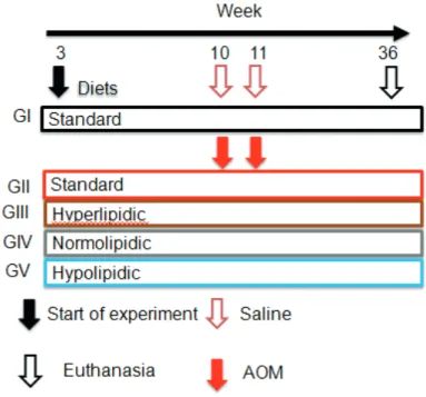
kg and distribution the following caloric: 59% carbohydrate, 29% protein and 12% lipid. At ten weeks, one dose a 1.0 ml 0.9% sterile saline solution (SS), intraperitoneal (IP), was administered once a week for two weeks. (Figure 1)
Group II - (GII): Control group with rats receiving a standard diet (SD) and exposed to AOM - The standard diet was Bio-tec Biobase Rats and Mice® with a total of 4444 kcal / kg and
distribution the following caloric: 59% carbohydrate, 29% protein and 12% lipid. At ten weeks, one dose a 20mg/kg AOM, IP, was administered once a week for two weeks. (Figure 1)
Group III - (GIII): Study group with rats receiving a hyperlipidic diet (DH) and exposed to AOM – The hyperlipidic diet was rich in unsaturated fatty acids (omega 3, 6 and 9) and composed of 11.8 g of dextrin Nutri + 9.5 g Oil (1.14g Oil Flaxseed + 4.18g Canola Oil + 2.75g Olive Oil + 1.42 g of Medium Chain Triglycerides (MCT) + 16.5g Protein Nutri HWP, totaling 4570 kcal/kg and distribution the following caloric: 25% carbohydrate, 30% protein and 45% lipid. At ten weeks, one dose a 20mg/kg AOM, IP, was administered once a week for two weeks. (Figure 1)
Group IV - (GIV): Study group with rats receiving a normolipidic diet (DH) and exposed to AOM - The normolipidic diet was rich in unsaturated fatty acids (omega 3, 6 and 9) and composed of 18g Nutri dextrin + 6,2 Oil g (0.744 g Oil Flaxseed + 2,728 g Canola Oil + 1,798 g Olive Oil+ 0.93 g TCM) + 16.5 g Protein Nutri HWP, totaling 4170 kcal/kg and distribution the following caloric: 40% carbohydrate, 30% protein and 30% lipídio. At ten weeks, one dose a 20mg/kg AOM, IP, was administered once a week for two weeks. (Figure 1)
Group V - (GV): Study group with rats receiving a hypolipidic diet (DH) and exposed to AOM - The hypolipidic diet was rich in unsaturated fatty acids (omega 3, 6 and 9) and composed of 27g of Nutri dextrin + 2.5 Oil g (0.3 g Linseed Oil + 1.1 g of Canola Oil + 1.725 g Olive Oil + 0.375 g TCM) + 16.5 g Protein Nutri HWP, totaling 3761 kcal/kg and distribution the following caloric: 58% carbohydrate, 30% protein and 12% lipid. At ten weeks, one dose a 20mg/kg AOM, IP, was administered once a week for two weeks. (Figure 1)
FIGURE 1 – Experimental design showing the groups and the respective periods during the study.
Carcinogenesis inducer
Azoxymethane (SIGMA-ALDRICH) was acquired at the presentation of 100mg and was diluted in sterile water for injection, to obtain a dose of 20mg/kg of body weight, administered by IP injections in two consecutive doses, at weekly intervals.
Clinical evalutaion
The animals were clinicaly evaluated, by two investigators, daily throughout the experiment. Changes were observed in weight, behavior (reactivity, immobility), involuntary contractions, breathing, diarrhea, anorexia, piloerection, loss of body hair and ulcerations. The weighing of the animals was carried out 4 times a week until the day of euthanasia.
Diets
The animals were fed with the standard, hyperlipidic, normolipidic and hypolipidic diet and water ad libitum (Tables 1
TABLE 1 – Nutrients composition.
Nutrient Quantity (g) Calories Carbohydrates (g) Proteins (g) Lipids (g)
Standard Diet
Biobase Bio-tec 27 120 17.6 8.8 1.6
Total 27 120 17.6 8.8 1.6
Total of calories (%) 70.4 (58.7%) 35.2 (29.3%) 14.4 (12.0%)
Hyperlipidic Diet
Nutri dextrin 11.8 44.84 11.21 0 0
Oils (mix) 9.5 78.92 0 0 8.77
Nutri Protein HWP 16.5 52.8 0 13.2 0
Total 37.8 176.57 11.21 13.2 8.77
Total of calories (%) 44.84 (25.4%) 52.8 (29.9%) 78.9 (44.7%)
Normolipidic Diet
Nutri dextrin 18 68.4 17.1 0 0
Oils (mix) 6.2 51.51 0 0 5.7
Nutri Protein HWP 16 51.2 0 12.8 0
Total 171.11 17.1 12.8 5.7
Total of calories (%) 68.4 (40.0%) 51.2 (29.9%) 51.5 (30.1%)
Hypolipidic Diet
Nutri dextrin 27 102.6 25.65 0 0
Oils (mix) 2.5 20.77 0 0 2.31
Nutri Protein HWP 16.5 52.8 0 13.2 0
Total 176.17 25.65 13.2 2.31
Total of calories (%) 102.6 (58.2%) 52.8 (30%) 20.8 (11.8%)
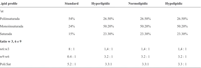
TABLE 2 - Lipid proile of the diets and ratio for omegas 3, 6 e 9.
Lipid proile Standard Hyperlipidic Normolipidic Hypolipidic
Fat
Poliinsaturada 54% 26.50% 26.50% 26.50%
Monoiinsaturada 24% 50.20% 50.20% 50.20%
Saturada 15% 23.30% 23.30% 23.30%
Ratio w 3, 6 e 9
w6:w3 8 : 1 1,4 : 1 1,4 : 1 1,4 : 1
Surgery procedings and euthanasia
Twenty six weeks after the irst AOM injection the animals were anesthetized with 80mg/kg ketamine and 8mg/kg xylazine by IP injections. They were positioned supine for surgery outset, undergoing laparotomy by midline xypho-pubic incision followed by protocolectomia. This was followed by measuring, weighing and evaluating for the presence of colon tumors. The intestinal segment was opened and washed with saline and immersed in a solution of 10% formalin for ixation. Then the animals were sacriiced by section of the abdominal aorta.
Statistical analysis
For statistical analysis the softwares GraphPadPrism 5.0 (California, USA) and Excel (New York, USA) were used. The normality test of Kolmogorov-Smirnov was also applied. Quantitative variables were presented in graphs with parametric trendlines or column charts showing mean and standard deviation (mean ± SD) and compared by t test Student, with a signiicance
level of 5% (p<0.05).
Results
Weight changes
FIGURE 2 – Mean of weight gain in each group in each month.
The begining (month 0), there was no statistically signiicant difference in weight among the ive groups (Student t test / p>0.05). By the 1st month of the experiment, it was observed that the weight of the animals in groups III, IV and V was signiicantly lower when compared to the weights of the animals of group I and II (Student t test / p <0.05 .) The p values obtained in the last month: GIII (p = 0.0045), GIV (p = 0.0110) and GV (p =
0.0018) compared to GI, and GIII (p = 0.0034), GIV (p = 0.0149) and GV (p = 0.0013) compared with GII.
Distribution of intake
FIGURE 3 – Mean of intake in each group during the experiment.
When comparing the intake of group I with group II there was no statistically signiicant difference (p = 0.997), but between group I and groups III, IV and V was signiicant difference with p <0.001, p <0.001 and p = 0.004, respectively. In the comparison of intakes of group II with group III, IV and V, there was also a signiicant difference with p <0.001, p <0.001 and p = 0.002, respectively.
Distribution of body weight of the rats with / without cancer groups at the end of the experiment
There was no difference in weight mean compared between rats with cancer and without cancer: GII (p = 0.9514), GIII (p = 0.0965), GIV (p = 0.7018) and GV (p = 0.8220).
Discussion
The cancer patients, as well as anorexia, exhibit metabolic abnormalities that impair anabolism causing severe weight loss leading to cachexia. The factor tumor to mobilization of lipids stimulates lipolysis, providing the loss of adipose tissue. The decrease in skeletal muscle mass is a result of decreased protein synthesis and increased protein degradation by activating the NF-kB pathway13. EPA acts by inhibiting protein degradation pathway and has a modulating effect on pro-inlammatory cytokines, the hepatic acute phase proteins, eicosanoids and tumor factors promoting a beneicial effect on body composition by modulating the loss of lean mass14.
Since the beginning of the experiment until euthanasia, it was observed that the weight of the animals in groups III, IV and V was signiicantly lower when compared to the weights of the animals of group I and II. The group I and II were fed with the same standard diet and no present signiicant difference in weight at the administration of AOM (p = 1.000), 20mg/kg weight, and at the end of the experiment (p = 1.000). Therefore AOM did not affect the food intake of the animals as well as the inal body weight. Possible explanation for this inding of low weight in the diets studied groups is that the amount of intake was greater in group I and II than in groups III, IV and V (Figures 2 and 3).
A recent study demonstrated that a saturated fat diet injured the hypothalamus and caused local inlammation, neuronal death and consequent loss of the ability of perception of timing the signal to burn or storage food. The intake of omega-3, for its anti-inlammatory action, restored neural control hunger and increased energy expenditure, promoting weight loss. It was also demonstrated that animals fed diets with lower concentrations of omega-3 and 9 presented even higher reduction in hypothalamic inlammation, restoring metabolic derangements regarding food and termogenics15. These indings were similar to those found in the group fed hypolipidemic diet in this research, which in absolute values received lower amounts of omega-3 and 9 although the relationship between the omegas (w6: w3 and w9: w6) remained
of fat mass16.
The beneicial effects of the PUFA are well-known in adipose tissue such as preventing hyperplasia and hypertrophy of adipose tissue, induction of mitochondrial biogenesis in adipocytes, inducing the secretion of adiponectin and reduction of adipose tissue inlammation. The action of adiponectin is more pronounced in the epididymal fat in the abdomen than in subcutaneous fat, contributing to increase the sensitivity of insulin17.
Omega-3 alter indicators related to the development of the metabolic syndrome and present beneicial effects on lipid proile and glucose, preventing excess weight gain. The mechanism by which the laxseed oil acts in reverse of abdominal obesity remains unknown. Evidence suggests that polyunsaturated fatty acids in the diet may act as modulators for the deposition of body fat18.
Another aspect is that the animals in groups of diets enriched with omegas presents diarrhea since the start of the experimentation contributing to the low body weight. It is known that high doses of omega-3 have few collateral effects, diarrhea recognized as the main one13.
There were signiicant differences in food consumption which was lower in the study group as compared to controls. This can be explained by the composition of the diets, which are rich in soluble iber. These ibers have a great ability to form gel. This leads to a decrease in the rate of gastric emptying, and prolongation of satiety, resulting in reduced food intake18.
The rats with CRC showed no decrease in weight in comparison with those without this condition independently of the offered diet (Figure 4). This inding is consistent with the literature, for colorectal cancer in early stage as it is asymptomatic and does not compromise the overall condition of affected individuals19.
Anthropometric data obtained from a recent study indicated the prevalence of normal weight (45.71%), overweight (35.71%), obesity (12.86%) and thinness (5.71%) in patients with colorretal cancer20. These indings are similar to the literature, because malnutrition is not common in this condition, which can not be explained by the no impaired food intake, malabsorptive disorders and obstructive factors.
Conclusions
References
1. Instituto Nacional do Câncer (INCA). Disponível em: http://www. inca.gov.br/conteudo_view.asp?ID=18.
2. Pereira LO, de Francischi RP, Lancha Jr AH. Obesity: dietary Intake,
sedentarism and insulin resistance. Arq Bras Endocrinol Metabol.
2003;47(2):111-27.
3. Feoli AM, Roehrig C, Rotta LN, Kruger AH, Souza KB, Kessler AM, Renz SV, Brusque AM, Souza DO, Perry MLS. Serum and liver lipids in rats and chicks fed with diets containing different oils.
Nutrition. 2003;19:789-93.
4. Ballard-Barbash R. The emerging evidence about the role of obesity
in cancer. NCI Cancer B. 2011;8(22).
5. Campos FG, Waitzberg DL, LogulloWaitzberg AF, Habr-Gama A, Kiss DR, Gama-Rodrigues J. Diet and colorectal cancer: current evidence for etiology and prevention. Nutr Hosp. 2005;20(1):18-25. 6. Simopoulos AP. Evolutionary aspects of diet, the omega-6/omega-3
ratio and genetic variation: nutritional implications for chronic
diseases. Biomed Pharmacother. 2006;60(9):502-7.
7. Steemburgo T, Dall’alba V, Gross JL, Azevedo MJ. Fatores dietéticos e síndrome metabólica. Arq Bras Endocrinol Metab. 2007;51(9):1425-33.
8. Burlamaqui IMB, Dornelas CA, Valença Jr JT, Mota DMC, Mesquita FJC, Veras LB, Vasconcelos PRL, Rodrigues LV. Effect
of a hyperlipidic diet rich in omegas 3, 6 and 9 on aberrant crypt
formation in rat colonic mucosa. Acta Cir Bras. 2012;27(1):30-6. 9. Rique ABR, Soares EA, Meirelles CM. Nutrição e exercício na
prevenção e controle das doenças cardiovasculares. Rev Bras Med
Esporte. 2002;8(6):244-54.
10. Brant LHC, Cardozo LFMF, Velarde LGC, Boaventura, GT. Impact of laxseed intake upon metabolic syndrome indicators in female Wistar rats. Acta Cir Bras. 2012;27(8):537-43.
11. Pellizzon M, Buison A, Ordiz F Jr, Santa Ana L, Jen KL. Effects
of dietary fatty acids and exercise on body-weight regulation and
metabolism in rats. Obes Res. 2002;10(9):947-55.
12. Belzung F, Raclot T, Groscolas R. Fish oil n-3 fatty acids selectively
limit the hypertrophy of abdominal fat depots in growing rats fed
high-fat diets. Am J Physiol. 1993;264(6):1111-8.
13. Carmo MCNS, Correia MITD. The role of omega-3 fatty acids in cancer. Rev Bras Cancerol. 2009;55(3):279-87.
14. Boddaert AS, Gerritsen WR, Pinedo HM. On our way to targeted therapy for cachexia in cancer? Curr Opin Oncol. 2006;18:335-40. 15. Cintra DE, Ropelle ER, Moraes JC, Pauli JR, Morari J, Souza
CT, Grimaldi R, Stahl M, Carvalheira JB, Saad MJ, Velloso
LA. Unsaturated fatty acids revert diet-induced hypothalamic
inlammation in obesity. PLoS One. 2012;7(1):e30571.
16. Simopoulos AP. Genetic variants in the metabolism of omega-6 and
omega-3 fatty acids: their role in the determination of nutritional
requirements and chronic disease. Exp Biol Med. 2010;235:785-95. 17. Kopecky J, Rossmeisl M, Flachs P, Kuda O, Brauner P, Jilkova
Z, Stankova B, Tvrzicka E, Bryhn M. N-3 PUFA: bioavailability and modulation of adipose tissue function. Proc Nutr Soc.
2009;68(4):361-9.
18. Rosa DD, Sales RL, Moraes LFS, Lourenço FC, Neves CA,
Sabarense CM, Ribeiro SMRo, Peluzio MCG. Flaxseed, olive
and ish oil inluence plasmatic lipids, lymphocyte migration and morphometry of the intestinal of Wistar rats. Acta Cir Bras. 2010;25(3):275-80.
19. Continente AJC, Pluvius CC, Martinez CV. Nutrición y neoplasias digestivas. Bras Nutr Clin. 2002;17:53-63.
20. Fortes RC, Recôva VL, Melo AL, Novaes MRCG. Hábitos dietéticos de pacientes com câncer colorretal em fase pós-operatória. Rev Bras Cancerol. 2007;53(3):277-89.
Correspondence:
Idália Maria Brasil Burlamaqui Avenida Beira Mar, 3680/2001 60165-121 Fortaleza – CE Brasil Tel: (55 85)3267-5780
Received: June 12, 2013 Review: Aug 14, 2013 Accepted: Sept 12, 2013 Conlict of interest: none
Financial source: Postgraduate Program in Surgery and Experimental Surgery Laboratory (LABCEX), Federal University of Ceara
1Research performed at Laboratory of Experimental Surgery, Department