LETTER TO THE EDITOR
Lipodystrophy during pegvisomant therapy:
a case report and review of the literature
Buyuktas D,ICelik O,IKantarcı F,IIKadioglu PI
IDivision of Endocrinology and Metabolism, Department of Internal Medicine, Cerrahpasa Medical School, University of Istanbul, Istanbul, Turkey. IIDivision of Radiology, Cerrahpasa Medical School, University of I˙stanbul, I˙stanbul, Turkey.
Email: [email protected]
Tel.: 90-216-462 31 14 / 90-532-404 10 40
INTRODUCTION
Acromegaly is a serious, chronic endocrine disorder cha-racterized by exaggerated growth hormone (GH) secretion and increased levels of insulin-like growth factor-1 (IGF-1). Treatment modalities include somatostatin analogues and pegvisomant and/or radiotherapy following pituitary tumor resection.1 Pegvisomant is a pegylated analogue of human
GH and functions as a GH receptor antagonist. Pegvisomant acts in the periphery and significantly decreases serum IGF-1 levels.2Various studies have shown that pegvisomant is safe and well-tolerated and is an effective therapy for acromegaly patients. Possible adverse effects of pegvisomant treatment include self-limited erthematous injection-site reactions, mild liver toxicity, influenza-like syndrome and headache.3Here, we describe an acromegalic patient who developed lipohy-pertrophy following initiation of pegvisomant therapy.
CASE REPORT
A 61-year-old female patient was diagnosed with acro-megaly eight years ago. The diagnosis of acroacro-megaly was confirmed by elevated serum IGF-1 levels accompanied by failure of GH suppression following a 75 g glucose load. The patient displayed a 22x17x16 mm pituitary adenoma, which was operated transsphenoidally two months after diagnosis. After surgery, the patient was treated with long-acting octreotide LAR for persistent symptoms. After 14 months of octreotide therapy, 0.5 mg cabergoline administered 3 days a week was added to the treatment regimen to address unremitting symptoms. The patient underwent a gamma-knife procedure three years after the initial surgery to treat the residual disorder, which was identified by persistently high levels of IGF-I and magnetic resonance imaging (MRI). At the 4thyear of follow up, the patient displayed an IGF-I level of 485 ng/ml. At this point (December 2009), carbego-line treatment was discontinued and the patient was given subcutaneous injections of 10 mg pegvisomant per day. The patient was advised to alternate the injection areas around the arms, thighs and the abdominal wall. After two months of treatment, the patient found soft, painful swellings on her arms, thighs and the abdominal wall. Clinical examination found a clearly visible thickening of the tissues in these
Copyrightß2010CLINICS– This is an Open Access article distributed under the terms of the Creative Commons Attribution Non-Commercial License (http:// creativecommons.org/licenses/by-nc/3.0/) which permits unrestricted non-commercial use, distribution, and reproduction in any medium, provided the original work is properly cited.
Figure 1 -Areas of lipohypertrophy on the anterior abdominal wall.
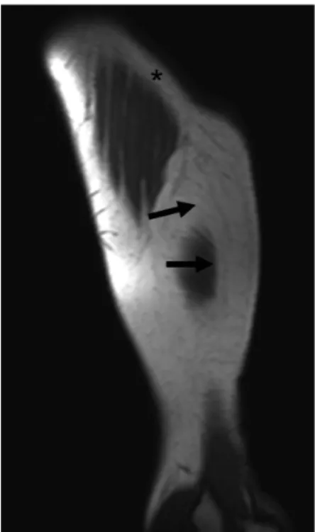
Figure 2 - Sagittal T1 weighted MRI of the left arm shows lipomatous hypertrophy (arrows) at the injection site. Note the normal subcutaneous fat (asterisks) where injections were performed.
CLINICS 2010;65(9):931-933 DOI:10.1590/S1807-59322010000900018
areas (Figure 1). No significant increase was detected in serum IGF-1 levels, indicating that the treatment was effective. Ultrasonography revealed a subcutaneous fat hypertrophy in the extremities. MRI of the left arm confirmed lipomatous hypertrophy at the injection site (Figure 2).
The accuracy of the injection sites and the drug applica-tions were controlled by a dedicated nurse (Somavert education service). The patient was dissatisfied with the lipohypertrophy at the sites of injection, and pegvisomant therapy was therefore discontinued.
DISCUSSION
Somatostatin analogues and pegvisomant are frequently used in the treatment of acromegaly. Pegvisomant is a pegy-lated analogue of human GH that blocks hepatic production of IGF-1. The adverse effects of pegvisomant therapy include self-limited erythematous injection-site reactions, mild liver toxicity, influenza-like syndrome, headache, asthenia, diar-rhea, arthralgia and hypercholesterolemia.3
Lipodystrophy is a disease of the adipose tissue. It can be localized and acquired by administration of particular drugs. If the adipose changes are related to depressed areas of skin, the disease is called lipoatrophy. Lipohypertrophy is an enlargement of fat tissue caused by subcutaneous deposition of adipose and is associated with parenteral drug administration. The pathogenesis of lipohypertrophy is unclear. It is known that growth hormone and IGF-1 both display lipolytic activity. Blockage of growth hormone action through pegvisomant therapy can affect adipose size and increase local adiposity.4-7Immunologic factors can also
contribute to lipodystrophy. An inflammatory reaction to substitutions of pegvisomant can often cause this disorder.8 However, a biopsy of a pegvisomant-induced lipohyper-trophy from an acromegalic patient showed normal-appearing mature adipocytes with no fat necrosis and no inflammatory cells. Additionally, the biopsy showed no anti-pegvisomant antibodies.7
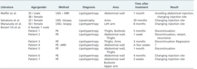
To our knowledge, the literature contains 4 reports describing 11 patients who developed lipodystrophy during pegvisomant therapy5-8 (Table 1). These reports describe lipohypertrophy in 10 patients and lipoatrophy in 1 patient.
Maffei et al. have reported one male and one female patient who suffered from lipohypertrophy on the abdominal wall, confirmed by ultrasonography and MRI, following 1 month of pegvisomant therapy. These patients were advised to avoid abdominal injections and alternate the injection sites.5 Salvatore et al. reported pegvisomant-induced lipoatrophy.6
Marazuela et al. described lipohypertrophy on the left arm of a female patient caused by pegvisomant therapy. This lipohypertrophy resolved once injections at the same site were ceased.7Bonert et al. identified seven different cases of lipohypertrophy induced by pegvisomant therapy; two of the patients began rotating the injection site, and four patients discontinued pegvisomant therapy.8
It is worth noting that 9 of the 11 patients described in the literature were female. Thus, pegvisomant-induced lipohy-pertrophy can be referred to as a gender-specific response. In most cases, lipohypertrophy was reversed after the injection site was rotated or treatment was discontinued. It is unknown whether the development of lipohypertrophy is associated with the dose or frequency of injection.5,7,8In our patient, pegvisomant therapy was administered together with a low dose of octreotide LAR. The treatment effectively normalized IGF-1 levels, but pegvisomant treatment was stopped due to discomfort caused by lipohypertrophy.
In conclusion, pegvisomant injection appears to have induced lipohypertrophy in our patient following two months of therapy. We recommend injection site rotation to different parts of the body, which should prevent the development of lipohypertrophy. Physicians should be aware of this complication and carefully monitor patients under pegvisomant treatment.
REFERENCES
1. Chanson P, Salenave S, Kamenicky P, Cazabat L, Young J. Pituitary tumours:acromegaly. Best Practise & Research Clinical Endocrinology & Metabolism. 2009;23:555-74, doi: 10.1016/j.beem.2009.05.010.
2. Kopchick JJ, Parkinson C, Stevens EC, Trainer PJ. Growth hormone receptor antagonists: discovery, development, and use in patients with acromegaly. Endocrin Reviews. 2002; 23: 623-46, doi: 10.1210/er.2001-0022. 3. van der Lely AJ, Hutson RK, Trainer PJ, Besser GM, Barkan Al,Katznelson L, et al. Lon-term treatment of acromegaly with peg-visomant, a growth hormone receptor antagonist. Lancet. 2001;358: 1754-9, doi: 10.1016/S0140-6736(01)06844-1.
4. Mersebach H, Feldt-Ramussen UF. Localized lipohypertrophy during growth hormone therapy. Ugeskr Laeger. 2002;164:1930-2.
Table 1 -Review of literature
Literature Age/gender Method Diagnosis Area
Time after
treatment Result
Maffei et al. 35 / male USG+MRI Lipohypertropy Abdominal wall 1 month Avoiding abdominal injection,
changing injection site 38 / female
Salvatore et al. 33 / female USG+biopsy Lipoatrophy Arms 20 months Changing injection site Marazuela et al. 50 / female USG+biopsy Lipohypertropy Left arm 8 months Changing injection site
Bonert VS et al. 6 female 1 male
Patient 1 PE Lipohypertropy Thighs, Buttocks 5 months Discontinuation Patient 2 PE Lipohypertropy Abdominal wall,
Thighs
1 week Discontinuation, restart, recurrence
Patient 3 PE Lipohypertropy Thighs, Arms 3 months Discontinuation Regression Patient 4 PE+MRI Lipohypertropy Abdominal wall A few weeks ?
Patient 5 PE Lipohypertropy Abdominal wall, Thighs
1 month Discontinuation Patient 6 PE Lipohypertropy Abdominal wall 4 months Changing injection site Patient 7 PE Lipohypertropy Abdominal wall
Buttocks Upper arm
3 weeks Changing injection site
PE: Physical examination
Lipodystrophy and pegvisomant
Buyuktas et al. CLINICS 2010;65(9):931-933
5. Maffei P, Martini C, Pagano C, Sicolo N, Corbetti F. Lipohypertrophy in acromegaly induced by the new growth hormone receptor antagonist pegvisomant. Annals of Internal Medicine. 2006;145:310-2.
6. Salvatore C, Giovanni R, Vittorio C, Claudia S, Patrizia CS. Subcutaneous lipoatrophy induced by long-term pegvisomant administration. Clinical Endocrinology (Oxf). 2009;70: 655-6, doi: 10.1111/j.1365-2265.2008. 03400.x.
7. Marazuela M, Dauden E, Ocon E, Moure D, Nattero L. Pegvisomant induced lipohypertrophy: report of a case with histopathology. Annals of Internal Medicine. 2007;147:741-3.
8. Bonert VS, Kennedy L, Petersenn S, Barkan A, Carmichael J, Melmed S. Lipodystrophy in patients with acromegaly receiving pegvisomant. Journal of Clinical Endocrinology & Metabolism. 2008;93:3515-8, doi: 10. 1210/jc.2008-0833.
CLINICS 2010;65(9):931-933 Lipodystrophy and pegvisomant
Buyuktas et al.