Rêgo RSN, QueiRoz FL, CoSta BXM, RaBeLo FeF, LaMouNieR PCC, SiLva LC, aLveS FiLho v, CaRMoNa Mz. evaluation of response to neoadjuvant treatment, by nuclear magnetic resonance, as a predictor of oncologic results and survival of patients with rectal cancer. J Coloproctol, 2011;31(4):334-338.
ABSTRACT: Introduction: Neoadjuvant chemoradiation promotes tumor size reduction and staging before the surgery, reducing the risk of involving the circumferential resection margin and local recurrence. For patients who have been submitted to the neoadjuvant therapy, the usefulness of a second nuclear magnetic resonance(MRI) after chemoradiation has not been clearly explained. Objective: Assess the degree of tumor regression and downstaging after chemoradiation using MRI, compared with the pathology, and its corre-lation with surgical outcomes and patient prognosis. Methods: This study investigated 13 patients. Their mean age was 52.3 years and 69.23% were male. Results: The agreement in T and N staging was 30.76%, between the second MRI and pathology, overestimated in 55.55% of the remaining. T staging agreement was 53.84% and N staging agreement, 61.53%. The circumferential resection margin was free of cancer in 100%. The survival rate was 92%, with 75% disease-free in a mean follow-up of 1-2 years. Conclusion: A second MRI after chemoradiation can evaluate the degree of tumor regression, but with low compliance in relation to pathology, with ten-dency to overstaging. More studies are required to conirm these initial observations.
Keywords: magnetic resonance imaging; rectal cancer; neoadjuvant therapy.
Evaluation of response to neoadjuvant treatment, by nuclear
magnetic resonance, as a predictor of oncologic results and survival
of patients with rectal cancer
RodRigo SoaReS NaPoLeão do Rêgo1, FáBio LoPeS de QueiRoz2, BReNo Xaia MaRtiNS da CoSta1, FeRNaNda eLiaS FeRReiRa RaBeLo1, PauLo CeSaR de CaRvaLho LaMouNieR3, LuCiaNa CoSta
SiLva5, vaLdiviNo aLveS FiLho4, MaRia zuLeiMe CaRMoNa4
1Resident, Service of Coloproctology of the Hospital Felício Rocho – Belo Horizonte (MG), Brazil. 2Coordinator of Residency, Service of Coloproctology of the Hospital Felício Rocho – Belo Horizonte (MG), Brazil. 3Coordinator, Service of Coloproctology of the Hospital Felício Rocho – Belo Horizonte (MG), Brazil. 4Preceptor, Service of Coloproctology of the Hospital Felício Rocho - Belo Horizonte (MG), Brazil. 5Professor, Department of Propedeutics of the Medical School of
the Universidade Federal de Minas Gerais – Belo Horizonte (MG), Brazil.
Study carried out at the Service of Coloproctology of the Hospital Felício Rocho – Belo Horizonte (MG), Brazil. Financing source: none.
Conlict of interest: nothing to declare.
Submitted on: 08/05/2011 Approved on: 09/06/2011
INTRODUCTION
Rectal cancer corresponds to 30 to 50% of all
colorectal tumors. Its prognosis is inluenced by sev -eral factors, such as lat-eral extension of tumor, lymph node involvement and presence of distant metastases. the rate of local recurrence, after isolated surgical treatment, ranges from 3 to 32%, with the presence of tumor less than 1 mm from the circumferential
extramural venous invasion with high accuracy,
iden-tifying factors of dificult prognosis1,5. For patients with rectal cancer submitted to neoadjuvant therapy, the usefulness of a second MRi after chemoradiation to assess the response to the treatment, and performed just before the surgical procedure, has not been
clear-ly explained. The potential beneits that have been re
-ported are: accurate identiication of tumor regression
for an adequate CRM, after a standard surgery with total mesorectal excision, and warn for possible points requiring more careful dissection or wider resection. in addition, some studies relate the response to the NaCR to the prognosis. Few studies have assessed the predictive value of MRi after NaCR as a predic-tor of oncologic results and survival of patients with rectal cancer.
this study has the purpose of assessing the de-gree of tumor regression and downstaging obtained after chemoradiation through MRi, comparing it to the anatomopathological (aP) study and its correla-tion with the patients’ surgical results and prognosis.
METHODS
this is a prospective observational study that included patients treated between august 2008 and december 2010. these patients were submitted to preoperative staging by thorax, abdomen and carci-noembryonic antigen (Cea) tomography, as well as a clinical evaluation. Locoregional staging was
per-formed through pelvic MRI. The patients classiied
to MRi as t3/t4 and/or with affected lymph nodes were submitted to chemoradiation (radiotherapy of 4500 to 5040 cgy for 5 weeks, associated with
chemoradiation with the combination of 5-luorou -racil 350 mg/m2/day and folinic acid 20 mg/m2/day for 5 days in weeks 1 and 5). the surgery was per-formed eight weeks after the neoadjuvant therapy, using the technique of total mesorectal excision. one week before the surgery, one more pelvic MRi was performed to assess the degree of tumor regression. Later, the preoperative MRi results were compared to the aP study of the surgical specimen. then, the predictive value of MRi was evaluated in relation to the response to the neoadjuvant therapy, as well as the relation between the response to neoadjuvant therapy and the prognosis of these patients.
t and N staging used in the study followed the uiCC (union for international Cancer Control)
classi-ication. The degree of tumor regression was assessed following the modiied Dworak classiication, which is
degree 0: no regression, similar aspect to the original tumor; degree 1: dominating tumor mass with small
areas of ibrosis/mucin; Degree 2: predominance of ibrotic or mucin alterations and visible intermediate sign; Degree 3: dense ibrosis, with no obvious resid -ual tumor; and degree 4: no evidence of tumor (com-plete response).
this study was approved by the Research eth-ics Committee of the hospital Felício Rocho, under Caae (Certiicado de Apresentação para Apreciação
Ética) protocol 0008.0.240.000-11.
RESULTS
this study analyzed 13 patients. table 1 shows the patients’ characteristics. Nine patients (69.23%) were male. their mean age was 52.3 years old. all pa-tients were submitted to the surgery of low anastomo-sis of colon with total mesorectal excision performed by the same surgeon. one patient presented hepatic metastases at the diagnosis, which was later resected.
all patients presented free circumferential resec-tion margins.
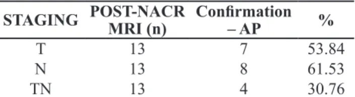
the aP study showed compliance in relation to t staging of 53.84% (7/13) estimated in the post-neoad-juvant therapy MRi, with tendency to overstaging of 83.33% (5/6) in the others. Regarding the lymph node status, the aP study agreed with the post-NaCR MRi in 61.53% (8/13). When assessing associated t and N staging, the compliance was 30.76% (4/13); also with tendency to overstaging of 55.55% (5/9) in the other patients (table 2).
in the follow-up period of the 13 patients, one patient died due to postoperative complications
(staged patient, T3N0 at the irst examination, with
he-patic metastases (one patient with metastases at the diagnosis, t3N2, with tumor regression estimated as degree 3, t3N0 at the second MRi and the aP study indicating txN1; and another t3N2 patient, of
tu-mor regression classiied as Degree 1, who remained
t3N2 at the post-NaCR MRi and the aP study show-ing t2N2). the nine other patients (69.23%) have had the disease under control so far, without recur-rence and/or metastases.
Regarding the two patients that presented local and/or distant recurrence, 1 (50%) showed tumor re-gression at the second MRi. and only one out of the nine patients showing tumor regression with reduction at staging presented recurrence (table 3).
DISCUSSION
MRi with emphasis on the rectum can classify rectal tumors according to prognostic factors and as-sess t and N staging with 85-90% accuracy3,6. For this reason, it enables a better surgical planning, show-ing the points of high vulnerability durshow-ing mesorectal dissection to the surgeon and leading to a lower rate of involvement of circumferential resection margins,
which is an important prognostic factor of local recur-rence and survival1,5.
this study shows initial observations indicating that the post-neoadjuvant therapy MRi could not es-timate the reduction at the post-neoadjuvant therapy staging, showing compliance in relation to the aP
Patient NACR MRIPRE- NACR MRIPOST- DWORAK r AP CRM Time to surgery Clinical status
1 aFS t2N2 t2N0 2 t2N0 Free 1-2 YeaRS No recurrence
2 agF t4bN2 t4bN1 3 t4N0 Free 1-2 YeaRS No recurrence
3 BNt t3aN0 t2N0 2 t2N0 Free 1-2 YeaRS No recurrence
4 dt t3bN0 t0N0 4 t0N1 Free 1-2 YeaRS No recurrence
5 dPC t3dN1 t3dN0 1 t3N2 Free >2 YeaRS No recurrence
6 KaS t2N0 t2N0 1 t1N1 Free >2 YeaRS No recurrence
7 MLd t3N0 t3N0 2 t2N0 Free 1-2 YeaRS death – surgical
complications
8 oaS t3N2 t3N2 1 t2N2 Free 1-2 YeaRS Resected hepatic Mtx
9 oRM t3bN0 t0N0 2 t2N0 Free <1 YeaR No recurrence
10 PFM t3aN0 t1N0 3 t1N0 Free 1-2 YeaRS No recurrence
11 Sh t3bN2 t3N0 3 t0N1 Free <1 YeaR Resected hepatic
metastases
12 tJLJ t3aN0 t3aN0 2 t0N0 Free 1-2 YeaRS No recurrence
13 vF t3cN2 t3cN0 3 t3N0 Free >2 YeaRS bone metastases – deathRecurrence + Lung and Table 1. Clinical characteristics of patients.
AP: anatomopathological study; CRM: circumferential resection margin; Mtx: metastases; MRI: magnetic resonance imaging; NACR: neoadjuvant chemoradiation.
STAGING POST-NACR MRI (n) Conirmation – AP %
t 13 7 53.84
N 13 8 61.53
tN 13 4 30.76
Table 2. Compliance index of the anatomopathological analysis in relation to the condition indicated in post-neoadjuvant therapy MRI.
AP: anatomopathological study; MRI: magnetic resonance imaging; NACR: neoadjuvant chemoradiation.
Recurrence Yes No Tumor
Regression
Yes 1 8
No 1 1
study of 30.76%. the other patients presented tenden-cy to overstaging. according to Barbaro et al., MRi
sensitivity and speciicity are approximately 80%, with
tendency to overtsaging6. When MRi shows staging higher than the aP results, it does not affect the onco-logic surgical quality, as the surgeon tends to consider a wider resection margin, for a free CRM. if, after the resection, the actual staging is lower than the value estimated in MRi, the non-involvement of margins is kept. this overstaging tendency occurs especially due
to the dificult differentiation of initial T2 to T3, and
the radiologist tends to classify as overstaging. in
ad-dition, post-neoadjuvant therapy ibrosis makes this
differentiation between tumor and cicraticial tissue
more dificult, which favors overstaging. When try -ing to avoid an undertreated patient, in an oncologic perspective, overstaging occurs in case of any doubt, leading to a more aggressive treatment, with higher morbidity, but oncologically adequate. in neoadjuvant NaCR, precise staging to assess the response to treat-ment is very important, as it can guide through sur-gical approach optimization, such as sphincter pres-ervation in low tumors, less aggressive resection of initially advanced tumors or intraoperative radiation therapy, according to the tumor response6,7.
NaCR results in reduced number and size of both benign and malign mesorectal lymph nodes. dow Mu-Koh et al.3 report that NaCR is useful in the assessment of lymph node response to neoadjuvant treatment, with 88% accuracy, but it is uncertain in terms of how much this response can be translated into survival3,8. the compliance found in lymph node sta-tus after NaCR was 61.53%. in the evaluation of me-sorectal lymph nodes, the utilization of morphological criteria (outline irregularity and sign heterogeneity) offers improved accuracy than the size to distinguish malign from non-malign lymphatic ganglia.
three patients (23.07%) presented complete pathological response, without evidence of tumor
tis-sue in the specimen, only ibrosis and inlammatory
alterations. only 1 (33,33%) of them presented post-NaCR MRi suggesting complete remission. in an-other case, MRi indicated complete remission, but the aP study showed neoplastic tissue. in the evaluation of complete pathological response, MRi presented the positive predictive value of 50.0% and the negative predictive value of 83.3%.
Pre-NACR MRI inluenced the proper surgi -cal planning, especially in larger tumors, resulting in 100% free CRM, which improves the prognosis, since a compromised CRM leads to local recurrence rate of 83.0%6.
Regarding the prognosis, two deaths occurred. one patient died of postoperative complications and one patient after the disease recurrence. Survival in this mean follow-up period of 1 to 2 years was 91.7% and 75.0% are free from the disease. these are initial observations and require longer follow-up periods for a better survival evaluation.
the patients with worse response, who pre-sented local recurrence or distant metastases, were those with tumors in advanced stage. at pre-NaCR
MRI, all patients were staged as T3N2, coniguring
a worsened prognosis, regardless of the response to NaCR when evaluated through MRi or the aP study.
The irst, with tumor recurrence and distant metas -tases diagnosed after 20 months, was t3N2, with
tumor regression classiied as Degree 3, but with
t3N0 staging after NaCR and the aP study indi-cated t3N0, and died six months after the salvage surgery due to recurrence. the second, with hepatic metastasis diagnosed when the clinical condition
ap-peared, was T3N2, with regression classiied as De -gree 3, post-NaCR MRi indicating t3N0 and the aP study indicating txN1, is now well, after metastasis resection, receiving clinical and oncologic follow-up care for seven months. the third, was t3N2, with re-gression degree 1, post-NaCR MRi still indicating t3N2 and the aP study showing t2N2, was submit-ted to hepatic metastasis resection and is receiving follow-up care. these patients, even after the lesion size reduction, with tumor regression after NaCR, still showed advanced staging at the second MRi (all were t3 and one was N2). the aP study of two pa-tients showed lymph node involvement despite the tumor regression and one showed no tumor regres-sion at the degree of wall invaregres-sion, only tumor size reduction (leading to patient’s death). the aP study
conirmed T staging with poor response indicated at
MRi in all these three patients.
degrees 2, 3 and 4. even a patient who was t4N2, presenting good response at NaCR and whose MRi indicated degree 3 regression, with the aP study showing t4N0, has the disease now under control, in 2-year follow-up. although the second MRi tends to overstage the lesion, these patients who were good responders, according to the radiologic criteria, and
later conirmed by the AP study, or the patients who
presented better pathological response, are those without disease recurrence.
CONCLUSION
our results indicate that the second MRi after the neoadjuvant therapy can show tumor regressions, if any, but it is of little use in the determination of downstaging, when compared to the aP study, tending to overstaging. Patients with tumors in advanced phases, lymph node involvement and poor response as evaluated through MRi, tend to show worsened prognosis. Further studies
are required to conirm these irst observations.
RESUMO: Introdução: A radioquimioterapia neoadjuvante promove redução do tamanho e do estadiamento dos tumores do reto antes da cirurgia, reduzindo o risco de acometimento de margem de ressecção circunferencial e da recorrência local. Para pacientes que se submeteram a neoadjuvância, a realização de uma segunda ressonância magnética (RNM) após a radioquimioterapia, para avaliação do resultado do tratamento, pode trazer dados relevantes para a programação cirúrgica e previsão do prognóstico, porém sua utilização ainda é controversa. Objetivo: Avaliar a capacidade da RNM prever o grau de regressão tumoral e o downstaging ob-tidos e a correlação entre o grau de regressão tumoral com o prognóstico dos pacientes. Métodos: Foram incluídos 13 pacientes até o momento; desses 69,23% eram do sexo masculino e a idade média foi de 52,3 anos. Resultados: O anatomopatológico (AP) mostrou conformidade em relação ao estadiamento T e N estimado pela RNM pós-neoadjuvância de 30,76%; nos demais pacientes, houve tendência ao superestadiamento em 55,55%. No estadiamento T houve concordância de 53,84% e quanto ao status linfonodal, concor-dância 61,53%. A margem de ressecção circunferencial foi livre de neoplasia em 100%. A sobrevida foi de 92%, com 75% de sobrevida livre de doença num seguimento médio de 1-2 anos. Conclusão: Uma segunda ressonância após neoadjuvância pode avaliar se houve regressão tumoral, porém com baixa conformidade em relação ao anatomopatológico, com tendência ao superestadiamento. Mais estudos são necessários para corroborar essas impressões iniciais.
Palavras-chave: imagem por ressonância magnética; câncer de reto; terapia neoadjuvante.
REFERENCES
1. Kulkarni t, gollins S, Maw a, hobson P, Byrne R, Widdowson d. Magnetic resonance imaging in rectal cancer downstaged using neoadjuvant chemoradiation: accuracy of prediction of tumour stage and circumferential resection margin status. Colorectal dis 2008;10:479-89.
2. Birbeck KF, Macklin CP, Tifin NJ, Parsons W, Dixon MF,
Mapstone NP, et al. Rates of circumferential resection margin involvement vary between surgeons and predict outcomes in rectal cancer surgery. ann Surg 2002;235:449-57.
3. dow-Mu K, Chau i, tait d, Wotherspoon a, Cunningham d, Brown g. evaluating mesorectal lymph nodes in rectal cancer before and after neoadjuvant chemoradiation using thin-section t2-weighted magnetic resonance imaging. int J Radiat oncol Biol Phys 2008;71:456-61.
4. o’Neill BdP, Brown g, heald RJ, Cunningham d, tait dM. Non-operative treatment after neoadjuvant chemoradiotherapy for rectal cancer. Lancet oncol 2007;8:625-33.
5. Burton S, Brown G, Daniels I, Norman A, Swift I, Abulai M, et al. MRI identiied prognostic features of tumors in distal
sigmoid, rectosigmoid, and upper rectum: treatment with
radiotherapy and chemotherapy. int J Radiat oncol Biol Phys 2006;65:445-51.
6. Barbaro B, vitale R, Leccisotti L, vecchio FM, Santoro L, valentini v, et al. Restaging locally advanced rectal cancer with MR imaging after chemoradiation therapy. Radiographics 2010;30:699-721.
7. Barbaro B, Fiorucci C, tebala C, valentini v, gambacorta Ma, vecchio FM, et al. Locally advanced rectal cancer: MR imaging in prediction of response after preoperative chemotherapy and radiation therapy. Radiology 2009;250:730-9.
8. Koh dM, Brown g, temple L, Raja Pa, toomey P, Bett N, et al. Rectal cancer: mesorectal lymph nodes at MR imaging
with USPIO versus histopathologic indings — initial
observations. Radiology 2004;231:91-9.
Correspondence to:
Rodrigo Soares Napoleão do Rêgo
Serviço de Coloproctologia do hospital Felício Rocho avenida do Contorno, 9530, Barro Preto