Arq Bras Cardiol 2004; 82: 191-3.
Pêgo-Fernandes et al
Left ventricular lipoma
1 9 1
Instituto do Coração do Hospital das Clínicas - FMUSP
Mailing address: Paulo Manuel Pêgo-Fernandes - InCor - Divisão Cirúrgica Av. Dr. Eneas C. Aguiar, 44 - 2º andar - Cep 05403-000 - São Paulo, SP, Brazil E-mail: [email protected]
Received 12/30/02 Accepted 4/29/03
English version by Stela Maris C. e Gandour
Arq Bras Cardiol, volume 82 (nº 2), 191-3, 2004
Paulo Manuel Pêgo-Fernandes, Carlos Alfredo Batagello, Fábio Fernandes, Fabio Biscegli Jatene, Sérgio Almeida O liveira
São Paulo, SP - Brazil
Left Ventricular Lipoma
Case Report
We report the case of a 21-year-old female referred to our institution complaining of dizziness when standing up, which improved in the dorsal decubitus position and at rest, after a few minutes. The symptom, which had lasted for years, was not accompanied by vertigo, syncope, or neuro-logical changes, but was gradually getting worse. Trans-thoracic echocardiogram showed a hyperechoic mass in the middle-apical region of the left ventricular posterior wall and normal pericardium. The cardiac nuclear mag-netic resonance allowed the diagnosis of the left ventricu-lar tumor suggestive of lipoma. Surgery was performed and the tumor was resected. The patient recovered well and is currently asymptomatic.
Primary cardiac tumors account for 5% to 10% of all neoplasias of the heart and pericardium, with an incidence in autopsies ranging from 0.0001% to 0.05% 1. Approximately 75% of the primary cardiac neoplasias are benign, 40% being myxomas and most of the remaining being lipomas, papillary fibroelastomas, and rhabdomyomas 1,2. Accor-ding to Fernandes et al 3, most tumors are located on the left side of the heart, myxoma being the most frequent histologi-cal type.
With the appearance of modern techniques of diag-nostic and surgical procedures, cardiac neoplasias have been diagnosed earlier, having, therefore, a greater chance of cure. The modern era of diagnosis began with the deve-lopment of angiography, which allows the visualization of cardiac tumors in vivo. Goldberg et al 4 reported the first an-giographic diagnosis of left atrial myxoma, and Crafoord 5 performed the first successful excision of an intracardiac tumor, a left atrial myxoma, using total extracorporeal circu-lation under direct visualization. Since then, successful
sur-gical excision of several cardiac tumors has become possi-ble, complete cure being obtainedin many cases 2.
Our case report illustrates the experience of the Surgi-cal Division of InCor of the Hospital das Clínicas of the Me-dical School of the University of São Paulo in the treatment of a rare cardiac tumor, the left ventricular lipoma. Kosuru et al 6, in a review of the literature, reported only 6 cases of lipo-ma in the interventricular septum.
Case report
1 9 2
Pêgo-Fernandes et al
Left ventricular lipoma
Arq Bras Cardiol 2004; 82: 191-3.
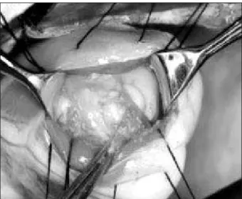
slightly elevated (29 mm) and the hemoglobin level was bor-derline (11.6 g/dL). Her hematocrit was 35%, serum leukocytes were 4,700 mm3, and platelets were 250.000 mm3. Her chest X-ray showed no alterations. The transthoracic echocardiogram showed the following characteristics: normal-sized left ven-tricular cavity (50 x 34 mm), delta D 32%, aorta 33 mm, left atrium 32 mm, mitral valve escape, and a hyperechoic mass measuring 45 x 33 mm in the middle-apical region of the left ventricular posterior wall with normal pericardium. The eso-phageal echocardiogram showed an intact interventricular septum with a 41 x 31mm hyperechoic mass attached to the middle-apical region with hyperrefringent spots inside. The computed tomography of the brain suggested ethmoidal si-nusopathy and no encephalic lesions. Nuclear magnetic re-sonance of the heart provided the diagnosis of cardiac tumor suggestive of lipoma due to the reduction in the magnetic sig-nal in the triple R sequence. The tumor measured 34 x 28 mm and was located in the inferior wall, close to the implantation of the posteromedial papillary muscle occupying part of the left ventricular cavity (fig. 1). Surgery was performed on 10/ 15/2001 via median sternotomy with extracorporeal circu-lation through cannucircu-lation of the aorta and venae cava, with hypothermia at 30 degrees and cardioplegia at 4 degrees. The ventricular cavity was approached through the left ven-tricular anterior wall, close to the apex (fig. 2), and the tumor, which looked like a lipoma (fig. 3) with a mild adherence to the left ventricular free wall and papillary muscle, was resected. The operation was uneventful. The patient was discharged from the anesthetic recovery room on the 2nd postoperative day. The esophageal echocardiogram in the immediate posto-perative period showed preserved cavity and function, mild to moderate mitral insufficiency, mild tricuspid insufficiency, and no intracardiac lesion. The patient has been followed up on an outpatient care basis, with no complaint of dyspnea, fatigue, dizziness, or any other cardiovascular symptom.
Discussion
Cardiac lipomas occur at any age and with the same frequency in both sexes. Most of them range from 1 to 15 cm in diameter, although tumors weighing more than 2 kg have been reported. Most tumors are sessile or polypoid and lo-cated in the subendocardium or subepicardium, although approximately 25% are completely intramuscular. The su-bendocardial tumors with intracavitary extension cause symptoms characteristic of their location, while the subepi-cardial tumors may cause compression of the heart and pe-ricardial effusion. The most commonly affected sites are the left ventricle, the right atrium, and the interatrial septum. In-tramural tumors may be asymptomatic or cause arrhyth-mias7, intraventricular or atrioventricular conduction disor-ders, or mechanical interference. Many tumors are clinically silent, being found only during necropsy or on routine chest X-rays 2. In our case, the imaging media were very helpful in the diagnosis of lipoma, as they were for Izumi et al8, whose infiltrative lipomatous tumor was suggested by the results of the echocardiogram, computed tomography,
Fig. 1 - Nuclear magnetic resonance - sagittal view - showing the tumor.
Fig. 2 - Surgical picture showing the open left ventricle and the lipoma being dissected.
Arq Bras Cardiol 2004; 82: 191-3.
Pêgo-Fernandes et al
Left ventricular lipoma
1 9 3
1. Miralles A, Bracamonte L, Souncul H, et al. Cardiac tumors: clinical experience and surgical results in 74 patients. Ann Thorac Surg 1991;52:886-95. 2. Colucci WS, Braunwald E. Tumores primários do coração. In: Tratado de
Medici-na Cardiovascular. p. 1551-63.
3. Fernandes F, Soufen HN, Ianni BM, Arteaga E, Ramires FJA, Mady C. Neoplasias primárias do coração: apresentação clínica e histológica de 50 casos. Arq Bras Cardiol 2001;76:231-4.
4. Goldberg HP, Glenn F, Dotter CT, Steinberg I. Myxoma of the left atrium: diagno-sis made during life with operative and postmortem findings. Circulation 1952;6:762-6.
5. Crafoord CL. Case report. In: Lam CR, editor. Proceedings. International Symposium on Cardiovascular Surgery. Philadelphia: WB Saunders, 1995: 202.
6. Kosuru SB, Mundayat G, Ramachandran M, Satyaprasad V. Lipoma of left ventri-cle. Asian Cardiovasc Thorac Ann 2002;10:64-5.
References
7. Hueb WA, Ramires JAF, Bellotti G, et al. Mixomas ventriculares e arritmias car-díacas: relato de 2 casos. Arq Bras Cardiol 1986;46:259-62.
8. Izumi T, Matsuoka A, Nagai K, et al. Massive lipomatous infiltration to the left ventricle mimicking a cardiac tumor. Jpn Heart J 1986;27:273-7.
9. Morikami Y, Higashi T, Isomura T, et al. Cardiac lipoma with changes of ST seg-ment and T wave on eletrocardiogram. Jpn Circ J 1994;58:733-6.
10. Silveira W L, Nery MW, Soares ECG, et al. Lipoma de átrio direito. Arq Bras Car-diol 2001;77:361-4.
11. Bradford JH, Nomier AM, Watts LE. Left ventricular lipoma: echocardiographic and angiographic features. South Med J 1980;73:663-5.
12. Centofanti P, Rosa ED, Deorsola L, et al. Primary cardiac tumors: early and late re-sults of surgical treatment in 91 patients. Ann Thorac Surg 1991;68:1236-41. 13. Kaza AK, Buchanan SA, Parrino P, Fiser SM, Long SM, Tribble CG.
Cardiosco-pe-assisted excision of a left ventricular tumor: a case report. Heart Surg Forum 2002;5:75-6.
thallium scintigraphy, and right ventriculography. Morikami et al 9 reported the presence of a cardiac lipoma based on the electrocardiographic alterations in the ST-T segment, sug-gestive of left ventricular hypertrophy. Silveira et al 10 diag-nosed a right atrial lipoma through computed tomography, which has a high specificity in identifying the tumor. Lipo-mas usually have low density ranging from -80 to 115 Hounsfield units 10.
Microscopically, the lesions are usually well encapsula-ted, composed of typical mature fat cells, and may occasional-ly have fibrous connective tissue (fibrolipoma), muscle tissue (myolipoma), or brown vacuolated fat similar to a hibernoma. Intraventricular lipoma was first described and succes-sfully removed by Bradford et al 11. Surgical excision, whe-never possible, is the treatment of choice for all primary car-diac tumors 12. Most patients with benign tumors are cured with resection and the tumors do not recur. Palliative treat-ment may be used for malignant tumors, but adjuvant the-rapies are required to improve the patient's prognoses 12. The great problem of the benign cardiac tumor does not reside in its histological characteristic, but in its intracavitary com-ponent, when the tumor invades cardiac cavities. The tumor becomes potentially lethal when it occupies the left ventri-cular cavity, as in our patient’s case, because it may alter car-diac output, which is clinically indicated by syncope 7, or may
simulate left ventricular insufficiency, peripheral embolism, and rhythm and conduction disorders. Therefore, surgery is mandatory after the determination of a diagnosis.
Although some epicardial tumors may be removed without the aid of extracorporeal circulation, most intramural and intracavitary tumors should be excised under direct vi-sualization with the use of artificial circulation, because, technically, it reduces cardiac manipulation and maneuvers, which may lead to release of parts of the tumor, causing em-bolisms. Kaza et al 13 excised a left ventricular lipoma with the aid of a video-assisted cardioscope inserted through the aortic valve through an opening in the aorta.
The major surgical considerations in the excision of ventricular tumors include the preservation of an adequate portion of the ventricular myocardium, maintenance of ade-quate atrioventricular valvular function, and preservation, as much as possible, of the conduction system.