110 Arq Gastroenterol v. 40 – no. 2 – abr./jun. 2003
REL
AT
O
DE
CA
SO
S
/ C
ASE
REPO
RT
S
MAGNIFYING ENDOSCOPY OF THE
DUODENUM WITH DYE
SCATTERING METHOD IN A CASE
WITH CELIAC DISEASE
Tetsuo MORISHITA1
, Toshiaki KAMIYA2
and Hiromasa ISHII3
ABSTRACT – Aim – To know the more detailed findings of the small intestinal mucosa with the use of a magnifying endoscope and a vital dye, and the efficacy of the both tools. Patient and Methods – A 54-year old female patient with celiac disease. The duodenal mucosa downward as far as the descending portion was observed with a magnifying endoscope (Olympus GIF HM) before and after spraying the mucosa with 0.1% indigo carmine. Results – The endoscopy clarified the atrophy and edema of each villus, and scattering of the dye revealed shorter villi with the relatively longer villi remaining in islands. Conclusion – The combination of magnifying endoscopy and the dye scattering method is useful for closer observation of the intestinal mucosa in celiac diseases.
HEADINGS – Duodenoscopy. Duodenoscopes. Dyes. Celiac disease.
ARQGA / 1067
1
Department of Internal Medicine, Tokyo Dental College, Chiba, Japan; 2Department of Gastroenterology, School of Medicine, Christian University of
Bolivia, Santa Cruz, Bolivia; 3Department of Internal Medicine, School of Medicine, Keio University, Tokyo, Japan
Address for correspondence: Tetsuo Morishita, MD – Department of Internal Medicine – Tokyo Dental College – 5-11-13 Sugano, Ichikawa-shi, Chiba - 272-8513 – Japan. e-mail: [email protected]
INTRODUCTION
There have been several papers on the effectiveness of conventional duodenal endoscopy in the diagnosis of celiac diseases(1, 2, 5, 10, 11, 14). Availability of the magnifying
endoscope(12, 13) and the dye scattering method(7) have made
it possible to observe the mucosa in intricate detail. This paper suggests the efficacy of the combination of magnifying endoscopy and the indigo carmine scattering method in a patient with celiac disease.
CASE REPORT
A 54-year-old white female from Canada, who had been living in Shizuoka, Japan for 2 years, complained of watery diarrhea three times a day for the previous month. She showed significant weight loss, 4 kg over a month, and felt exhausted. Although she denied ever having suffered from any diarrheal disease as a child, she developed intractable diarrhea immediately after giving birth at the age of 26 to
v. 40 – no. 2 – abr./jun. 2003 Arq Gastroenterol 111 Barium examination of the small intestine revealed the so-called
“malabsorption pattern”, i.e. dilatation of the small intestine, thickening of the mucosal folds, segmentation of the ileum, and flocculation of the barium.
Magnifying endoscopy of the upper gastrointestinal tract was performed following the patient’s informed consent. After the patient fasted, the duodenal mucosa was visualized through a magnifying fiberendoscope (Olympus GIF HM)(12, 13), inserted downward as far as
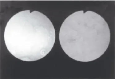
the descending portion of the duodenum. Without magnification, the entire bulbus showed a flat mucosal pattern with reddened spots located immediately below the pylorus ring (Figure 1 left). The mucosa showed a mosaic pattern with edema and a reduction in number of Kerckring’s folds in the descending portion (Figure 1 right). Under lower magnification, about x20, the mucosa showed unevenness of its surface with minute nodules (Figure 2). Under higher magnification, the villi seemed to be fused with each other (Figure 3 left, about x30) and each
villus was flat and edematous with an indistinct margin (Figure 3 right, about x40). After spraying the mucosa with 0.1% indigo carmine, good contrast was obtained in the mucosal unevenness. The mucosa was extensively flattened; the shorter villi lay flat while the relatively longer villi remained in islands (Figure 4, about x10).
FIGURE 1 – The endoscopy without magnification shows a flat mucosa with reddened spots in the bulbus (left) and a mosaic pattern with edema and decreased Kerckring’s folds in the descending portion of the duodenum (right).
FIGURE 2 – Unevenness of the mucosal surface with minute nodules observed with magnification x20.
Morishita T, Kamiya T, Ishii H. Magnifying endoscopy of the duodenum with dye scattering method in a case with celiac disease
FIGURE 4 – The relatively longer villi remain in islands in the sea of the dye over the shorter villi after spraying of indigo carmine (x10).
FIGURE 3 – The villi appear to be fused (left, x30) and each villus was flat and edematous (right, x40).
112 Arq Gastroenterol v. 40 – no. 2 – abr./jun. 2003
FIGURE 5 – Biopsy specimen reveals subtotal villous atrophy with cellular infiltrate (5A, x100) and cuboidal epithelial cells and intraepithelial lymphocytes (5B, x200).
Morishita T, Kamiya T, Ishii H. Magnifying endoscopy of the duodenum with dye scattering method in a case with celiac disease
A B
Further evaluation disclosed an increased level of antigliadin antibodies of IgG and IgA isotypes. Tissue typing in the patient detected the HLA antigens B8, DR3 and DR7.
The patient was diagnosed as having celiac disease, and was immediately placed on a strict gluten-free diet. Within a week her bowel habits improved, and she began to regain her energy. Her laboratory results returned toward normal. Her weight increased dramatically, and she gained 12 kg in 5 months.
DISCUSSION
Celiac disease is one of the major cause of malabsorption in the west. The prevalence is 1:250-300 in western countries(15). In contrast
with the high incidence of the disease among whites, there have been very few cases among the Japanese. Six cases of celiac disease have been reported by endoscopists(4, 6, 8, 16, 17, 20) in Japan.
Celiac disease is now understood to be a disorder with a wide range of presenting manifestations of variable severity, as well as the classic malabsorptive symptoms of diarrhea and weight loss. Over 50% of patients with celiac disease have no gastrointestinal symptoms. Less than 50% present with diarrhea. One fifth are underweight and 1/3 are overweight. Glossitis, angular stomatitis, dental enamel defects, hair loss, calcium and vitamin D malabsorption, neurologic syndromes, reduced fertility and anemia have been reported in patients with celiac disease.
Celiac disease is associated with IgA deficiency, diabetes mellitus, thyroid disease, dermatitis herpetiformis, primary biliary cirrhosis and autoimmune hepatitis(15).
Endoscopy of the duodenum and endoscopic biopsy are useful tools for the diagnosis of celiac disease. The endoscopic markers of the small intestine, such as loss or decrease of duodenal folds, a mosaic pattern, scalloping of the folds in the descending portion(1, 5, 10, 11, 14) and
multiple lesions with a prominent and roundish aspect in the bulb(2)
have been identified.
In this paper, we reported on the magnified endoscopic images of the duodenum in addition to staining with a dye.
Magnifying endoscopy revealed the atrophy and edema of each villus, showing flat and uneven mucosa with minute nodules. Only one report has appeared on the use of magnifying endoscopy of the intestinal tract in celiac disease. HIRATSUKA et al.(4)reported on the
mucosal changes of the terminal ileum using the Machida magnifying endoscope for the small intestine, FIS-MS III (maximum magnification x30, depth of field 2 mm~∞ ). They described the fusion of atrophic villi forming edematous sausage-like protrusions. It is not yet known if our magnifying endoscopic findings are typical or not for celiac disease, because they have not been described in other diseases. GILLBERG and AHRÉN(3)observed the duodenal villous atrophy on
a close-up view with an Olympus GIF type D2 (depth of view field 5 mm∞ by reducing the focal length of the viewing lens. No paper on the utility of magnifying endoscopy for the early diagnosis of celiac disease has been reported.
The indigo carmine dye scattering method(7) made clearer the
mucosal change observed with the magnifying endoscopy, especially the unevenness. STEVENS and McCARTHY(18) reported that severe
atrophy of the mucosal surface could be demonstrated throughout the duodenal cap using indigo carmine scattering with an Olympus GIF type D2 endoscope.
Magnifying endoscopy is useful for the observation of fine mucosal patterns and the detection of minute mucosal lesions(12, 13). By
v. 40 – no. 2 – abr./jun. 2003 Arq Gastroenterol 113 the method may make the early detection of mucosal changes possible,
and present a morphological classification of the changes. For example, the diagnostic classification of colorectal tumorous lesions was made based on the pit patterns of the mucosa seen with magnifying colonoscopy coupled with indigo carmine and cresyl violet staining(20).
The combination of the magnifying and dye scattering methods is effective for more-advanced diagnostic study of the intestinal mucosa in patients with celiac disease.
Morishita T, Kamiya T, Ishii H. Magnifying endoscopy of the duodenum with dye scattering method in a case with celiac disease
Morishita T, Kamiya T, Ishii H. Endoscopia com magnificação e coloração por dispersão no duodeno em paciente com doença celíaca. Arq Gastroenterol 2003;40(2):110-113.
RESUMO – Objetivos – Ampliar os achados na mucosa intestinal com o uso da endoscopia com magnificação e coloração ao vivo e verificar a eficácia de ambos os métodos. Paciente e Métodos – Paciente de 54 anos, do sexo feminino, com doença celíaca. Observou-se a mucosa duodenal mais distal possível com endoscópio com magnificação (Olympus GIF HM), antes e depois da dispersão por jato de índigo carmim a 0,1%. Resultados – A endoscopia acentuou a atrofia e o edema dos vilos, e a dispersão do corante revelou vilos longos, remanescentes em ilhas, em meio a vilos encontrados.Conclusão – A combinação da endoscopia com magnificação e a coloração por dispersão mostrou-se método útil para observação próxima da mucosa intestinal na doença celíaca.
DESCRITORES – Duodenoscopia. Duodenoscópios. Tinturas. Doença celíaca.
ACKNOWLEDGEMENT
The authors are indebted to late Dr. Masaharu Tsuchiya (Keio University, Japan) and Dr. Hitoshi Asakura (Niigata University, Japan) for their guidance, and to Dr. Koushi Ando (Hamamatsu Red Cross Hospital, Japan), Dr. Atsuko Morishita (Tokyo Dental College Ichikawa General Hospital, Japan) and Ms. Wendy Lenner (Calgary, Canada) for their assistance.
REFERENCES
1. Brocchi E, Corazza GR, Caletti G, Treggiari EA, Barbara L, Gasbarrini G.
Endoscopic demonstration of loss of duodenal folds in the diagnosis of celiac disease. N Eng J Med 1988;319:741-4.
2. Brocchi E, Corazza GR, Brusco G, Mangia L, Gasbarrini G. Unsuspected celiac
disease diagnosed by endoscopic visualization of duodenal bulb micronodules. Gastrointest Endosc 1996;44:610-1.
3. Gillberg R, Ahrén C. Coeliac disease diagnosed by means of duodenoscopy and
endoscopic duodenal biopsy. Scand J Gastroenterol 1977;12:911-6.
4. Hiratsuka H, Gocho K, Tanaka M. Examination of the magnified mucosal surface
and pathological changes of the human intestine. Stomach and Intestine, 1978;13:615-24.
5. Jabbari M, Wild G, Goresky CA, Daly DS, Lough JO, Cleland DP, Kinnear DG.
Scalloped valvulae conniventes: an endoscopic marker of celiac sprue. Gastroenterology 1988;95:1518-22.
6. Kihara T, Kukida S, Ichikawa Y. Ultrastructual studies of the duodenal epithelium
of Japanese celiac sprue. J Clin Electron Microscopy 1977;10:383-4.
7. Kohli Y, Nakajima M, Ida K. Minute endoscopic findings of duodenal mucosa
using the dye scattering method. Endoscopy 1974;6:1-6.
8. Kosaka K, Kihara T, Ueda K. Per oral biopsy of the intestinal mucosa. Naika
1961;7:1061-6.
9. Kudo S, Tamura S, Nakajima T, Yamano H, Kusaka H, Watanabe H. Diagnosis
of colorectal tumorous lesions by magnifying endoscopy. Gastrointest Endosc 1996;44:8-14.
10. Magazzu G, Bottani M, Tuccari G, Bottari M, Tuccari G, Arco A, Pallio S, Lucanto
C, Tortora A, Barresi G. Upper gastrointestinal endoscopy can be a reliable screening tool for celiac sprue in adults. J Clin Gastroenterol 1994;19:255-8.
11. Mauriño E, Capizzano H, Niveloni S, Kogan Z, Valero J, Boerr L, Bai JC. Value
of endoscopic markers in celiac disease. Dig Dis Sci 1993;38:2028-33.
12. Morishita T, Asakura H, Miura S. Magnifying endoscopic findings of the
duodenum in a case of intestinal lymphangiectasia. Gastroenterol Endosc 1981;23:133-41.
13. Morishita T, Kamiya T, Akagi K. Magnifying endoscopy of the upper
gastrointestinal tract in Japan and its application in olive oil tolerance test. Acta Gastroenterol Bol 1982;2:115-20.
14. Oderda G, Forni M, Morra I, Tavassoli K; Pellegrino P; Ansaldi N. Endoscopic
and histologic findings in the upper gastrointestinal tract of children with celiac disease. J Pediatr Gastroenterol Nutr 1993;16:172-7.
15. Olds G, McLoughlin R, O’Morian C, Sivak MV. Celiac disease for the
endoscopist. Gastrointest Endosc 2002;56:407-15.
16. Ooba T, Doi T, Kobayashi H. A case suspected of celiac disease [abstract].
Nippon Shokakibyo Gakkai Zasshi 1984;81Suppl:2473.
17. Sakazaki S, Toudou T, Kitamura T. A case of chronic intestinal pseudo-obstruction
(CIP) syndrome suspected of celiac sprue. Nippon Shokakigeka Gakkai Zasshi 1983;16:2129-33.
18. Stevens FM, McCarthy CF. The endoscopic demonstration of celiac disease.
Endoscopy 1976;8:177-80.
19. Tada M, Misaki F, Kawai K. Endoscopic observation of villi with magnifying
enteroscopes. Gastrointest Endosc 1982;28:17-9.
20. Yamagata S, Ishikawa M, Yamagishi G. Case of primary sprue followed by
reticulosarcoma. Gastroenterol Jap 1966;1:1-4.